X-ray of the sinuses for sinusitis
An examination is prescribed as soon as the first signs of inflammation appear. Its goal is to assess how affected the nasal cavity is and to find out what caused the sinusitis. For adult patients, the basis for prescribing an examination is:
- The appearance of unpleasant sensations in the nasal cavity after an infectious disease.
- Headaches that get worse with every movement of the head.
- Chronic runny nose, bleeding.
- Deterioration of sense of smell.
- Increased body temperature, lacrimation, photophobia.
All these symptoms may indicate the onset of inflammation. In addition to sinusitis, x-rays make it possible to determine ethmoiditis, frontal sinusitis, sphenoiditis, as well as to identify a curvature of the nasal septum, the appearance of tumor and cystic formations, and purulent-necrotic processes in bone tissue.
To get the most informative picture of sinusitis, two projections are chosen for shooting: direct and lateral. When laying in the first case, the chin and nose are placed at the same level. When shooting using the second method, they are placed at an angle. Sometimes it may be necessary to check the condition of the sinuses while lying down. The radiologist will be able to get the result and describe the sinusitis in the image within a few minutes.
No preliminary preparation is required before the procedure. The patient will be advised to remove all objects and jewelry from the area being examined.
The procedure is contraindicated during pregnancy, since radiation has a negative effect on the fetus. Research may only be permitted if absolutely necessary. In this case, the woman must be provided with additional protection. When breastfeeding, the test can be done at any time.
The procedure is prescribed with caution to children and patients with oncology. This type of research is not recommended if the patient has metal-containing dentures or other foreign objects in the area being examined. Due to their ability to reflect rays, test results may be distorted.
Sinusitis on an x-ray with a description
The interpretation of the radiograph is carried out only by a specialist radiologist or doctor monitoring the patient. If there are no deviations in the image, the following will be displayed:
- Nasal cavity. It looks like a light triangle with a partition in the middle.
- The nasal passages have the appearance of shadows located symmetrically on both sides of the nasal cavity.
- Maxillary sinuses. They are displayed in the form of triangular clearings with clear outlines located on the side of the nasal cavity.
- Frontal sinuses. They appear above the eye sockets as clear spots of different shapes and sizes.
Thanks to the features that each image has, it is possible to recognize the nature of the inflammation and determine its location:
- The appearance of dark spots above the nose indicates the development of inflammation in the frontal sinuses.
- Darkening in several zones will indicate an inflammatory process in several places.
- Round-shaped shadows with extending threads will indicate the presence of polyps.
- Allergies are manifested by additional darkening of varying intensity.
- Catarrhal inflammatory processes of a chronic nature are depicted as thickening of mucous tissues.
Signs of sinusitis on x-ray
The accumulation of purulent contents in the sinuses during sinusitis will look in the photo as a light spot against the background of a black sinus. If the content is large, you can see its horizontal border.
The image of healthy sinuses looks like dark semi-oval niches, which are located on both sides of the nose. Their colors and eye sockets match. This situation indicates the absence of any pathologies. The appearance of even small light areas will signal the onset of inflammatory phenomena. The stronger the inflammation, the brighter the white spots. In addition, the image of the sinuses will change. The sinuses with sinusitis will appear on an x-ray with uneven edges and thickened walls.
The development of pathology begins with inflammation of the lateral sinuses. Then the inflammatory process expands to the frontal areas. This will appear on the image as darkened areas above the nose. Often, inflammatory phenomena develop simultaneously in the frontal and lateral sinuses. Such signs indicate the development of several diseases - sinusitis and sinusitis.
During the diagnosis, the doctor examines and evaluates the condition of the ethmoid bone. Sinusitis can cause inflammation to spread into the maxillary maxillary sinuses. Liquids of a different nature can accumulate in them: catarrhal, mucous or purulent. On an x-ray photo, it will be visible as a light spot with clear outlines against the background of the dark nasal sinus.
If, when photographing the nose during sinusitis, the photo records the presence of clearly defined circles with smooth borders in the sinuses, then this indicates the presence of cystic or other neoplasms. With the development of pathology, a repeat study will be required, which will record an increase in their size, change in shape and degree of growth.
Varieties of the disease in the picture
Since an inflammatory pathology can cause serious harm to the patient’s health, the doctor needs to make a correct diagnosis and also write out a conclusion about the disease. Based on the type of disease, sinusitis occurs (these types can be easily detected on an X-ray):
- Catarrhal. On an x-ray, this type of disease can be determined due to the strong thickening of the walls of the nasal cavity. Sometimes at their base you can see exudate located horizontally. In addition to X-rays, the patient will also be prescribed an MRI, which will help to timely identify the transition of the disease to the skull and brain.
- Parietal-hyperplastic. Manifests itself in the form of slight darkening at the borders of bone tissue. Sometimes it may look like severe swelling, with a wavy contour of the surface of the sinuses, as well as unevenness of the mucous membrane. During this type of sinusitis, it will not be possible to determine the boundaries of the nose.
- Polypous. It will not be possible to hide its course, since this type of pathology looks like a protrusion of the maxillary walls. It is not difficult to identify the disease in the image, since polypous growths will be immediately visible.
- Purulent. It appears as a strong darkening of one or both sinuses, making it difficult for the doctor to determine the contour of the nasal walls. If left untreated, the patient may develop frontal sinusitis, which poses a significant danger to the patient's health.
It is important to identify the type of sinusitis, since the complex treatment of inflammation, as well as the prescribed medications, depends on it.
Since sinusitis is harmful to health, it is necessary to get rid of it soon after the symptoms of inflammation are detected - this is the only way to quickly normalize the general condition of the patient, as well as improve breathing.
Source: ProNasmork.com
Is it necessary to take an x-ray for sinusitis?
Diagnosis of the disease is carried out by an ENT doctor. The presence of pain in the nasal area is determined by palpation. During a visual examination, changes in the mucous membrane of the inner surface of the nasal passages are assessed.
If there is a suspicion of the onset of inflammation, the doctor definitely recommends taking an X-ray for sinusitis. The radiologist deciphers, describes the image obtained in the picture and gives his conclusion. Based on this, the doctor draws up a treatment plan.
With the development of sinusitis, blockage of the paranasal sinuses often occurs. Swelling may appear in the soft tissues, and congestion and congestion may also develop. Deformed nasal canals make it difficult to remove mucus. Its increased concentration contributes to the further spread of infection.
Under these conditions, inflammation can cause various complications. The hearing aid is most often affected. Inflammation of the inner ear develops. This is explained by anatomical features. The maxillary sinuses and middle ear are located close to each other.
Due to complications caused by sinusitis, the following appear:
- Pain in the ear, often of a convulsive nature.
- Nasal congestion, soft tissue swelling.
- Headache.
- Increased body temperature, chills, weakness.
In addition, hearing acuity is impaired, a large volume of thick exudate is formed, and sometimes pus appears in it. Diagnosis of pathology and determination of the extent of damage require a number of studies, including x-ray.
How often can an x-ray be taken for sinusitis?
X-rays of the sinuses for sinusitis must be taken at least twice. For the first time, filming is carried out at the initial stage of pathology. It is needed to confirm or refute the diagnosis and draw up a treatment plan. The second time - 10-12 days after the start of therapeutic measures. Based on how sinusitis looks on an x-ray photograph, a specialist will be able to assess the effectiveness of the therapy and, if necessary, change or supplement it.
In severe cases, when constant monitoring of the condition of the ENT organs is needed, fluoroscopy can be performed more often, since the harm from an unscheduled process may not be as great as from improper treatment.
If drug therapy does not give the desired result, surgery is prescribed. After this, an X-ray examination may be recommended to assess the patient's condition.
Diagnosis of sinusitis using x-ray of the sinuses
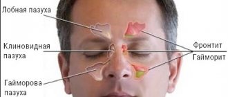
Many people suffer from various sinus-related diseases. The fact is that a lot of processes associated with viral diseases occur in them; various neoplasms can also appear there and many disorders can occur. For this reason, x-ray of the sinuses is a very important diagnostic procedure that must be performed for many diseases, especially sinusitis. This examination method is primarily aimed at identifying deviations from the normal state of the sinuses, as well as identifying infectious diseases and pathological processes. An x-ray of the sinuses with sinusitis, a photo of which you can see below, is capable of determining the degree of damage and changes in the mucous membranes, as well as tumors and cysts.
It is worth noting that x-ray examinations of this kind are very different from computed tomography and MRI diagnostics, since x-rays are not a safe method. Its advantages are low prices (free in government agencies) and greater availability, because photographs of the nasal sinuses are taken even urgently, that is, immediately after injury. This procedure most often takes less than 20 minutes and can only be performed by radiologists. An x-ray of the nose for sinusitis is necessarily done in two projections (standing position and lying position). Both images should be examined by a doctor, and on their basis a decision on further treatment will be made.
Let us immediately mention that this method does not even require special preparation, and, as mentioned earlier, it can even be carried out urgently! Sometimes it is necessary to perform an x-ray procedure of the sinuses in the presence of sinusitis several times. The fact is that sometimes treatment is ineffective, as a result of which symptoms persist and some complications arise. In severe cases, everything can even go as far as inflammation of the membranes of the brain, as well as the eye sockets. Bone changes can be seen if you take a classic panoramic photo.
CT scan
CT scan for sinusitis is also a common method that allows you to get a more detailed picture of what processes are occurring in the patient’s nasal passages. The procedure is prescribed for suspected unilateral or bilateral sinusitis, as well as when the course of the disease only worsens and there is every reason to believe that the person has a chronic form of the disease.
CT scans may be performed before and after surgery to remove pus from the sinuses
Computed tomography helps do the following:
- Thoroughly study the structures of the walls of the sinuses.
- Identify increased levels of mucus or pus in the maxillary sinuses.
- Determine mechanical changes in the paranasal sinuses.
- Timely detect the presence of neoplasms in the form of polyps or cysts in the nose.
CT scanning should be performed using modern and high-quality medical equipment, the diagnostic results of which can be trusted.
Computed tomography helps to thoroughly examine the structures of the walls of the sinuses
Indications and contraindications for the procedure
It is important to familiarize yourself with the list of indications and contraindications for this procedure. The fact is that you can completely harm your health with an x-ray if you ignore the rules regarding admission to the procedure. But first, let's look at the indications for the procedure, here is a list of them:
- various clinical manifestations reminiscent of sinusitis, for example, severe nasal congestion, facial pain, etc.;
- tumors (both benign and malignant);
- penetration of any foreign bodies into this area;
- the appearance of bleeding;
- injuries;
- new headaches for unknown reasons;
- cysts;
- development of caries of the upper teeth, as well as the need for surgery associated with implantation;
- polypous diseases affecting the nasal concha.
Important! An x-ray of all the maxillary sinuses can reveal darkening characteristic of sinusitis. If you have a bacterial infection, then most likely the specialist will see purulent fluid in the images. If total or, for example, subtotal, darkening of all walls related to the maxillary sinuses is detected, the specialist concludes that you are experiencing proliferative changes caused by hyperplastic sinusitis.
As for contraindications that limit the possible situations of the procedure, x-rays are prohibited if you are pregnant. The fact is that scientists have long proven the negative impact of such ionizing X-ray radiation on the fetus. It is worth understanding that x-rays are prohibited not only in the initial stages, but also in later stages. You cannot take x-rays even in situations where you have already exceeded the permissible number of procedures per year!
In what cases is MRI necessary?
The examination of the maxillary sinuses using a tomograph is resorted to in the following situations:
- the patient has been complaining of pain in the sinuses for a long time,
- prolonged rhinitis and nasal congestion cannot be treated, but only gets worse,
- breathing through the nose is impossible during the day, but relief occurs at night,
- X-ray and CT confirm the presence of a pathological process in the paranasal sinuses,
- headaches persist for a long time,
- the possibility of a runny nose as a symptom of an allergic reaction is excluded,
- with deviated nasal septums,
- after nasal injuries,
- if you suspect a cyst or other malignant or benign formations of the nasal sinuses.
Tomography allows you to obtain a three-dimensional picture of the paranasal cavities and the processes occurring in them. Accordingly, this method allows you to accurately determine not only the pathology itself, but also the extraneous factors that became its cause.
It is worth noting that this technique allows not only to diagnose sinusitis, but also to assess the condition of the nasal cavity as a whole. A detailed study helps determine the most appropriate treatment method, that is, direct all funds to eliminate the original cause of the pathology.
What diseases can be detected using MRI?
The description of MRI does not indicate the definition of the disease, since this is just an additional research method, the results of which must be reviewed by the doctor and based on them make a diagnosis.
Among the sinus diseases that can be identified using MTP are:
- Rhinitis of allergic and non-allergic nature.
- Polypous and cystic formations on the mucous membranes of the nasal sinuses and nasopharynx.
- Malignant and benign tumors.
- Adenoid growths.
- Hypertrophy of the nasal tonsils of varying degrees.
The MRI method is quite informative, but it is used extremely rarely. This is primarily due to the fact that the tomograph and its maintenance are quite expensive, which is why this research technique remains inaccessible to many patients. In addition, the duration of the examination is about 50 minutes, this time is not comparable to the widely used x-ray of the nasal sinuses. It is worth concluding that MRI represents a real breakthrough in medicine, but at this stage of development, unfortunately, it is not widely used.
Symptoms
Here are the main symptoms of various types of sinusitis; if they are present, you should immediately contact a specialist who will examine you and write a referral for an x-ray:
- headaches that appear constantly and without obvious reasons;
- pain that occurs in the bridge of the nose, as well as the temples, eyebrows or forehead;
- constant dizziness;
- problems associated with general well-being, decreased performance;
- problems with smell;
- labored breathing;
- excessive sweating;
- loss of appetite;
- tearfulness;
- sleep-related disorders;
- swelling that appears on the face (most often it is localized in the cheeks or eyelids).
Note! If you notice these symptoms, contact a qualified healthcare professional immediately. This way you can diagnose the disease in the early stages, then the treatment will be as effective and efficient as possible. Otherwise, if you neglect the disease, there will be a risk of developing serious complications!
Carrying out the procedure
X-ray of the sinuses is an incredibly effective method for diagnosing sinusitis. As mentioned earlier, no special preparation is required for this procedure, but it is important to never forget that there are contraindications, the list of which was presented earlier. We also mention that children are not recommended to have x-rays, or the number of procedures should be kept to a minimum. The fact is that a child’s body is much more susceptible to harmful ionizing radiation, which can cause serious harm to health at an early age. Here are the main steps in the process of this procedure:
- First, the radiologist will explain to the patient in detail the essence of the entire diagnostic process; this is necessary to ensure that everything happens in accordance with the requirements, then the images will be clear and informative.
- The person will have to lean such parts of the face as the nose and chin into the stand of the device, which will be adjusted in advance to the patient’s height. After this, the doctor will leave the room and begin setting up the equipment.
- From the next room, a speakerphone specialist will tell you what to do next. Just follow his instructions, then no problems will arise. The essence of such instructions will be to hold your breath when you need to take a photo. The duration of holding your breath will be short, maximum 10 seconds. When the photo is taken, it will be recorded and signed, then it will need to be dried.
- It is important to understand that in some situations it is necessary to take pictures in several projections, namely two. The fact is that sometimes it is necessary to monitor the condition of the sinuses even in a lying position. Thus, we can accurately conclude that there is sinusitis. The description will be given to you, after which you will have to take it to your doctor.
Description of sinusitis in the picture
Let us immediately mention that under no circumstances should you decipher the X-ray image yourself! The fact is that only a qualified specialist can do this correctly.
In the photographs, soft tissues are practically impossible to notice; only bones, which have a lighter shade, are visible on them. As for the sinuses, they have a characteristic oblong oval shape, and in the photographs they are expressed in a dark color. A conclusion about the presence of sinusitis and other problems with the sinuses can be made based on a simple comparison with the eye sockets. If the color is the same, then everything is normal, that is, there are no pathological processes.
What abnormalities are not visible in the photograph?
If the inflammation of the maxillary sinuses has a catarrhal or purulent form, it is difficult to determine the type of fluid even for a specialist, because these fluids look almost the same on an x-ray. In this case, the doctor needs to carry out additional procedures to determine the type of sinusitis and prescribe the correct treatment. Additional procedures involve collecting fluid through a puncture.
Another problem often arises with determining the difference between swelling of the mucous membrane and the accumulation of some amount of pus in the sinuses; in the pictures these phenomena look very similar.
If the patient provides an image in only one projection, it is difficult for the specialist to determine what is causing the discomfort: a cyst or a tumor. This is one of the reasons why the patient needs to have several radiographs taken in different projections.
To avoid possible errors in determining the diagnosis and treatment program, the patient is prescribed additional tomography. Sometimes children also receive similar studies, because it is extremely difficult to distinguish between rhinitis and sinusitis in X-ray images of small patients.
CT and x-ray of the maxillary sinuses
X-ray examination is an effective method for diagnosing the human body, including the condition of the ENT organs. If a person has a stuffy nose, there is discharge from it, or the timbre of his voice has changed, then this is an indication for an examination. X-rays are considered one of the most effective ways to detect sinusitis. With its help, it is possible to diagnose various pathological changes even at an early stage of their development, which is extremely important when prescribing appropriate treatment.
An examination is prescribed if the following symptoms are present:
- Nasal discharge (sometimes with pus).
- Problems with nasal breathing. As a rule, only one nostril is blocked.
- Frequent headaches.
- General fatigue, trouble sleeping and loss of previous activity.
- Nosebleeds.
- Soreness and swelling of the forehead.
If these symptoms are present, the doctor may suspect the presence of an inflammatory process, but it is necessary to determine the exact cause of this inflammatory process, because similar signs exist with polyps and a deviated septum. Therefore, an x-ray of the maxillary sinuses is a mandatory procedure, which allows us to determine the degree of development of the pathology, the presence of neoplasms and other aspects.
Content
Why do you need an x-ray of the nose for sinusitis?
Vivid symptoms will help the doctor make an appropriate diagnosis, but without an accurate diagnosis, further treatment will be impossible. It is important to first examine the condition of the sinuses, and then determine the medications and physiological procedures that can get rid of the disease.
When diagnosing, it is extremely important to know what sinusitis looks like on an x-ray. With this examination method, soft tissues are not visible, but bone structures are clearly visualized. If in the picture the shade of the paranasal sinuses and eye sockets is identical, then there is no inflammation. If there is purulent content, then visually it looks like large darkened areas.
An x-ray of the sinuses with sinusitis allows you to determine a number of pathological changes:
- Localization of lesions. They appear as dark spots.
- Presence of cystic formations. They have fairly clearly defined boundaries.
- The inflammatory process and the degree of its development. In the picture they are visible as white spots. Accordingly, the brighter they are, the stronger the inflammatory process.
- Various changes. You can see the level of filling of the nasal sinuses with pus, thickening of the mucous membrane and other pathological changes.
How can you see sinusitis on an x-ray?
Below you see a picture of sinusitis before and after treatment. The radiograph is interpreted by the attending physician or radiologist.
In the absence of pathological changes and abnormalities, the image will show the following information:
- The nose is in the form of a triangle of a light shade, in the middle of which there is a septum.
- On the side of the nasal cavity, the maxillary sinuses are located in the form of triangular clearings with not blurred boundaries.
- Symmetrical arrangement of the nasal passages on both sides of the nasal cavity.
- The frontal sinuses are located above the eye sockets. In the picture they look like clearings of various sizes.
The pathology develops from inflammation of the lateral sinuses. Further, it is localized in the region of the frontal lobes. This will be clearly visible in the darkened areas above the eye sockets and nose. Simultaneous inflammatory processes in the frontal and lateral sinuses indicate not only the presence of sinusitis, but also frontal sinusitis.
A specialist who takes pictures of the nose for sinusitis always determines the condition of the ethmoid bone. Infiltrative fluid often accumulates in the maxillary sinuses:
It is quite easy to notice - the liquid appears as a clearly formed white area. This is a clear sign of pathology, which is one of the grounds for making a final diagnosis.
How is an x-ray of the maxillary sinuses performed?
X-ray of the nasal sinuses for sinusitis is a completely standard diagnostic procedure. Before the examination, you must remove all metal objects and removable dentures. If implants, crowns or pins are installed, the radiologist should be notified about this.
To obtain an accurate picture of the disease, a photograph of the nasal sinuses with sinusitis is taken in several projections:
- Chin. Based on this image, the specialist analyzes the actual condition of the maxillary sinuses.
- Nasomental. This projection clearly shows the presence of an inflammatory process in the mucosa.
- Nasofrontal. In this picture you can see the state of the lattice labyrinth.
The examination takes 10-20 minutes, after which the doctor can clearly see the presence or absence of pathology.
X-ray in childhood
Experts agree that X-ray examinations are advisable from the age of 14. Here we are not talking about potential harm to the child’s body, because a single dose of radiation during an examination is absolutely safe for the child. The fact is that at an early age the paranasal sinuses are not yet fully formed, so the examination result cannot be 100% accurate.
In addition, x-rays require maintaining complete stillness throughout the entire examination, which, for obvious reasons, is very difficult to achieve in childhood.
How to determine sinusitis in an adult patient from an image?
Pictures of sinusitis in adult patients are quite characteristic, especially when compared with radiographs of people who do not have this pathology. X-ray will show the following changes:
- The eye sockets and sinuses will be of different shades.
- The presence of infiltrative fluid (a clear white spot against a darkened background).
- Thickened walls and blurred boundaries of the sinuses.
- Cystic formations of varying sizes with clearly defined boundaries.
The only thing that cannot be seen in a photo of the nose with sinusitis is the type of fluid that has accumulated in the sinuses. The study only shows its presence in the form of a white spot, but cannot show what kind of infiltrate (purulent, mucous or catarrhal fluid).
Referral for x-ray and procedure
If a patient complains of the following symptoms and signs of inflammation, this indicates the course of sinusitis in acute or chronic form:
- swelling of the nasal cavity;
- redness and slight enlargement of the wings of the nose;
- pain on the face when turning or tilting the head;
- pain or discomfort in the bridge of the nose, forehead, temples or a feeling of pressure on them;
- difficulty breathing;
- congestion of a permanent nature;
- increased body temperature;
- thick discharge that comes out weakly or not at all from the nose, which will contain purulent impurities.
If such symptoms appear soon after the cure of a cold and attack the patient for 7 or more days (the usual runny nose completely disappears during this time), you must definitely go to an appointment with an otolaryngologist, as this indicates the progression of sinusitis.
The doctor will definitely prescribe the patient to undergo fluoroscopy, which will help confirm the development of inflammation, as well as find out about the condition of the mucous membrane.
Indications for X-rays for maxillary sinusitis are:
- clarification of the diagnosis made by the doctor based on the clinical picture of the disease;
- identifying the degree of neglect of the disease;
- identification of the general condition of the maxillary cavities;
- suspicion of the development of polyps or tumors of any type in the nasal cavity, which usually appear based on the development of inflammation;
- monitoring treatment;
- if therapy does not help, a photograph of sinusitis will help to understand that the patient requires a puncture to accurately identify the type of pathogen;
- re-examination of the maxillary cavities, which will help to identify the lack of effectiveness of complex treatment.
Sinusitis in the picture looks quite clear, which means that after the examination it will be possible to immediately make the correct diagnosis, and then begin comprehensive treatment of the disease.
To correctly identify sinusitis on an x-ray, the patient does not need to prepare specially before the examination. The patient must be placed on a stand, the size of which can be adjusted to the height of any person (woman, man or child). Then the doctor will ask the patient to rest his nose on this stand, and then hold his breath for a couple of seconds.
In a minute the picture will be ready, and after half an hour the result can be taken and given to the doctor so that he can decipher it. The main thing is that it is completely dry, otherwise the result of this diagnosis cannot be called truthful.
What should a photo of the procedure look like in an adult and a child? Usually it is done in a direct projection, but if necessary, the doctor may require a lateral x-ray.
If a woman needs an x-ray of her nasal sinus during pregnancy, she must notify her doctor about this, since this procedure is not always considered safe. The fact is that radiation has a negative effect on the fetus, which means that X-rays are required only if necessary and with maximum protection for the baby. Also, in childhood, it is necessary to carefully take the picture, since it is often necessary to repeat the examination due to image distortion.
How to determine sinusitis from an image: what an x-ray of the sinuses shows
In the presented x-ray image, the paranasal sinuses (PSN) are highlighted as ovals. Normally, they are not pneumatized, so they appear on the image as dark semi-oval formations on both sides of the nose. To determine the intensity of the “coloring,” radiologists compare them with the shade of the eye sockets. If the color shades match, then the picture shows the norm.
The x-ray passes through the airy tissues and is not reflected from them, so they do not appear clearly on the x-ray. When the cavity is filled with liquid, intense darkening with an upper horizontal level can be observed. As an example, below is a description of an x-ray by a radiologist.
In the presented x-ray, PPN-pneumatization is not noticeably reduced, the cells of the ethmoid bone are not changed. Conclusion: There are no X-ray signs of sinusitis.
In the photo you can clearly see dark cavities on both sides of the nose, similar to those located in the projection of the frontal part of the skull. The maxillary or maxillary sinuses are affected by the inflammatory process more often than the frontal sinuses.
Sinusitis in the picture is displayed as a darkening with an upper horizontal level, just like frontal sinusitis.
X-ray: darkening with an upper horizontal level in the right maxillary and frontal sinus. On the left – a shadow can be traced in the lower third with an upper wavy contour due to chronic proliferative sinusitis
This photo is a variant of pathology. If readers compare it with the one that was brought to their attention a little earlier, they will be able to distinguish pathological X-ray symptoms from normal ones.
What is sinusitis (the “milk in a glass” effect)
Sinusitis is inflammation of the paranasal sinuses. Infiltrative fluid can be of the following types:
When analyzing the x-ray picture for pathology, you can notice the black color of the sinuses with white infiltrative contents. The sharp contrast allows you to clearly separate normal from pathology. The “milk in a glass” effect is a diagnostic symptom that allows the radiologist to establish the correct conclusion.
Attention readers. We teach you how to identify inflammatory changes in the accessory cavities from an image, not so that you self-medicate. The information in the article should be used exclusively for early diagnosis of pathology.
The X-ray symptom “milk in a glass” on a direct image is not a direct confirmation of inflammatory changes in the sinuses, although it has a high degree of reliability. If it is detected on the X-ray of the PPN in frontal and lateral projections, then the specialist can calmly write a conclusion that the patient has sinusitis.
Photo of an x-ray for right-sided maxillary sinusitis.
Obviously, sinusitis and frontal sinusitis can be traced on an x-ray by intense white shadows against the background of a black cavity around the nose. However, not all so simple.
In the above photo, pathological shadows are marked with an oval. The radiologist described a similar picture as X-ray signs of sinusitis. The specialist is partly right, but he did not notice a round darkening of medium intensity with an even contour about 1 cm in diameter.
If a purulent or catarrhal infiltrate accumulates, rounded pathological shadows will not be visible on the radiograph. A round darkening on an X-ray image appears with tumors and cysts.
With a malignant neoplasm of the paranasal sinuses, you can see the growth of the formation over time. Cysts can also increase in size, but proportionally in different directions.
Ultrasound examination
To perform an ultrasound for sinusitis, no special preparation is required. The study is carried out using a sensor that transmits information about the size of the cartilage and tissues of the nose, the structural features of the nasal cavity and sinus cavities.
Ultrasound helps detect fluid in the cavity of the paranasal sinus, notice abnormal thickening of the mucous membrane and other changes characteristic of a disease such as sinusitis. The advantage of this method lies in the following points:
- Instant receipt of research results.
- There are practically no contraindications.
- The ability to detect changes that are not visible on an x-ray.
- A completely painless and safe procedure.
Ultrasound of the sinuses helps identify changes that are not visible on an x-ray