Ear cancer: causes of the disease in adults and children
Ear cancer occurs when a cell undergoes malignant degeneration—the ability to divide uncontrollably. The presence of benign tumors in the ear canal area or factors predisposing to malignancy significantly increases the risk of oncology.
Factors that increase the risk of developing common ear cancers include:
- pathologies that provoke skin changes in the area of the outer part of the hearing organ (eczema, psoriasis, systemic lupus erythematosus);
- scars formed as a result of trauma, burns, inflammation or other damage to the outer ear;
- insolation;
- high doses of ionizing radiation;
- hereditary predisposition to ear cancer;
- chronic, adhesive and purulent form of otitis, chronic inflammation of the ENT organs (laryngitis, sinusitis);
- precancerous conditions of the tissues of the outer and middle ear;
- polyps in the nasopharynx and ear canal;
- treatment of granulations with silver compounds.
The occurrence of benign tumors (atheromas, lipomas, chondromas) is caused by pathological processes that cause rapid cell division in cartilage, vascular, adipose and other tissues. For example, a fatty cyst behind the ear and in the conch area is the result of the function of the sebaceous gland.
Outer ear cancer
Tumors of the outer part of the ear account for up to 95% of the total number of benign and malignant neoplasias of the hearing organ. Concha neoplasia is observed in 80% of cases of ear tumors, and tumors of the external ear - in 15%.
The provoking factors for oncology of the outer ear are pathological changes in the skin (diseases, scars), large doses of ionizing and ultraviolet radiation.
Epithelial tumors can progress quickly, so if a suspicious growth has formed in the lobe, earlobe or ear canal, you should immediately consult a doctor.
Middle ear cancer
Middle ear tumors develop as a result of an inflammatory process (chronic and adhesive otitis media, inflammation of the eardrum).
In some cases, the material for the growth of malignant neoplasia becomes mesenchymal tissue, which remains in the epitympanic space of newborns.
Inner ear cancer
Unlike the outer and middle ear, in which pathological lumps and growths may appear, the inner ear (labyrinth) is not a possible location for primary tumors. Benign and malignant formations in the labyrinthine area develop from structures located next to the inner ear or are metastatic in nature.
Clinical cases of inner ear tumors are extremely rare.
Lump in the ear cavity and its removal
The phenomenon in which a lump forms in the ear can act as a normal process or indicate a large-scale pathology in the body. Numerous phenomena can visually have a similar appearance.
For example, general and local inflammation, tumors of benign and malignant nature.
All doubts can be put to rest only by an ENT specialist who has interviewed the patient, studied the test results and sorted out the situation.
Causal factors of the phenomenon
A tumor in the ear, which is accompanied by pain and has a bright red color, can act as a sign that a boil has formed. It is a purulent inflammatory process in the area of the hair follicle or sebaceous gland. As a result of this phenomenon, a necrotic core is formed, surrounded by purulent contents.
The causative agent of the disease is Staphylococcus aureus, which begins to become active when a favorable environment is created for it.
A lump on the ear can manifest itself due to a number of causative factors that are worth paying attention to:
- neglect of the simplest requirements and hygiene standards;
- traumatic phenomena in the area of the hair follicle or skin;
- frequent illness with purulent otitis media;
- hormonal surges;
- metabolic problems;
- progression of viruses and colds;
- excessive overheating or hypothermia;
- reduced immune system defense.
Thus, an ear tumor can arise as a result of simple phenomena that require only minor correction, or as a result of serious diseases.
What does a lump in the ear area mean?
An ear tumor is a small-sized seal on the auricle, which is traditionally round in shape and has soft contents. Usually this phenomenon is not accompanied by feelings of discomfort and does not cause inconvenience to the person.
In some cases, the formation of a lump above the ear may be evidence of a serious illness and pose a threat to health.
After all, the appearance of a lump above, in or behind the ear can be evidence of a harmless cold or a serious phenomenon.
In no case should you engage in independent treatment; you should contact an ENT specialist. There are several common situations that involve the appearance of a hard lump in or above the ear.
Acute otitis of infectious nature
This pathology is accompanied by an acute pain sensation, which is popularly called “shooting.”
In this case, body temperature rises to 37.5 degrees, and a transparent or yellow secretion is observed from the ear canal.
Depending on the form of the disease, there are several characteristic symptomatic signs that accompany a tumor in the ear:
- slight itching or pain - this indicates the fungal nature of the lesion;
- the secretion has an unpleasant aroma - most often this indicates the progression of staphylococcus or streptococcus bacteria;
- prolonged development of symptoms and a gradual increase with the feeling that water is gurgling in the ear - we can talk about the allergic nature of otitis media.
If lumps have formed above the ear or in its cavity, otitis media is the first phenomenon that should be paid special attention to.
Furunculosis or folliculitis
Usually, bumps do not appear above the ear; in this case, they prefer to be localized in the ear canal or in the sink area. This process can be detected by pressing on the lower shell wall.
This formation already hurts when pressed, but a more reliable diagnosis is carried out if the boil is characterized by canonical elevation. To examine it, the lobe is usually pulled back; this phenomenon also entails sensations of pain.
Lumps in the ears in this case require proper treatment.
Wen
At the first stage of its manifestation, this disease resembles a pimple in appearance; an ear tumor forms only later. Shiny skin rises above the wen; during the inflammatory process, a local increase in temperature is observed, and during development, signs of general intoxication appear.
The ear tumor is small in size and slightly interferes with life.
Entry of foreign bodies
An ear tumor may appear due to foreign bodies entering the passage. They traditionally cause the patient to form a painful type of formation, and all this can be detected during a visual examination using a mirror.
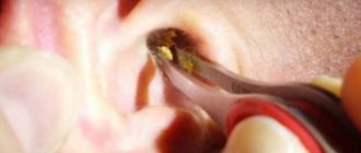
Plug in the ear
In this case, not only the phenomenon of swollen ears and pain is observed, but also additional symptoms. Most often, these include deterioration of auditory function, the formation of congestion, and painful sensations of a pulling nature.
Lymphadenitis
If there is a lump in the auricle and it hurts, this may indicate this pathology. If the size of the parotid lymph nodes increases, increased pressure appears. This process is also accompanied by a deterioration in general health indicators, pain in the head, and weakness.
Other diseases
When there is a seal in the ear, you should pay attention to several other unpleasant phenomena. This could be a progressive sore throat, laryngitis, mastoiditis, neuritis or other diseases of a neuralgic nature.
The doctor usually uses examination of the patient for diagnosis, collects anamnesis and, if necessary, prescribes examinations and consultations with other specialists.
Thus, phenomena inside the ear are accompanied by general complications and cause several side effects and complications.
How to treat a lump in the ear area
If your ear is swollen and painful, you should pay attention to proven therapy methods; there are special instructions for this.
- The first thing to do is to determine the causative factor in the formation of an ear lump. If the manifestation of a ball near the ear does not itch or is accompanied by pain, most likely we are talking about an enlarged lymph node. The phenomenon indicates that perhaps the body is experiencing a severe runny nose, which does not always mean that a ball has appeared and it hurts. Sometimes otitis media becomes a causative factor. Sometimes patients feel a tingling sensation, but the inflammatory process continues.
- If lumps appear above the ear, and you are not sure that they act as an inflammatory process of the lymph node, you should not touch them. The fact is that only the causative factor in the occurrence of the phenomenon, and not the pain, is treated. If the painful sensation when pressing is bothersome, it is recommended to drip special anti-inflammatory compounds into the ears, and vasoconstrictor drops into the nose.
- If a ball appears on the ears, and the body temperature remains normal, you should refrain from using antibiotic compounds. However, in any case, it is worth visiting an ENT specialist who is competent in the rational selection of therapeutic tactics and will be able to prescribe the necessary medications. If the ear is swollen, this must be done without fail, otherwise the advanced process may lead to the phenomenon of chronic otitis media, which can pose a serious threat to hearing.
- If the formation appears gradually, is red in color and has an increased level of pain, most likely it is a boil formed in the outer ear. You should not squeeze it out, since the process is designed to lead to the formation of an infectious process. What to do is to moisten a piece of cotton wool with Vishnevsky’s special ointment and place it in the area of the external auditory canal. This action will ensure rapid maturation of the boil and drainage of pus.
- If a lump appears on the cartilage or in the area of the ear canal, which hurts when pressed, it is strictly forbidden to rub it with alcohol and other homemade products.
If a person’s ear is swollen, and it doesn’t matter whether the formation that appears hurts or not, this is a clear reason to pay a visit to a competent treating specialist. Only an otolaryngologist will be able to determine the exact causative factor due to which the lump appeared and the balloon inflated, and will also prescribe the appropriate remedy.
Remember that if you try to overcome swelling on your own, you may encounter diseases that will lead to serious inflammation that threatens hearing loss. Through diagnosis, it is possible to determine why a disease may appear and how to prevent the phenomenon.
Lump in the ear cavity and its removal Link to main publication
Source: https://lorgid.ru/ear/ushnie-simptomi/shishka-v-uhe
Classification of ear tumors
Tumors of the middle and outer ear are divided into benign and malignant. In addition to the classification that is most important for the patient, there are others:
- by structure (solitary-vegetative, ulcerative-infiltrative or ulcerative neoplasia);
- by histological structure (sarcoma, basal cell carcinoma, spinocellular epithelioma).
If upon palpation of the ear it seems that a ball, ulcer or convex scar has appeared inside, you need to contact an otolaryngologist and oncologist to determine the size, type and structure of the tumor.
Benign
The group of benign ear tumors includes the following neoplasias:
- Nevus (moles) - occur during the active division of pigment cells (melanocytes). Insolation and trauma can lead to malignancy of nevi.
- Papillomas are the most common benign neoplasia of the ear. They are formed due to infection with HPV. Papillomas are localized only on the outer part of the hearing organ and are small in size.
- Adenomas develop from glandular tissues and have the appearance of small nodular neoplasms.
- Atheromas - occur when the function of the sebaceous glands is impaired (blocked). Over time, these tumor-like formations transform into an ulcer or a hard spherical tumor. Malignancy of atheroma is possible, but is observed only in rare cases.
- Fibroids are formed from connective tissues with a fibrous structure. They are localized mainly on the earlobe, where punctures for earrings are located. Less often, this formation appears on other parts of the auricle. The size of fibroids can reach 1-2 cm.
- Lipomas are formed in adipose tissue and are localized in the subcutaneous tissue. They are characterized by their small size and tendency to recur frequently.
- Osteomas are tumors of this type that are formed from the bone structure of the ear canal and auricle. Osteomas often recur after treatment.
- Neuroma, or schwannoglioma, is a neoplasia that develops from the cells of the myelin sheath of the nerve fiber. The auditory nerve is the most common site of neuroma.
- Chondromas develop in the cartilage tissue of the ear. This type of neoplasia is observed quite rarely.
- Glomus neoplasia (paraganglioma) - is formed from glomus bodies, which are located on the tympanic cavity.
- Hemangioma. Soft vascular tumors develop from endothelial cells and are located in the outer ear.
See also
What to do if your ears hurt when you have a cold and how to treat them
Read
Paraganglioma and hemangioma are conditionally benign neoplasias. As they grow, they destroy surrounding tissue and extend beyond the auricle and middle ear.
Malignant
Malignant auricular neoplasias include the following tumors:
- Basalioma. Neoplasia developing from basal epithelial cells is characteristic of elderly patients with skin pathologies. Basaliomas are characterized by slow growth and reluctance to form metastases. Neoplasia of this type often recurs and ulcerates.
- Spinocellular epithelioma (squamous cell carcinoma) is the most common neoplasia of the ear. It is characterized by rapid growth, bleeding, and a tendency to metastasize and ulcerate. The neoplasm develops against the background of pathological or age-related changes in the skin. Without treatment in the first stages of the disease, epithelioma quickly grows into the salivary glands, ear canal, middle ear and cranial cavity.
- Sarcoma. Tumors of this type develop from immature connective tissue cells. They grow slowly, do not cause damage to the lymph nodes and ulcerate late. Sarcomas occur predominantly in children and elderly patients.
- Melanomas are formed from malignant melanocyte cells. They can grow both above the surface of the skin and deep into the tissues. Neoplasia of this type is prone to early formation of metastases in the lymph nodes (less often in internal organs), which significantly worsens the prognosis.
Malignant neoplasia varies in extent of spread:
- 1st degree (size up to 5 mm, localized on the surface of the skin);
- 2nd degree (spread of malignant disease to cartilage tissue);
- 3rd degree (involvement of the entire structure of the ear in the cancer process, adenopathy);
- 4th degree (tumor growth to regional lymph nodes).
Lump in the ear - what to do if the auricle is swollen inside and hurts 2020
The phenomenon in which a lump forms in the ear can act as a normal process or indicate a large-scale pathology in the body. Numerous phenomena can visually have a similar appearance.
For example, general and local inflammation, tumors of benign and malignant nature.
All doubts can be put to rest only by an ENT specialist who has interviewed the patient, studied the test results and sorted out the situation.
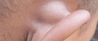
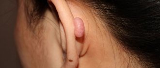
Wen (lipoma)
The ball under the ear or under the lobe near it can be an ordinary wen, mobile and soft, which forms at the site of the growth of adipose tissue. The size of lipomas is no more than 1.5 cm, they do not develop into tumors, and very rarely increase significantly. The reasons for their appearance may be:
- hereditary predisposition;
- violation of fat metabolism;
- sudden changes in the fat layer.
In this case, the seal is mainly a cosmetic problem. Wen often resolves on its own. A person, after consulting with a doctor, will decide for himself whether to remove it or not. The operation is performed using a laser under local anesthesia. The skin is incised over the clot, which is then also excised with a laser beam, while coagulation of the vessels is carried out to avoid bleeding.
It often takes a long time before the lump is discovered. Lipoma is painless, at first has a soft consistency and grows slowly. However, if the wen is damaged, pain may also occur.
The growth of a tumor can also lead to the same consequences if the formation is localized near the nerves. Then, with its increase, the lipoma begins to put pressure on the nerve processes. This condition can also be perceived by the patient as if something is swollen in the ear and hurts.
Treatment and its stages
To alleviate the condition and accelerate the maturation of the boil, fairly simple conservative manipulations help:
Simple conservative manipulations help to alleviate the condition.
- Bandages and tampons with Vishnevsky's liniment, ichthyol and zinc ointment, fixed with adhesive tape. A good effect allows you to get compresses from a concentrated saline or soda solution at night on the inflamed element.
- For many decades, smearing the bump behind the ear with an asterisk and gluing the pulp of an aloe or Kalanchoe leaf have proven themselves to be excellent.
- When visiting a doctor, the patient is prescribed a complex of antipyretic drugs, analgesics and antibiotics.
Despite the apparent simplicity of removing a lump inside the ear, you should not attempt to perform an autopsy on your own. Usually, at home, it is not possible to completely clear the purulent sac, which leads to the development of new spilled lesions.
Perichondritis, a lesion of the perichondrium of the ear cartilage, is much less commonly diagnosed. The ear is swollen, red and shiny. A severe pain syndrome appears, characteristic of a lump inside the ear.
Treatment is prescribed exclusively by an ENT specialist, since self-medication at home leads to such a serious complication as melting of the ear cartilage.
What symptoms are observed?
At an early stage of the cancer process, no characteristic symptoms may be observed. An ulcer, a small spot or growth resembling a wart, forms in the area of the auricle or external auditory canal. At this stage, some patients turn to a dermatologist because a dense pimple has swelled on the ear, painless when pressed. The cancerous nature of the neoplasm is detected during differential diagnosis.
The asymptomatic stage does not last long. Immediately after it, severe ear pain occurs, the intensity of which increases every day. Pain sensations radiate to the temple and jaw area.
Signs of a progressing cancer tumor also include:
- dizziness, imbalance (with germination into internal structures);
- conductive and sensorineural hearing loss (when the ear canal is blocked by a tumor and grows into the labyrinth area, respectively);
- itching in the ear canal;
- putrid odor and purulent discharge, reminiscent of signs of purulent otitis (with a tumor in the area of the eardrum);
- swollen lymph nodes, swelling and swelling of the salivary glands affected by cancer cells.
With tumor formations in the ear canal and deeper structures of the hearing organ, the tragus of the ear hurts when pressed.
See also
Causes, symptoms and treatment of inflammation of the lymph node behind the ear in a child
Read
In the later stages of ear cancer, when malignant neoplasia grows into the facial canal, skull, nasopharynx, ethmoid bone and large internal vessels, more severe disorders appear:
- facial paralysis;
- neuritis of the vestibulocochlear nerve fiber;
- dizziness;
- trigeminal neuralgia;
- sensory and movement disorders;
- carcinomatous meningitis;
- heavy bleeding.
Why is it important to visit a doctor?
More often, the appearance of a tumor on the earlobe is caused by blockage of the sebaceous glands, which leads to the formation of a wen. The latter, even in the case of a secondary infection, creates only discomfort for the person and does not pose a serious threat to life.
However, sometimes a neoplasm arising from the epithelium or fatty tissue becomes malignant. And it is possible to determine the type of tumor if a histological examination of the contents of the capsule is performed.
Therefore, when painful balls appear on the ears, and the skin in the problem area turns red, you need to seek medical help.
Tumor diagnosis
If you find a lump, ulcer or granulation in the area of the auricle or ear canal, you should contact an otolaryngologist or dermatologist. If a malignant nature of the formation is suspected, the patient is scheduled for a consultation with an oncologist.
The following methods are used to diagnose malignant ear diseases:
- microotoscopy;
- MRI, CT scan of the brain;
- radiography;
- angiography;
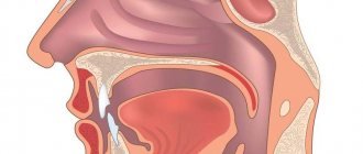
- endoscopy of the nasopharynx;
- probing the bottom of a tumor ulcer;
- biopsy and histological examination of tissue.
If tumor tissues have grown into the meninges, the patient is prescribed additional examination in neurology.
Treatment of neoplasm
Self-medication of a lump behind the ear can worsen the situation and lead to harmful consequences. If you are confident in the viral nature of the swollen area, attention should be paid to treating the underlying disease, most often a cold.
If the follicle is inflamed, it is recommended to use salicylic acid or alcohol to cleanse the affected area.
The sore spot should not be heated or rubbed, especially with warming ointments.
In other cases, treatment should begin with a visit to the therapist.
Depending on the preliminary diagnosis, the therapist (general practitioner) will be able to determine whether the intervention of more specialized specialists is required. Perhaps he will refer the patient for additional diagnostics to an otolaryngologist, surgeon, neurologist, dentist, and so on. It is possible that you will need to carry out hardware diagnostics - CT, MRI, X-ray or ultrasound. In doubtful cases, a tissue biopsy will be required to rule out malignancy of the tumor.
A lump behind the ear can appear for many reasons. Some of them require treatment, others do not cause discomfort and soon disappear. But you shouldn’t wait for the tumor to go away on its own, because this may not happen. If such a problem occurs, you should definitely consult a doctor. If you have any doubts about which doctor to contact, you can visit a therapist. He will prescribe the necessary examinations and refer you to the right specialist.
Pimples don't only appear on the face. They often form behind the ear. Dirt and sweat often accumulate in this area. In this case, the lump is small, red, with purulent contents in the center. Drying agents and alcohol will help quickly eliminate the defect. To avoid such problems in the future, special attention should be paid to cleanliness in this area.
Treatment
The goal of therapy for malignant tumors of the outer and middle ear is the complete elimination of tumor tissue and restoration of the patient’s hearing. The choice of treatment method depends on the severity of the patient’s condition, the histological structure of the neoplasia, the stage of the cancer process, the presence of secondary tumor foci (metastases) and other factors.
Surgical method
Surgical intervention for ear cancer involves complete excision of the tumor, partial removal of the temporal bone and surrounding tissues involved in the pathological process (cervical and submandibular lymph nodes, salivary glands, cartilage).
For small tumors, surgeons can resort to a less traumatic, but more risky method: curettage of tumor tissue.
Contraindications to surgery are:
- serious condition of the patient;
- germination of neoplasia into vital internal organs;
- the presence of multiple secondary foci of cancer.
Radiation therapy
Radiation therapy is used as an auxiliary or primary method of treatment. It helps to slow down the proliferation of pathological cells and stop the manifestations of the disease in the later stages of oncology.
Chemotherapy
Chemotherapy is used for the same indications as radiation therapy. It is classified into several types:
- Neoadjuvant - performed immediately before surgical treatment. The introduction of cytotoxic and cytostatic drugs is intended to reduce tumor size and reduce the risk of metastasis after surgery.
- Adjuvant - prescribed after resection of pathological tissues. It prevents the recurrence of cancer.
- Induction – used for palliative treatment. It does not affect the operability of neoplasia, but improves the patient’s quality of life.
The use of several courses of systemic chemotherapy is also indicated in the presence of distant metastases.
Cryodestruction of the tumor
Cryodestruction involves cauterizing the ear tumor with a cryo-tip containing liquid inert gas. Cooled nitrogen causes necrosis of cancer cells.
This technique is effective only in the presence of a localized tumor of small size.
Radiotherapy
Radiation therapy is carried out using a cyber or gamma knife, which focuses ionizing radiation. A high-energy beam allows you to eliminate tumor cells without affecting surrounding healthy tissue. Radiotherapy treats not only superficial but also internal cancer.
Cauterization
When cauterizing (electrocoagulation) of tumor formations, the affected tissues are destroyed by high-frequency electric current. This treatment method is effective for neoplasia of the outer ear and auricle. Its advantage over surgery is the lower risk of recurrence and the absence of postoperative bleeding.
Treatment and home remedies
Treatment for a swollen earlobe depends on the underlying cause. People with swollen earlobes due to an allergic reaction should avoid allergens in the future. This may include nickel jewelry.
They should see a doctor if their symptoms become severe.
Infections or other problems caused by bacteria usually require treatment with antibiotics. Mastoiditis or cauliflower ear will require emergency medical attention.
A doctor or dermatologist must make the correct diagnosis and treatment to reduce the risk of complications.
Some home remedies may also help reduce symptoms if the cause of your swollen earlobe does not require medical attention.
Home remedies for swollen earlobe include:
Warm or cold compresses: A cold compress can numb the pain, while a warm compress can increase circulation to the area to reduce swelling.
Over-the-counter pain relievers: Medicines to treat pain and inflammation, such as ibuprofen (Advil), acetaminophen (Tylenol), and naproxen (Aleve), may reduce pain and swelling.
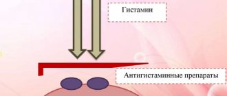
Antihistamine: Oral or topical antihistamines may help reduce the symptoms of bites and allergic reactions. Calamine lotion may also have a soothing effect on rashes.
Astringents: Astringents such as witch hazel can help tighten tissue and reduce swelling in the earlobe.
Oatmeal baths: Allergic reactions, pain, or itching can be soothed in an oatmeal bath. A person can keep their ear in a small bowl of warm water and finely ground oatmeal, or apply the mixture directly to the ear before rinsing it off.
Tea Tree Oil: This essential oil can speed up the healing process of wounds and fight bacteria.