What is an eardrum?
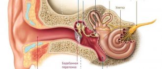
The membrane that separates the cavity of the middle ear and the ear canal is called the eardrum .
The membrane has a high level of sensitivity, which allows it to capture sound waves. When sound waves enter the ear canal, the membrane begins to vibrate and activates the auditory ossicles, which transmit sound to the cochlea and a person can recognize what they hear.
The membrane is located in the temporal zone, and is attached with one edge to the temple bone. The second edge of the membrane is not secured. In adults, the membrane has an oval shape; in childhood, the membrane has the shape of a circle.
What does a membrane in the ear look like?
The tympanic region is necessary in the structure of the ear organ. It helps not only in converting sound waves into nerve impulses, but also protects the hearing organ from the penetration of water and foreign objects, and also protects the auditory ossicles and labyrinth from various viruses, harmful bacteria and dangerous infections.
Anatomically, the tympanic region is located in front of the auditory ossicles , which are located in the middle region of the ear and end in the inner part of the ear.
This area is particularly sensitive, since the thickness of the membrane is only 0.1 millimeter. By the way, the membrane itself is pearl-colored and has a very sensitive structure. Therefore, this area can pick up sounds and various noises with frequencies from 15 hertz to 25 kHz.
In case of disease of the ear organ, for example, otitis media or other inflammatory processes in the ears, the upper indicator is significantly reduced. In the absence of proper treatment and ignoring the symptoms of inflammation, absolute partial hearing loss or absolute deafness may occur.
It is known that the membrane itself undergoes age-related changes. Thus, in infants the membrane has a round shape, in adolescent children it is more oval. In mature people, the diameter of this area changes.
In its largest dimension it can be around ten millimeters , while its smallest circumference can measure up to eight millimeters.
The strength of the membrane, its diameter and thickness in an elderly person varies greatly.
As a result, many people experience hearing problems as they age.
In this case, if hearing problems develop, doctors prescribe hearing aids.
The membrane consists of several tissues:
- The outer region consists of the epidermis of the auditory canal.
- Next is the main layer of the membrane, which consists of various fibers of the radical and circulation tissue.
- The final layer, called the inner layer, is formed from the mucous membrane of the tympanic cavity.
With the help of the parts described above, sound waves, converted into nerve impulses, pass from the tympanic region to the inner ear, and then to the brain using the glossopharyngeal nerve.
Behind the eardrum itself is the tympanic region . It is the main one in the middle ear cavity and occupies up to one centimeter. It is believed that this area is the smallest in the structure of the ear organ, but despite this, it has a complex structure.
This cavity is localized in the temporal bone of the skull , and is attached to this area using special rings. Therefore, the membrane is quite tense. Because of this, when viruses penetrate this part, a person experiences severe shooting pains not only in the ears, but also in the head.
The upper area is attached to the bone part by an inset, but it is located quite freely.
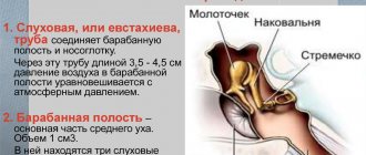
The two remaining parts of the tympanic membrane are bordered by the malleus folds, which are divided into two types:
- Front fold.
- Rear end.
The smallest part of the malleus is attached to the membrane of the eardrum. Thanks to this structure, a person picks up various sound vibrations.
From inside the middle part of the ear there is
constant pressure on the eardrum.
This pressure is equal to atmospheric pressure, so when air comes into contact with the membrane, it bends.
At this time, sound impulses are transmitted to the sound ossicles, in particular to the malleus.
A person, being in an aquatic environment, experiences a strong resonance, since the frequency of sounds in this atmosphere does not reach even twenty Hz.
A person has the ability to hear due to the balance of external and internal pressure . The Eustachian tube, which normalizes blood pressure, is responsible for this possibility.
But in case of dysfunction of this organ and with a strong change in ambient pressure, the membrane is subject to severe trauma .
This usually occurs with strong pops, explosions and other sharp sounds, which can severely damage and even rupture the eardrum. At this time, a person experiences severe and sharp pain, which can significantly worsen hearing acuity and even deprive the ability to hear forever.
Sounds that can damage the membrane must be incredibly strong, since this area is durable and can withstand pressure up to one hundred millimeters of mercury.
If the eardrum is damaged, urgent treatment is required. In some cases, the membrane recovers on its own. In more complex cases, if the main layer of the membrane is damaged, surgical intervention is required.
In severe cases of damage, the tympanic cavity ruptures and damages the inner ear. In this case, the prognosis is extremely unfavorable. A person may lose their hearing forever.
Look at the photo of the eardrum in various states:
More information about the eardrum
As we have already found out, the eardrum in the ear is distinguished by its delicate structure. The membrane located in this cavity does not allow air flows, as well as various viruses and infections, . Small insects and foreign objects will not be able to pass through this area.
A healthy and intact membrane has a translucent color, giving off a pearly color. Through it, when examined with special instruments, you can see a hammer. This is a fairly important criterion for otoscopy and examination of the ear organ, since in the case of inflammation of the ear, the tympanic area is greatly modified.
Therefore, the appearance of this area is of great importance when diagnosing various inflammatory processes in the organ of hearing.
The malleus , attached to the eardrum, transmits sound waves further to the incus and stapes. After processing the received sounds, nerve impulses pass to the inner ear, in particular to the cochlea.
The cochlea is a coil of canals. It is divided into several compartments using membranes.
In addition to these elements, the membrane contains a transformer . The main task of the described area is the translation and processing of received information into a nerve impulse containing electrical potential. In the medical field, this part of the eardrum is called the organ of Corti.
During inflammatory processes or in the event of a violation of the integrity of the eardrum, a person experiences severe pain . This causes loss of appetite and the patient is deprived of sleep. In isolated, but extremely rare cases, dizziness, nausea, bloody or purulent discharge are noted.
During this period, the patient must be provided with timely and comprehensive treatment, since a rupture in this area is considered quite dangerous.
In the event of a rupture of the eardrum or the formation of various holes in this area, a person experiences hearing loss, loss of sound acuity and other symptoms.
Often perforations of the membrane appear in the case of purulent otitis or acute inflammatory process. Another common cause of holes in the membrane is otitis media.
In any case, whether it is otitis media, infection, or a ruptured membrane due to very loud sounds, urgent treatment is necessary.
What does the membrane consist of?
The eardrum has a strong structure, thanks to which it can withstand pressure significantly higher than atmospheric pressure. It has a three-layer structure and consists of:
- squamous epithelial cells, which are a continuation of the integumentary tissues of the ear;
- interweaving of fibrous fibers;
- single-layer mucous membrane.
Thanks to the interweaving of fibrin fibers, the eardrum has a strong and tensile structure. Radial fibers are well developed and extend from a common center to the edges of the membrane. Circular (circular) are located only along the edge and pass into the tissue of the periosteum of the tympanic ring.
Its elastic properties are maintained by constant temperature and humidity, maintained due to the structure of the ear canal. These indicators in the ear, near the eardrum, do not change when weather conditions outside fluctuate.
Location and innervation
This membrane is located at the end of the external auditory canal, and on the reverse side it is adjacent to the tympanic chamber. In adults, it is attached at an angle to the bone of the ear canal. In the upper part, the angle is about 140° and the slope goes towards the inner ear, and in the lower part it is 27°, the slope goes towards the outer ear. In newborns the slope is greater than in adults, and in the human embryo it is inclined almost horizontally.
On the side of the tympanic chamber, a complex hinge system is adjacent to the membrane - the malleus, the incus and the stirrup.
To make it easier to describe the structures, the drum membrane is divided into squares. The vertical axis runs along the handle of the hammer from top to bottom, the horizontal axis goes through the protrusion (umbilicus) of the membrane. In the posterior-superior quadrant of the membrane, the handle of the malleus and the long process of the incus are visible. A stirrup can be seen behind them.
The innervation of the membrane is carried out by branches of the auditory nerve. Dividing into several branches, the nerve endings form a network that penetrates both epithelial layers of the membrane.
Blood supply occurs on both sides of the ear membrane. Externally, a network of capillaries comes from the deep auricular artery, running along the handle of the malleus. The internal network consists of vessels arising from the stylomastoid and tympanic arteries. The outflow of blood occurs through veins, which are also located in the epithelial and mucous tissue of the membrane. The lymphatic network, lying under the layer of epidermis and mucous membrane, has anastomoses with each other.
What happens in the tissues of the eardrum during injury?
Under the influence of a traumatic agent, the integrity of both the entire thickness of the eardrum and its individual layers or elements may be damaged. With low-force impacts, only congestion of the membrane vessels is noted; with more intense ones, the vessels rupture, forming hemorrhages in the membrane tissue; with the most pronounced impacts, the eardrum ruptures along its entire length, connecting the external auditory canal with the tympanic cavity.
In case of gunshot wounds, the rupture of the membrane itself is accompanied by destruction of the tissues surrounding it.
In the case of a chemical burn, the eardrum is often completely destroyed, allowing a toxic substance to enter the deep parts of the ear, which leads to the destruction of their structures and permanent impairment of the functions of the hearing organ.
Biophysical characteristics
The structure and location of the eardrum determine its functions. The funnel-shaped shape of the membrane enhances its resonant properties. The mesh structure of the intertwined fibers of the middle layer of the membrane also plays an important role. Its meaning was first described and studied by G. Helmholtz. Each radial fiber is stretched between the center and edge of the eardrum, the middle of the fiber is movable.
Fibers along their length vibrate with varying strength and frequency. The middle of the radial fibers trembles with greater frequency and less force than the ends. There the vibration force is greater with a smaller amplitude. The combination of radial and circular fibers constitutes a transforming system. As a result of the research, it was found that the inelastic part of the tympanic membrane, which lies at the center of the convergence of the radial fibers, vibrates with the same amplitude at a sound intensity of up to 2400 Hz; if the sound intensity exceeds this value, then the vibrations of the hammer handle lag behind the vibrations of the underlying membrane by almost 3-3. 4 times.
Sound vibrations transmitted to the lever system and further to structures in the inner ear decrease in amplitude but increase in strength. The elastic properties of the eardrum quickly return it to its original position. If the pressure increases gradually, the eardrum can withstand pressure of up to 2 atmospheres. In childhood, the membrane not only has a round shape, but also high elasticity. When breathing, the membrane vibrates with a slight amplitude, which is caused by the opening and closing of the opening of the Eustachian tube during inhalation and exhalation. The same mechanism reduces the formation of echo during conversation.
The size of the receptive surface of the membrane significantly exceeds the area of the oval window located in the inner ear. Accordingly, the pressure on the oval window increases. The lever mechanism of the auditory ossicles reduces the amplitude of sound waves and increases their strength. In addition, the structure of the outer ear increases the strength of sound. Thus, the impact on the oval window is enormous. Special protective mechanisms protect the inner ear from damage. There are two muscles in the middle ear: the tensor tympanic membrane and the stapes fixator. As the sound intensity increases, the muscles contract and reduce vibrations of the membrane and stapes, which protects the receptor apparatus of the auditory analyzer from destruction.
The balance between external and internal pressure in the ear is maintained by the Eustachian tube. When the balance is disturbed, a person not only experiences unpleasant sensations, but also hears worse.
In the inner one there is a secondary membrane covering the oval window of the cochlea. Its function is to reduce vibrations of the scala tympani perilymph.
Functionality of the eardrum
The functions of the eardrum in a sound analyzer are not only to conduct sounds, but also to protect the receptors of the inner ear. The unique structure of the human ear amazes with its perfection. The auricle helps focus sound into the long (21-27 mm) ear canal, which is a resonator and has its own vibration frequency. If the sound matches the ear canal's own resonance, then strong pressure is applied to the eardrum. Therefore, some sounds are perceived as unpleasant.
The natural frequency vibrations of the ear canal are, on average, 3000-4000 Hz. The structure of the external auditory canal allows the sound on the eardrum to be amplified several times. In this case, not only the resonating properties are important, but also the reflection of sound from the eardrum. With age, the diameter of the ear canal changes, the elasticity of the eardrum decreases, and accordingly, the sound strength changes.
The structure of the hearing apparatus allows not only to perceive sounds in the range from 16-20 Hz to 15-20 kHz, but also to determine the direction of the sound.
With age, the upper range of sound perception decreases. There is also the ability to perceive sound without the participation of the eardrum due to its transmission through the bones of the skull directly to the cochlea.
When the fibers of the middle layer of the eardrum rupture, its integrity is not restored and the oscillatory ability is impaired, which leads to decreased or complete loss of hearing.
How does hearing work?
Sound vibrations are partially collected by the pinna, which in humans has a very limited function. If you watch dogs raise their ears in response to a sound that interests them, you will notice that erect ears help dogs not only hear better, but also determine the direction from which the sound is coming. In humans, these tortuosities of the auricle help very little to either one, but are still able to determine the direction and direct the sound to the auditory canal. Therefore, a person without an ear will hear several decibels worse and will not be able to determine the exact direction.
The external auditory canals not only protect the eardrum from direct damage, but also help you hear better. Due to the unique structure of the auditory tubes, which are open on the outside and closed by the eardrums on the inside, sounds are amplified only in a certain range as they move toward the eardrum. The most understandable example of resonance is when you blow into an empty bottle to produce a note. If the bottle is partially filled, the note will change in pitch because the resonance has changed. For the size and structure of the human ear, this amplification of sound is most noticeable in the range from 1500 to 6000 Hertz. This is quite enough to hear speech and distinguish it from other noise.
Most of the eardrum collects sounds due to its elastic structure. At the same time, it bends slightly to help concentrate the energy of the sound waves. The malleus, incus, and stapes transmit this sound energy into the small opening of the oval window.
This system, consisting of an eardrum connected to the auditory ossicles, which act as a lever to amplify sound, is extremely effective as a transducer of airborne sound waves into waves that propagate in the fluid medium of the inner ear, converting them. As a result of this mechanical system, approximately fifty percent of the sound waves that reach the eardrum enter the inner ear, which converts them into electrical signals. They then travel along the auditory nerve to the brain, which can convert them into audible sounds.
For the normal functioning of the eardrum, it is necessary that the air pressure on it on both sides be equal. Pressure on the eardrum equal to atmospheric pressure provides air flowing through the Eustachian tubes. Infectious diseases of the middle ear may cause blockage of the Eustachian tubes . Due to negative pressure in the cavity, retraction of the eardrum occurs. This causes the membrane to retract more inward.
With prolonged dysfunction, a retraction pocket of the eardrum occurs. A complication of this may be such a dangerous disease as a choleastomy tumor, which destroys the surrounding tissues in the middle and inner ear, which can only be treated surgically.
Causes and prevention of rupture
The most common cause of perforation of the eardrum is advanced purulent otitis media.
If the accumulation of pus is too large, then it exerts strong pressure from the inside, stretching it and causing unbearable pain. A puncture correctly performed in a medical facility helps to get rid of pain. After perforation, a thin shunt is inserted into the hole. It allows the pus to come out, and after it is removed, the eardrum is restored. But this is not the only reason why the membrane in the ear hurts. Painful sensations can be caused by:
- ordinary sulfur plug;
- foreign body in the ear;
- fluid from the inner ear due to barotrauma;
- caused mechanical damage.
Mechanical rupture can occur even with a strong kiss on the ear, which creates a vacuum. Often the eardrum is damaged when cleaning the ears with hairpins or cotton swabs. It can also burst with a sharp sneeze, if the nose is blocked.
People working with technological explosions and artillerymen are advised to open their mouths at the moment of explosion to compensate for the difference in pressure on both sides of the eardrum.
You can suspect a ruptured eardrum if a person first feels a sharp pain, and then a sudden loss of hearing. There may be some slight bleeding and discharge. Often rupture and perforation are accompanied by dizziness and noise or ringing in the ears.
Causes of damage to the integrity of the tympanic membrane
Based on the nature of the factor that caused its rupture, damage is distinguished:
- Mechanical. A rupture of the eardrum occurs when various objects impact it locally, as well as during traumatic brain injuries.
- Physical. These include injury caused by changes in ambient air pressure, exposure to high or low temperatures (burn, frostbite).
- Chemical. If active substances (acids, alkalis and others) come into contact with the skin of the ear canal and tympanic membrane.
- Biological. This conditionally selected group includes a violation of the integrity of the tympanic membrane, as a result of the progression of purulent otitis media and its melting.
Most often, the influence of mechanical factors is associated with:
- General trauma of the skull, when the temporal bone with the middle ear cavity enclosed in it is damaged;
- Entry of a foreign body into the external auditory canal;
- Violation of the rules for cleaning the ear canals. The latter is more often observed in children, especially infants, when roughly cleaning the ears with cotton swabs. Also, children often experience self-injury with sharp objects.
Manifestations of eardrum injury
Patient complaints:
- Sharp pain in the ear at the time of injury, which soon subsides;
- A feeling of congestion in the hearing organ and noise in it of varying degrees of intensity;
- Decreased ability to hear up to complete deafness.
Severe trauma affecting the balance apparatus is accompanied by impaired coordination, nausea, and dizziness.
Objectively, there may be the following symptoms of damage to the eardrum:
- Release of air from the affected ear canal when coughing, sneezing, or strong exhalation;
- Leakage of clear fluid (perilymph) from the damaged organ of balance.
Trauma to the skull and a sharp change in atmospheric pressure (barotrauma) may be accompanied by bleeding from the ear canal.
If the membrane rupture is complicated by otitis or labyrinthitis, purulent discharge will be detected.
In young children under 2 years of age, diagnosis is complicated by the lack of meaningful complaints and parents’ silence about a previous ear injury or ignorance about it. Usually they go to the doctor with suspicion of congenital deafness in the baby.
Prevention of pathology
Since it is traumatic in nature, this problem can be prevented by general injury prevention and by following the rules of hearing care. Children should be supervised by strictly prohibiting inserting objects into natural openings and limiting dangerous games that may cause excessive sound waves and head injury.
Classification of external otitis and symptoms
The choice of treatment for otitis externa and its accompanying symptoms will be determined mainly by the type of disease and the extent of its spread.
Furuncle
A furuncle of the external ear is a highly localized inflammation that occurs on the sebaceous gland or hair follicle.
This is a rather specific form of external otitis, the symptoms of which include the following:
- Patients complain of acute pain in the organ of hearing, radiating to the jaw, temples and back of the head;
- the chewing process significantly increases painful symptoms;
- if you try to pull the auricle to the side or lightly press on the tragus of the ear, the patient will experience sharp pain;
- Sometimes there is a slight increase in temperature.
The boil in the outer ear matures for about a week, then bursts. The pus accumulated in the head gradually comes out, and the patient’s well-being immediately improves.
Diffuse otitis media
Diffuse acute external otitis is an inflammation that spreads to the external auditory canal.
The disease is characterized by the following symptoms:
- First, an annoying itching occurs in the outer ear area;
- the more the tissues swell, the more pain increases when pressing on the tragus at the entrance to the ear canal;
- the formation of pus in the affected tissues causes a significant narrowing of the lumen of the ear canal;
- the development of the inflammatory process leads to an increase in temperature in patients;
- pus is discharged from the ear canal.
The functionality of the auditory organ in diffuse otitis of the external ear is not impaired, since the eardrum, as a rule, is not affected.
Erysipelas
This form of bacterial otitis is caused by streptococci, which is accompanied by the following symptoms:
- Itching and severe pain occur in the ear;
- the skin in the area of the auricle swells noticeably;
- the skin begins to become inflamed, turns red in spots with clear contours;
- the local temperature in the area of inflammation increases significantly;
- the patient’s body temperature also rises greatly and remains at 39-40 degrees;
- Quite rarely, erysipelas of tissue is accompanied by the formation of watery blisters.
Otomycosis
Inflammation of the outer ear can also be caused by a fungal infection. Symptoms of otomycosis appear gradually and increase as mycobacteria penetrate the tissues and produce toxins:
- Itching, peeling and pain in the ear;
- fungal infection can cause congestion and tinnitus;
- flaky crusts form on the affected tissues of the auricle, and flakes of dead epidermis actively emerge from the ear canal along with sulfur masses;
- persistent headaches occur on the side of the affected ear.
Perichondritis
This type of otitis externa usually occurs after injury to the ear, which causes an infection to enter the tissue. The skin over the cartilage of the auricle suffers from the inflammatory process; in the absence of timely treatment, it can affect areas of soft tissue adjacent to the wound.
Symptoms of the development of perichondritis:
- Increasing pain in the affected areas;
- swelling of the affected tissues;
- accumulation of pus in the soft tissues above the cartilage;
- after a few days, cavities with liquid are felt in the swollen areas when palpated;
- the purulent process leads to an increase in the patient’s body temperature.
In the absence of timely treatment, the cartilage tissue “melts” under the influence of a purulent inflammatory process, the ear decreases in size and seems to shrink.
Diagnostics
To identify membrane damage, you should contact an otolaryngologist to diagnose and determine the necessary type of treatment. If membrane damage occurs as a result of skull trauma, the patient is initially referred to a traumatologist.
When visiting a specialist, the patient will be asked in detail about the presence of symptoms. After which the doctor will collect the necessary tests and examine the damaged area using special devices.
Anamnesis
Before starting the diagnosis, the doctor must interview the patient and identify the following information:
- information about passport data for entering into the medical history;
- date of onset of the disease and signs of occurrence;
- information about possible diseases of a similar type that were previously suffered;
- elucidation of possible allergic reactions to medicinal substances.
Taking an anamnesis is the first procedure that allows you to correctly determine the type of problem and prevent possible consequences.
External inspection
A visual examination of the ear cavity is carried out immediately after contacting a specialist. The doctor will palpate the ear area and, if necessary, clear the ear cavity of possible secretions for further examination.
During an external examination, the doctor may reveal:
- detect damage to the outer ear;
- detect swelling and the formation of inflammation;
- identify possible disorders of the facial nerve;
- examine the condition of the auditory canal.
The external inspection technique cannot provide complete information and the complexity of the damage.
Otoscopy
To carry out the examination, a special device, an otoscope, is used. The device has the shape of a small cone, which is carefully inserted into the damaged ear and with the help of light, the specialist can examine the area of damage. Before the device is inserted, the ear is pulled back to realign the ear canal.
This diagnostic method allows you to determine:
- size of the gap;
- shape;
- presence of several breaks.
The procedure is carried out not only when making a diagnosis, but also after treatment to determine the stage of scarring of the damaged area.
Laboratory research
To identify the presence of an inflammatory process in the body, laboratory tests are prescribed.
If the tympanic membrane is damaged, the patient must undergo the following types of tests:
- clinical blood test;
- blood chemistry;
- Analysis of urine.
These types of tests can be prescribed throughout treatment to continuously monitor the patient's condition.
An external examination of the ear cavity is not enough to make a correct diagnosis. If the membrane is damaged, the leaking fluid from the patient's ear is taken for analysis. This type of study allows you to determine the presence of infection in the damaged area.
Audiometry
A procedure that involves determining the patient’s hearing qualities.
For example, in order to determine how much the patient’s hearing has decreased. They use a special device into which sounds of varying intensity are gradually fed, an audiogram is formed, on the basis of which specialists decide on further treatment methods.
Eardrum treatment
There is no reliable way to determine the integrity of the eardrum yourself.
This can only be done by an experienced specialist after a visual examination using an otoscope and a series of special tests. If there is a partial perforation rather than a tear, hearing can be restored with a paper patch. The procedure is quite simple and virtually painless. After thoroughly cleansing the external auditory canal, the damaged area is treated with an antiseptic and then with a special drug that stimulates cell regeneration.
The perforation site is covered with a small flap of the thinnest paper, which quickly becomes overgrown with epithelial cells.
Completely ruptured eardrums cannot be restored in this way. This will require a serious surgical operation, in which it is replaced with a thin flap of skin taken from the patient himself. A flap of skin is sutured with modern absorbable sutures to the edges of the hole, closing the gap. It takes root in about a month. However, such a membrane has less elasticity and sensitivity. Therefore, hearing is only partially restored.
Therapy for damage to the tympanic membrane
Some time after the hole is formed, spontaneous restoration of the eardrum is possible with virtually no disruption of its functions. This can happen with shallow damage affecting no more than 25% of the organ area.
The regenerative capabilities of connective tissue are relatively high, which allows the auditory membrane to heal even with more severe injuries, but in such situations a scar forms on it and calcium salts are deposited. Scarring and calcification tighten the membrane, changing its shape and configuration, which affects the quality of its functioning as an organ.
If the doctor, having assessed the extent of the damage, sees that spontaneous regeneration of the membrane is impossible without subsequent disruption of its functions, then he immediately offers plastic surgical treatment methods. Both own tissues (fascia, muscle flaps) and foreign ones (chicken embryo amnion) are used as material.
Conservative therapy
Important! It is prohibited to use ear drops when the eardrum is perforated, since it can cause infection in the “open” middle ear.
If the lesion is not severe, the patient is instructed not to do anything, just keep the ears and the outer part of the ear canals clean. If there is blood in the ear canal, it should be carefully removed with a cotton swab moistened with alcohol, without penetrating deep into the ear. A foreign body, if found in the passage, is also removed. This should be done by a doctor. If necessary, he will place a sterile cotton swab in the affected ear canal to protect the tympanic membrane and underlying tissue. The doctor also decides on the need for surgical intervention (suturing the hole in the membrane) in cases where conservative treatment for some time has not produced results and the damaged membrane has not healed.
When purulent inflammation develops, systemic antibiotics are used, selected taking into account the sensitivity of microbes to them.
In most cases, young children, even with uncomplicated rupture of the auditory membrane, are required to be hospitalized to avoid inflammation and other consequences.
In patients with complicated damage to the tympanic membrane in the event of sensorineural or conductive hearing loss, surgical interventions are performed to restore hearing (implantation of high-tech hearing devices). Modern hearing aids are also used.
Retracted eardrum in a child
Today we went to the ENT specialist for a fee. Ours is already old and the answer to everything is Otipax, Vibrocil. No explanation. So we went and looked at different things. They say it was a retracted membrane, supposedly it could be from our bronchitis. We can’t cure the snot in two weeks. And my ear hurt recently. I told him not to drip protargol. It does only one harm. Anauran in the ear, rhinorin in the nose. Then vasoconstrictor for 5 days. Then IRS-19. and preparation Bion 3 kids. But I think who had a retracted membrane in children. What did the ENT say ?
no. Fomichev had a long time ago. He was so young. We went to the sun. We went to Babkin 2 times. That’s what he said after a cold. I thought who had this
the first time I've heard. the membrane is either damaged by inflammation or not. and the treatment seems to be competent. I don’t treat snot, I just rinse my nose with saline solution constantly and inhale it. If the snot turns yellow or green, then I drip Miramistin 3 times a day. It rarely swells, then when there is congestion, I apply vasoconstrictor drops. I do almost everything in terms of nose and cough according to Komarovsky’s book. As soon as I started following his recommendations, the child stopped getting sick.