Mechanism for distinguishing sound vibrations
Scientists have found that the perception of sound, which is essentially air vibrations in the auditory analyzer, is transformed into the process of excitation.
Responsible for the sensation of sound stimuli in the auditory analyzer is its peripheral part, which contains receptors and is part of the ear. It perceives vibration amplitude, called sound pressure, in the range from 16 Hz to 20 kHz. In our body, the auditory analyzer also plays such an important role as participation in the work of the system responsible for the development of articulate speech and the entire psycho-emotional sphere. First, let's get acquainted with the general plan of the structure of the hearing organ.
Human ear. Sound transmission and transformation
All of the above structures of the ear are interconnected links, each of which performs its own important function. Let's see how this complex system works.
So some vibration caused the sound. The sound vibrations were amplified by the auricle, and then they were received by the eardrum. Through the ossicular system of the middle ear, the sound wave is transmitted to the perilymph of the scala vestibularis of the cochlea. At the apex, at the junction, the wave goes down to the base of the cochlea along the scala tympani. These vibrations are transmitted through the thin septum to the endolymph of the membranous canal of the cochlea. And then the membrane covering the hairs of the receptor cells reacts.
Sensory hairs bend, a bioelectric potential arises, which is captured by the processes of neurons and transmitted to the ganglion. Then the nerve impulse travels along the pathways to the brain. The final stage of sound processing is the temporal cortex of the brain. There are centers of auditory, musical and speech perception.
It is interesting that on the way to the auditory analyzer of the cerebral cortex, some of the nerve endings go to the motor nuclei of the spinal cord. They are responsible for involuntary movements. A protective mechanism is associated with this - running, covering your face with your hands, flinching, closing your eyes in response to a sharp sound.
In the organ of Corti of the inner ear, sound differentiation occurs in terms of pitch and volume. The higher the vibrations of the endolymph, the more hairs of sensory cells react, the stronger the nerve impulse and the perception of sound.
Sound vibrations differing in height are perceived by different parts of the organ of Corti. High frequencies cause vibrations in the lower parts of the cochlea, low frequencies - in the upper ones, which is associated with the peculiarities of hydrodynamic phenomena during the cochlea.
This is how the human ear works interestingly. Thanks to its unique structure, we can not only detect sound and determine its source, but also distinguish its pitch, volume and timbre.
Sink
It is known that the human ear is a paired organ located in the temporal part of the human skull. However, we cannot study the structure of the auricle ourselves, since our auditory canal is located too deep. We can only see the ears with our own eyes. The ear has the ability to perceive sound waves with a length of 20 m or 20 thousand mechanical vibrations per unit of time.
The ear is an organ that is responsible for a person’s ability to hear. And so that it can correctly perform this function, the following parts of it are used:
- outer ear - includes the auricle itself and the passage;
- middle - it contains the eardrum, part of the middle ear cavity, the ossicular system and the Eustachian tube;
- internal - this part is formed by a mechanical sound transducer; the internal part also includes cochleae (nerve electrical impulses) and labyrinth systems (this concept refers to regulators responsible for the position and balance of a person).
The ear also includes the following parts:
- lobe;
- tragus;
- antitragus;
- antihelix;
- curl.
The auricle is attached to the temple with the help of special muscles, which are called vestigial.
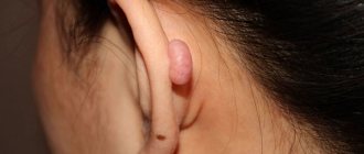
This structure of this organ exposes it to many negative influences from the outside; the ear is also susceptible to inflammatory processes or otohematomas. There are pathological conditions, some of which are congenital in nature and may be reflected in the underdevelopment of the auricle.
The auricle can have different shapes and sizes in different people. But its structure is the same for everyone. This is a cartilaginous zone covered with skin, in which there are many nerve endings. Cartilage is absent only in the earlobe, where the fatty tissue is located in a kind of skin pouch.
Compound
The outer ear consists of 3 main parts:
- The auricle.
- Eustachian tube.
- Eardrum.
Let us consider in detail all the components of each organ.
- The auricle consists of:
- Darwin's tubercle is the outermost convex cartilaginous formation of the ear.
- The triangular fossa is the inner recess of the shell closer to the temporal part.
- The rooks are the depression after the ear tubercle on the outside.
- The pedicles of the helix are cartilage on the auditory opening closer to the face.
- The cavity of the auricle is a tubercle above the opening.
- Antihelix - cartilage protruding above the auditory opening from the outside.
- The curl is the outer part of the shell.
- The antitragus is the lower convex cartilage above the lobe.
- The ear lobe is the earlobe.
- The intertragal notch is the lower part of the auditory opening.
- The tragus is a protruding cartilage closer to the temporal area.
- The supracoscal tubercle is a semicircular cartilage above the auditory opening.
- The helical-tragus groove is the upper part of the ear arch.
- The crus of the antihelix are the depressions and elevations in the upper part of the shell.
- Eustachian tube
- Eardrum
The canal connecting the external concha and the eardrum is the Eustachian tube or auditory tube. It is through it that sound travels, which causes certain impulses in the thin membrane of the outer ear. The middle ear system begins behind the eardrum.
Consists of mucous membrane, squamous epithelial cells, fibrous fibers. Thanks to the latter, the membrane is plastic and elastic.
Where is the middle ear located - Wow!
The middle ear is part of the human auditory system. It is a small space between two other parts of the organ: the external auditory canal and the labyrinth (inner ear).
Middle ear, its functions and structure
- Structural features
- Main functions
The middle ear is a component of the ear. Occupies the space between the external auditory organ and the eardrum. Its structure involves numerous elements that have certain features and functions.
Structural features
The middle ear consists of several important elements. Each of these components has structural features.
Tympanic cavity
This is the middle part of the ear, very vulnerable, often subject to inflammatory diseases. It is located behind the eardrum, not reaching the inner ear. Its surface is covered with a thin mucous membrane. It has the shape of a prism with four irregular faces and is filled with air inside. Consists of several walls:
- The outer wall with a membranous structure is formed by the inner part of the eardrum as well as the bone of the ear canal.
- The inner wall at the top has a recess in which the window of the vestibule is located. It is a small oval hole, which is covered by the lower surface of the stapes. Below it there is a cape along which a furrow runs. Behind it is a funnel-shaped dimple in which the cochlear window is placed. From above it is limited by a bone ridge. Above the window of the cochlea there is a tympanic sinus, which is a small depression.
- The upper wall, which is called the tegmental wall, as it is formed by hard bone substance and protects it. The deepest part of the cavity is called the dome. This wall is necessary to separate the tympanic cavity from the walls of the skull.
- The lower wall is jugular, as it participates in the creation of the jugular fossa. It has an uneven surface because it contains drum cells necessary for air circulation.
- The posterior mastoid wall contains an opening that leads into the mastoid cave.
- The anterior wall has a bone structure and is formed by substance from the carotid artery canal. Therefore, this wall is called the carotid wall.
Conventionally, the tympanic cavity is divided into 3 sections. The lower one is formed by the lower wall of the tympanic cavity. The middle is the larger part, the space between the upper and lower borders. The upper section is the part of the cavity corresponding to its upper border.
Auditory ossicles
They are located in the area of the tympanic cavity and are important, since without them sound perception would be impossible. These are the hammer, anvil and stirrup.
These elements connect to each other to form real joints. They have limited mobility, but allow you to change the position of the elements. They are connected to each other as follows:
- The hammer has a rounded head connected to the handle.
- The anvil has a rather massive body, as well as 2 processes. One of them is short, rests against the hole, and the second is long, directed towards the handle of the hammer, thickened at the end.
- The stirrup includes a small head, covered on top with articular cartilage, which serves to articulate the incus and 2 legs - one straight and the other more curved. These legs are attached to the oval plate contained in the fenestra vestibule.
The main function of these elements is the transmission of sound impulses from the membrane to the oval window of the vestibule . In addition, these vibrations are amplified, which makes it possible to transmit them directly to the perilymph of the inner ear.
This occurs due to the fact that the auditory ossicles are articulated in a lever manner. In addition, the size of the stapes is many times smaller than the eardrum. Therefore, even small sound waves make it possible to perceive sounds.
Muscles
The middle ear also has 2 muscles - they are the smallest in the human body. The muscle bellies are located in the secondary cavities. One serves to tension the eardrum and is attached to the handle of the hammer. The second is called the stirrup and is attached to the head of the stapes.
These muscles are necessary to maintain the position of the auditory ossicles and regulate their movements. This provides the ability to perceive sounds of varying strengths.
Eustachian tube
The middle ear connects to the nasal cavity through the Eustachian tube. It is a small canal, about 3-4 cm long. On the inside it is covered with a mucous membrane, on the surface of which there is ciliated epithelium. The movement of its cilia is directed towards the nasopharynx.
Conventionally divided into 2 parts. The one that is adjacent to the ear cavity has walls with a bone structure. And the part adjacent to the nasopharynx has cartilaginous walls.
In the normal state, the walls are adjacent to each other, but when the jaw moves, they diverge in different directions.
Thanks to this, air flows freely from the nasopharynx into the hearing organ, ensuring equal pressure within the organ.
Due to its close proximity to the nasopharynx, the Eustachian tube is susceptible to inflammatory processes, since infection can easily enter it from the nose. Its patency may be impaired due to colds.
In this case, the person will experience congestion, which brings some discomfort. To deal with it, you can do the following:
- Examine the ear. An unpleasant symptom may be caused by an ear plug. You can remove it yourself. To do this, drop a few drops of peroxide into the ear canal. After 10-15 minutes, the sulfur will soften, so it can be easily removed.
- Move your lower jaw. This method helps with mild congestion. It is necessary to push the lower jaw forward and move it from side to side.
- Apply the Valsalva technique. Suitable in cases where ear congestion does not go away for a long time. It is necessary to close your ears and nostrils and take a deep breath. You should try to exhale it with your nose closed. The procedure should be carried out very carefully, as during it the blood pressure may change and the heartbeat may accelerate.
- Use Toynbee's method. You need to fill your mouth with water, close your ears and nostrils, and take a sip.
If after carrying out the above manipulations the symptom does not go away, you should consult a doctor. Otherwise, complications may develop.
Source: https://buk-journal.ru/istoriya/gde-nahoditsya-srednee-uho.html
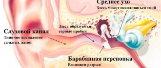
The auditory canal is the structure of the outer ear.
music, birdsong, the sound of the sea surf. In the process of phylogenesis of the biological species “Homo sapiens,” the organ of hearing played a vital role, as it ensured the manifestation of such a phenomenon as human speech. The sections of the hearing organ were formed during human embryonic development from the outer germ layer - the ectoderm.
Further. The auditory canal is a tube consisting of cartilage and partly bone tissue. It is covered with epithelium containing modified sweat glands that secrete sulfur, which moisturizes and disinfects the passage cavity. The muscles of the auricle in most people are atrophied, unlike mammals, whose ears actively respond to external sound stimuli.
The visible part of our hearing aid is the auricle. It is here that sound waves enter, which then go into the Eustachian tube and are brought to the eardrum - a thin membrane that reproduces sound impulses and sends them further - to the middle and inner ear.
The auricle is the part that we see from the outside. Its main function is sound perception. Therefore, it must always be clean and allow sound waves to pass through without obstacles.
If the auricle becomes clogged with wax or pathogenic microelements during the inflammatory process, then a visit to an otolaryngologist is necessary. External damage to the auricle may be associated with:
The Eustachian tube performs several functions:
- Conducts sound.
- Protects the inner ear from damage, infections, and foreign objects.
- Stabilizes blood pressure.
- Drainage – spontaneous cleaning of the pipe from excess cells and tissues.
- Provides ventilation of the auditory organ.
Frequent diseases of this organ are inflammatory processes, in particular tubo-otitis. For any discomfort in the ear area or partial temporary hearing loss, contacting an otolaryngologist is mandatory. The eardrum performs the following functions:
- Sound conductivity.
- Protecting inner ear receptors.
Eardrum problems
Another middle ear disease is a ruptured eardrum. This damage can be caused by infection or injury. A ruptured membrane causes pain, bleeding, hearing loss, and noise or ringing in the ears. The presence of perforation is determined using an otoscope. A small tear heals on its own; large damage requires surgical intervention.
The most common cause of non-traumatic tympanic membrane rupture is otitis media. Some of the traumatic causes include:
- A sudden increase in pressure caused by an explosion, a blow to the ear, or diving to great depths.
- A sudden decrease in pressure caused by flying in an airplane or using various devices that draw air near the ear canal.
- Severe head trauma, especially if the bones at the base of the skull are damaged.
A rupture in the tympanic membrane can be caused by the penetration of foreign objects into the ear canals (for example, cotton swabs, a pencil, a low-hanging tree branch). An object that penetrates the eardrum can dislodge or damage the bones in the middle ear, which connect the eardrum to the inner ear. In this case, pieces of broken bones or fragments of a penetrated object can even end up in the inner ear and cause irreversible hearing loss.
Features of the anatomy of the inner ear
The ear is a paired organ located deep in the temporal bone. The structure of the human ear allows it to receive mechanical vibrations in the air, transmit them through internal media, transform them and transmit them to the brain.
The most important functions of the ear include analysis of body position and coordination of movements.
General structure
The anatomical structure of the human ear is conventionally divided into three sections:
- external;
- average;
- internal.
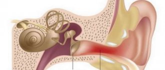
When making a diagnosis for this or that reason, otolaryngologists, first of all, have to find out in which part of the ear the focus of the disease arose. Often patients complaining of pain cannot determine exactly where the inflammation occurs. And all because they know little about the anatomy of the ear - a rather complex organ of hearing, consisting of three parts.
Below you can see a diagram of the structure of the human ear and learn about the features of each of its components.
There are quite a few diseases that lead to ear pain. To understand them, you need to know the anatomy of the ear. It includes three parts: the outer, middle and inner ear. The outer ear consists of the pinna, the external auditory canal, and the eardrum, which is the boundary between the outer and middle ear.
The middle ear is located in the temporal bone of the skull. It includes the tympanic cavity, the auditory (Eustachian) tube and the mastoid process.
The photo above shows a diagram of the structure of the human ear: inner, middle and outer.
Let's start with the anatomy of the external ear: it is supplied with blood through the branches of the external carotid artery. In addition to the branches of the trigeminal nerve, the innervation involves the auricular branch of the vagus nerve, which branches in the posterior wall of the auditory canal. Mechanical irritation of this wall often contributes to the appearance of the so-called reflex cough.
The structure of the outer ear is such that the outflow of lymph from the walls of the ear canal enters the nearest lymph nodes located in front of the auricle, on the mastoid process itself and under the lower wall of the ear canal. Inflammatory processes that occur in the external auditory canal are quite often accompanied by a significant increase and the appearance of pain in the area of these lymph nodes.
If you look at the eardrum from the side of the ear canal, you can see a funnel-shaped concavity in its center. The deepest place of this concavity in the structure of the human ear is called the navel.
The anterior and posterior folds diverge from it anteriorly and posteriorly. They separate the relaxed part of the eardrum from the tense part.
The anatomy of the middle ear includes the tympanic cavity, mastoid process and eustachian tube, which are interconnected. The tympanic cavity is a small space located inside the temporal bone, between the inner ear and the eardrum.
The structure of the middle ear has the following feature: in front, the tympanic cavity communicates with the cavity of the nasopharynx through the Eustachian tube, and in the back, through the entrance to the cave, with the cave itself, as well as with the cells of the mastoid process.
The tympanic cavity contains air that enters it through the Eustachian tube.
The anatomy of the structure of the human ear up to three years of age differs from the anatomy of the adult ear: newborn children do not have a bony auditory canal, as well as a mastoid process.
In the upper regions, where there is no bony ring, the eardrum is attached directly to the lower edge of the scale of the temporal bone, which is called the Rivinian notch. When a child turns three years old, his external auditory canal is fully formed.
The structure of the inner ear includes the bony and membranous labyrinths. The bony labyrinth surrounds the membranous labyrinth on all sides, looking like a case. The membranous labyrinth contains endolymph, and the free space remaining between the membranous and bony labyrinth is filled with perilymph, or cerebrospinal fluid.
The bony labyrinth includes the vestibule, cochlea and three semicircular canals. The vestibule is the central part of the bony labyrinth.
On its outer wall there is an oval window, and on the inner wall there are two impressions necessary for the vestibular sacs, which have the appearance of membranes.
The anatomy of the inner ear is such that in the interconnected sacs of the vestibule there are otolithic devices, or terminal devices of statokinetic reception. They consist of a specific nerve epithelium, which is covered on top by a membrane. It contains otoliths, which are crystals of phosphate and carbon dioxide of lime.
The semicircular canals are located in three mutually perpendicular planes. The external canal is horizontal, the posterior is sagittal, the upper is frontal. Each of the semicircular canals has one dilated and one simple, or smooth, pedicle. The sagittal and frontal canals have one common smooth pedicle.
In the ampulla of each of the membranous canals there is a comb. It is a receptor and is a terminal nervous apparatus composed of highly differentiated nerve epithelium. The free surface of the epithelial cells is covered with hairs that perceive any displacement or pressure of the endolymph.
The cochlea is a bony canal that forms two whorls around a bony shaft. The external resemblance to the common garden snail gave this organ its name.
This article has been read 67,807 times.
Unlike the outer and middle ear, the inner ear has the most complex structure; otolaryngologists call it a labyrinth. This part of the ear includes:
- vestibule;
- snails;
- semicircular tubules.
Then the division occurs according to the anatomical forms of the labyrinth.
In the vestibule, the cochlea, saccule and utricle unite into the endolymphatic duct. Here is the clinical form of the receptor fields. Next are the semicircular canals:
- front;
- rear;
- lateral.
Each of these canals has a pedicle and an ampullary end.
The inner ear has the shape of a cochlea, its parts are:
- staircase vestibule;
- duct;
- scala tympani;
- organ of Corti.
Pillar cells are located in the organ of Corti.
This part of the peripheral auditory sensory system is located deep in the temporal bone. It consists of semicircular canals related to the organ of balance and the bony labyrinth. The last structure contains the cochlea, inside which is the organ of Corti, which is a sound-receiving system.
Along the spiral, the cochlea is divided by a thin vestibular plate and a denser basilar membrane. Both membranes divide the cochlea into canals: lower, middle and upper. At its wide base, the upper canal begins with an oval window, and the lower one is closed with a round window. Both of them are filled with liquid contents - perilymph.
It is considered a modified cerebrospinal fluid - a substance that fills the spinal canal. Endolymph is another fluid that fills the canals of the cochlea and accumulates in the cavity where the nerve endings of the balance organ are located. Let's continue to study the anatomy of the ears and consider those parts of the auditory analyzer that are responsible for transcoding sound vibrations into the process of excitation.
What is the middle ear and how to treat diseases associated with it?
The middle ear is part of the human auditory system. It is a small space between two other parts of the organ: the external auditory canal and the labyrinth (inner ear).
Structure of the middle ear
The middle ear contains:
- tympanic cavity;
- auditory (Eustachian) tube;
- a cave surrounded by mastoid cells.
Let's take a closer look at the structure of the middle ear. Each cavity is filled with air. The tympanic cavity of the middle ear is shaped like a tambourine, standing on its edge and strongly inclined towards the external auditory canal. It is small in volume - only about 1 cm³.
The middle ear contains three auditory ossicles: the malleus, the incus and the stapes. They got their name from their appearance. The auditory ossicles are located directly behind the eardrum. They are connected by a pair of real joints of limited mobility. They are also strengthened by a number of individual ligaments, so they represent a more or less movable chain.
However, in the direction from the malleus to the stapes, the mobility of the auditory ossicles gradually decreases. In this way, the spiral organ of the inner ear is protected from shocks and the negative effects of loud sounds.
Between the tympanic cavity and the nasopharynx is the Eustachian tube, through which the pressure in the middle ear is equalized. If it does not correspond to a and the person reflexively begins to yawn.
Functions of the middle ear
The function of the middle ear is sound conduction. Wave-like vibrations of air create sound waves that vibrate the eardrum and auditory ossicles. These vibrations, slightly modified, are transmitted to the inner ear.
The structure of the middle ear allows it to perform the following functions:
- maintaining the eardrum and the chain of auditory ossicles in good shape;
- adaptation of the acoustic apparatus to sounds of varying strength and pitch;
- protection from harsh sounds.
When pressure in the middle ear increases, the amplitude of vibration of the auditory ossicles decreases.
As a result, the sensitivity of the acoustic device decreases. About 10 ms after the appearance of a sound of more than 40 dB, two muscles begin to reflexively contract.
One of them, attached to the handle of the hammer, increases the tension of the eardrum and reduces the amplitude of its vibrations. The other limits the vibrations of the stapes.
Thanks to this, the human auditory system adapts to intense sounds that can harm the body.
However, the protective function does not operate in the event of unexpected sounds. For example, a sudden explosion can damage the acoustic apparatus, since the reflex contraction of muscles in the middle ear is delayed.
Middle ear diseases
Middle ear diseases include a number of pathological conditions. All of them are called otitis. The diseases are equally common among both adults and children.
Often, otitis media leads to hearing loss, which reduces social activity and professional suitability. Neglected cases threaten intracranial complications and even death. That is why it is so important to diagnose the disease in time and begin treatment.
Otitis is divided into acute and chronic. Moreover, the acute form easily turns into chronic. There are also serous and purulent otitis media.
These diseases are rarely primary and almost always develop with inflammation of the upper respiratory tract. When you have a cold, bacteria and viruses travel from the nasopharynx into the auditory tube, and then into the middle ear.
Thus, provoking factors are diseases that make nasal ventilation difficult:
- adenoids;
- nasal polyps;
- abnormal structure of the nasal septum;
- hypertrophy of the nasal concha;
- sinusitis.
The prevalence of inflammation and the possibility of full recovery after the disease depend on the stage of damage to the auditory tube, the virulence of viruses and bacteria, and the resistance of the patient’s body.
Symptoms of otitis media
Symptoms of otitis consist of the following signs:
- pain in the ear and surrounding tissues.
- headache, in rare cases - vomiting;
- hearing impairment;
- feverish conditions;
- noise in ears;
- sensation of a foreign body in the ear cavity.
When the first symptoms appear, you should consult a doctor, since untimely or incorrect treatment is fraught with complications.
For a patient with acute otitis, the doctor will first of all prescribe bed rest. Medicines prescribed include antibiotics, sulfonamides, vasoconstrictor nasal drops, compresses and ear warmers. Ear drops relieve pain well.
An inflamed human ear must be protected from drafts. It is useful to warm it up with blue light or a Sollux lamp. Procedures can be performed at home, but only as a supplement to medical prescriptions.
In the case of otitis media, self-medication is strictly contraindicated. With inflammation complicated by the formation of pus, the infection often penetrates into the cranial cavity.
In this case, the risk of developing meningitis, abscesses of the temporal lobe of the brain and cerebellum, sinus thrombosis and even sepsis (blood poisoning) increases.
If the disease is advanced, the doctor will have to make an incision in the eardrum in order to provoke the outflow of pus. If the periosteal tissue is damaged, only surgery can preserve the person’s hearing.
Diagnosis and treatment
Only a qualified otolaryngologist can make an accurate diagnosis of otitis media. First, the doctor examines the patient's ear using an otoscope.
Very often, the signs of the disease are unclear or only partially present, so additional time is required to confirm the diagnosis.
In addition, examination of the ear cavity may be difficult due to the accumulation of earwax. To continue diagnostics, it must be removed.
A comprehensive examination consists of determining the following signs:
- is there inflammation in the tympanic cavity;
- are there any complications (pus, hearing loss, thinning of the eardrums);
- what bacteria or virus are the causative agents, their resistance to antibiotics;
- what is the stage of the disease and is there a need for drug therapy.
When treating otitis, the patient is usually at home; round-the-clock medical supervision is not required. Hospitalization is carried out only if there is a suspicion of severe purulent complications, for example, meningitis.
Drug therapy consists of antibiotics, antipyretics, painkillers (individually or all in combination). Improvement in the patient's well-being, as a rule, occurs within 1 - 2 days. Otherwise, it is necessary to urgently appear for examination by a doctor.
Prevention of otitis media
Prevention of otitis media consists of maintaining careful personal hygiene, timely treatment of diseases of the nose and pharynx, and combating chronic infections.
For the health of the middle ear, it is necessary to treat inflammation of the outer ear in a timely manner. If a person comes into contact with chemicals at work, personal protective equipment must be used.
To exclude acoustic trauma, it is necessary to undergo annual medical examinations. If pathologies are detected, doctors advise changing jobs. In production, it is necessary to use earplugs, swabs, helmets and other protective equipment. The room must be soundproofed.
The structure of the tympanic cavity implies its sensitivity to changes in atmospheric pressure, and there is a risk of barotrauma. Therefore, it is necessary to take precautions when jumping with a parachute, flying in an airplane, or diving to depths. In case of injury, you should not wash your ear yourself, as there is a high risk of infection of the tympanic cavity.
Prevention of vibration injuries in the ear cavity includes vibration isolation, vibration absorption and vibration damping.
If there are any symptoms indicating a pathology of the auditory analyzer, you should immediately contact a specialist. Preventing a disease is always easier than curing it. It is important to realize that damage to the middle ear often results in deafness.
Source: https://vashlor.ru/anatomiya/srednee-uho.html
Physiological characteristics of human ears
Our hearing organ in the body has two key purposes:
- forms and maintains balance of the human body;
- receives and converts noise and vibrations into sound forms.
In order for us to be in balance even during rest, and not just when moving, the vestibular apparatus must work constantly. But not everyone knows that our ability to walk on two legs in a straight line lies in the structural features of the inner ear. This mechanism is based on the principle of communicating vessels, which have the shape of an auditory organ.
This organ includes the semicircular canals, which maintain fluid pressure in our body. When a person changes body position (changes rest for movement and vice versa), the clinical structure of the hearing organ is able to adapt to one or another physiological state and regulates intracranial pressure.
Research methods
The main method of studying S. at. is otoscopy (see). Direct otoscopy with an intact tympanic membrane makes it possible to indirectly judge the condition of S. at. This reveals thinning and blueness of the tympanic membrane in otosclerosis, localization of its hyperemia and infiltration, as well as overhang of the posterior superior wall of the external auditory canal in acute and chronic cases. purulent otitis media, the presence of exudate in the tympanic cavity with exudative otitis media. With limited, subtotal and total defects of the tympanic membrane, otoscopy can be used to directly assess the nature and degree of S.'s pathology. For more detailed otoscopic diagnostics, optical instruments are used - a Siegle funnel (see Siegle funnel), special magnifiers (see Magnifier), and an operating microscope (see). One of the diagnostic procedures is tympanopuncture (see).
Functional studies of the sound-conducting system of S. are the determination of hearing acuity using whispered and spoken speech, tuning fork tests, suprathreshold and subthreshold audiometry (see).
Rice. 2. Schematic representation of head placement for radiography of the temporal bone and diagram of radiographs in three projections: I - lateral (according to Schuller); II - oblique (according to Stenvers); III - axial (according to Mayer); a - front view; b — top view (projections of the foramen magnum and pyramids of the temporal bones are indicated in black); C - sagittal plane; B - vertical axis; H — cassette with X-ray film (the arrow indicates the direction of the X-ray beam): 1 — auricle; 2 - internal auditory canal; 3 - external auditory canal; 4 - head of the lower jaw; 5 - top of the pyramid; 6 - mastoid process; 7 - mastoid cells; 8 - occipital-mastoid suture; 9 - groove of the sigmoid sinus; 10 - parietal-mastoid suture; 11 - cells of the temporal bone scales; 12 - mastoid cave; 13 - lateral semicircular canal; 14 - anterior semicircular canal; 15 - vestibule; 16—snail; 17 - auditory ossicles; 18 - canal of the carotid artery.
An important research method for S. is radiological - radiography of the temporal bone in the lateral (according to Schuller), oblique (according to Stenvers) and axial (according to Mayer) projections (Fig. 2). Schüller's method provides information about the condition of various parts of the ear and is produced as an overview image. From it one can judge the pneumatization of the mastoid process and the scaly part of the temporal bone, the mastoid cave, the size of the openings of the internal auditory canal, its walls, etc. The oblique projection according to Stenvers is an additional way to study only certain details of the pyramid (petrous part) of the temporal bone ; it is not very informative for middle ear pathology. The Mayer axial projection also serves as an additional method, but is used much more often in diseases of S. u. than the Stenvers method. In this position, X-ray radiation is directed along the axis of the pyramid, which makes it possible to obtain an image of important anatomical details of the tympanic cavity - the auditory ossicles, the supratympanic recess (attic), and the sigmoid sinus. The most informative for imaging the tympanic cavity is tomography (see) in a direct (posterior) projection, with both temporal bones visible on the tomogram.
Middle ear cavity
A person receives most of the information about the world around him through vision and hearing. Moreover, the structure of the ear is very complex.
Any disturbances in the middle ear or other parts of the hearing system can lead not only to hearing loss, but also to the creation of a situation where a person’s life is in danger.
Let's figure out what the functions and structure of the middle ear are, what diseases affect this part of the hearing system and how to prevent their occurrence.
The middle ear is located between the inner and outer ears. The main purpose of this part of the hearing aid is to conduct sounds. The middle ear consists of the following parts:
- Auditory ossicles. They are the stirrup, hammer and anvil. It is these details that help transmit sounds, and distinguish them by strength and height. The peculiarities of the auditory ossicles help protect the hearing aid from sharp and loud sounds.
- Eustachian tube. This is the passage connecting the nasopharynx with the tympanic cavity. Its mouth is closed when a person swallows or sucks something. In newly born children, for some time the auditory tube is wider and shorter than in adulthood.
- Tympanic cavity. It is this part of the middle ear that contains the auditory ossicles described above. The location of the tympanic cavity is the area between the outer ear and the temporal bone.
- Mastoid. This is the convex part of the temporal bone. It contains cavities that are filled with air and communicate with each other through narrow holes.
The middle ear is a device that conducts sound vibrations, consisting of air cavities and complex anatomical formations. The tympanic cavity is lined with mucous membrane and is separated from the rest of the skull by an upper wall. All auditory ossicles are also covered with mucous membrane. The middle and inner ear are separated by a bony wall. They are connected to each other only by two holes:
- round window;
- oval window in the ear.
Each of them is protected by a flexible and elastic membrane. The stapes, one of the auditory ossicles, enters the oval window, located in front of the water-filled inner ear.
Air cavities and other anatomical formations located in the middle ear provide sound passage. The main functions of the middle ear are:
- maintaining the functionality of the eardrum;
- transmission of sound vibrations;
- protection of the inner ear from sharp and too loud sounds;
- ensuring the sensitivity of sounds of a wide variety of strength, pitch and volume.
Experts call the following signs and conditions of a person the main symptoms of problems in the middle ear area:
- pain in the ear area of varying intensity (mostly very severe);
- feeling of stuffiness;
- decreased or complete loss of hearing;
- discharge of fluid or pus from the ear canal;
- increased body temperature;
- decreased appetite and poor sleep;
- change in color of the eardrum to a redder color.
Among the most common diseases of the middle ear are the following:
- Purulent otitis media of the middle ear. This is an inflammation in which purulent and purulent-bloody discharge from the ear canal is observed, the person complains of unbearable pain, and hearing deteriorates significantly. The disease affects the middle ear cavity and eardrum, and can spread to other parts of the hearing system.
- Cicatricial otitis. In this case, the inflammatory process led to the formation of scars and decreased mobility of the auditory ossicles. Because of this, severe hearing loss is observed.
- Mesotympanitis. The disease is similar in symptoms to purulent otitis. In this case, the eardrum is affected, and the person notices decreased hearing and purulent discharge.
- Epitempanitis. During this disease, inflammation of the epitympanic space of the middle ear occurs; a protracted course of the inflammatory process can disrupt the structure of the middle and inner ear, which will entail a decrease and a sharp deterioration in hearing.
- Mastoiditis. Most often, this is a consequence of purulent otitis not treated correctly and in a timely manner, which affects not only the middle ear, but also the mastoid process.
- Qatar of the middle ear. The disease usually precedes purulent otitis and affects the auditory tube.
- Bullous otitis media. The disease occurs against the background of influenza and has symptoms similar to other otitis media. The focus of the inflammatory process is located in the supratympanic air cavity.
Wear a hat during the winter season
To prevent the development of middle ear diseases, experts recommend that children and adults adhere to the following rules:
- Treat diseases of the upper respiratory tract, nose and ears in a timely manner. If treatment is incorrectly selected or absent, the infection quickly spreads from the nasopharynx or outer ear further and disrupts the functioning of the hearing aid. Always follow the recommendations of doctors during the treatment of diseases of the ENT organs. Do not stop therapy, even if you feel great, do not change the dosage and treatment regimen of drugs, do not extend the period of their use.
- If a person has congenital abnormalities of the ear structure, then they should be resolved with the help of a specialist, if possible. Sometimes it is necessary to undergo surgery, and in some cases it is enough to take certain medications.
- Maintaining hygiene. Accumulation of wax, dirt or water entering the ear canal can lead to inflammation. Therefore, try to clean your ears and your children’s ears with cotton wool pads in a timely manner. When swimming or bathing, use special caps and earplugs, and avoid direct water jets entering the ear canal.
- Make sure your ears are not injured. The entry of a foreign body, the use of sharp and hard objects when cleaning the ears, as well as some other reasons can cause inflammation and provoke infection in the middle ear.
- In winter, wear a hat. Protect yourself from drafts and hypothermia, sudden changes in temperature and humidity. It is best for young children to wear special thin caps, even if the room temperature is comfortable.
- In childhood, as a preventive measure for frequently occurring otitis media and other inflammatory processes due to overgrown or greatly enlarged adenoids, their removal is sometimes recommended.
Remember, middle ear diseases are very dangerous for a person’s hearing and life. If you have any disturbing symptoms, you should immediately consult a doctor. Self-medication for otitis media and other inflammatory processes is prohibited either in childhood or in adulthood.
This can lead to serious complications, including the spread of infection beyond the middle ear, penetration into the brain, as well as reduction and complete loss of hearing.
The sooner you see a doctor and start treatment, the lower the risk of complications and the higher the chance of eliminating the disease as soon as possible without any consequences.
Loading…
StopOtit.ru » Additional information - tips and tricks
The auditory canal ends with an elastic film that separates the outer ear from its middle part. This is the eardrum. It receives sound waves and begins to vibrate, which causes similar movements of the auditory ossicles - the hammer, incus and stapes, located in the middle ear, deep in the temporal bone.
The hammer is attached to the eardrum with its handle, and its head is connected to the incus. It, in turn, with its long end closes with the stapes, and it is attached to the window of the vestibule, behind which the inner ear is located. Everything is very simple. The anatomy of the ears has revealed that a muscle is attached to the long process of the malleus, which reduces the tension of the eardrum. And the so-called “antagonist” is attached to the short part of this auditory ossicle. A special muscle.
Transition Department
The middle part of the ear is a complex part of the system for transmitting sound vibrations. The eardrum is located here, it ends the designated auditory canal, as well as the smallest details - 3 bones.
Sounds arriving at a certain intensity cause the drumhead to move at different frequencies. After this, the vibrations are transmitted to the “hammer”, the handle of which originates from the membrane. This part of the hearing aid hits the “anvil,” which in turn directs the resulting sound to the “stapes,” but its base connects to the inner ear.
The middle section can be called a transmission mechanism. It does not perceive noise, but is only transmitted to the inner ear, amplifying it 20 times. In terms of total area, the average department occupies no more than 1 square. cm, the location is the temporal bone on the skull.
The perception of incoming and transmitted noise is carried out only by the inner ear. It is separated from the previous parts by oval and round holes, behind which the cochlea is located. There are also small niches here that the body seeks to fill with lymphatic fluid. Such a liquid medium is capable of receiving vibrations coming from the external environment. The received signal enters the brain through special nerve endings. The structure of the ear of a child and an adult is composed of the following parts:
- external auricle;
- ear canal;
- drum record;
- “hammer”, “invil”, “stirrup”;
- round and oval holes;
- vestibule;
- cochlear zone;
- auditory nerve.
Human sound sensations and their nature
Is a person capable of feeling all vibrations in the air? Not really. A person can transform air vibrations only from 16 to thousands of hertz, but we are no longer able to hear infra- and ultrasounds. Thus, infrasounds in nature can appear in the following cases:
- lightning strike;
- earthquake;
- Hurricane;
- storm.
Elephants and whales are especially sensitive creatures to infrasound. They seek shelter when a hurricane or storm approaches. But ultrasounds can be heard by moths, bats and some species of birds. The perception of this type of vibration in nature is called echolocation. It is used in such areas as:
- cosmetology;
- medicine;
- different types of production.
So, we have learned that the structure of the ear includes three main parts:
- external;
- average;
- internal.
Each part has its own anatomical features, which determine their functions. The outer part includes the auricle and the external meatus, the middle part includes the auditory ossicles, and the inner part contains sensitive hairs, respectively. Together, their work ensures that sound vibrations enter the receptors, converting them into nerve impulses, then they are transmitted through neural processes to the central part of the human sensory system.
It is very important to include ear care in your daily hygiene, because if its functional levers are impaired, this can cause hearing loss or a number of diseases related to problems of the middle, inner or outer ear.
Hearing loss leads a person to partial isolation from the outside world, naturally, which is not the same as with vision loss, but the psychological component here is also very strong. Therefore, regularly taking care of your hearing organs and consulting a doctor if something bothers you in this regard is very important for each of us.
The structure of the auricle
Blood circulation of the hearing organs
Special attention should be paid to these functions, especially for those who want to study in detail how the ear and the circulatory system function, which, by the way, is ensured by the trigeminal nerve and the cervical plexus. The auricular nerves provide blood supply to the pinna muscle. The main blood supply is via the external carotid artery.
The structure of the ear is a unique and complex mechanism. Thanks to it, we can perceive various sounds, hear the interlocutor, sing, write music and much more. The organ of hearing helps us communicate and forms our speech correctly. In addition, it is with its help that we can maintain a certain position and maintain balance. Don’t forget to look after this important organ, carry out hygiene procedures, protect yourself from negative external factors, and consult a doctor in time for help.
The site contains exclusively original and author's articles. When copying, place a link to the original source - the article page or home page.
Eustachian tube
The middle ear is connected to the pharynx through a canal named after the scientist who described its structure, Bartolomeo Eustachio. The pipe serves as a device that equalizes the atmospheric air pressure on the eardrum on both sides: from the external auditory canal and the middle ear cavity. This is necessary so that vibrations of the eardrum are transmitted without distortion to the fluid of the membranous labyrinth of the inner ear.
The Eustachian tube is heterogeneous in its histological structure. The anatomy of the ears has revealed that it contains more than just a bone part. Also cartilaginous. Descending down from the middle ear cavity, the tube ends with the pharyngeal opening, located on the lateral surface of the nasopharynx. During swallowing, the muscle fibrils attached to the cartilaginous part of the tube contract, its lumen expands, and a portion of air enters the tympanic cavity.
Structure, functions of the middle ear and its age characteristics
The middle ear is a system of air cavities in the thickness of the temporal bone and consists of the tympanic cavity, the auditory tube and the mastoid process with its bone cells (Fig. 3.)
Rice. 3. Middle ear cavities:
1 —
auditory tube;
2—
tympanic cavity;
3—
cave;
4
- cells of the mastoid process
Tympanic cavity
is the central part of this system and is a narrow space in the thickness of the temporal bone with a volume of about 1 cm3.
There are six walls in the tympanic cavity. The outer wall
along most of its length is the eardrum.
The remaining walls are bone. Inner wall
separates the tympanic cavity from the inner ear.
In this wall there are two openings called fenestra: the oval
or
fenestra
(long diameter 3-4 mm) and
the round
or cochlear window (diameter 1-2 mm).
Inserted into the oval window, as if in a frame, is the foot plate of the stapes, attached to the edges of the oval window by means of an annular ligament.
The round window is covered with an elastic thin membrane, which is called
the secondary tympanic membrane.
The upper wall, or roof of the tympanic cavity, separates
tympanic cavity from the cranial cavity. Bottom wall
borders on a large blood vessel - the bulb of the jugular vein.
In the posterior wall
below there is a hole connecting the tympanic cavity with the mastoid cave.
The upper and lower walls of the tympanic cavity are often very thin, and often, especially in early childhood, there are holes in these walls.
Then the mucous membrane of the tympanic cavity is adjacent directly to the meninges or to the bulb of the jugular vein, which poses a significant danger in the sense of the possible transition of the inflammatory process from the tympanic cavity to the meninges or to the walls of the jugular vein. In the thickness of the inner and posterior walls of the tympanic cavity there is a canal of the facial nerve.
Due to the close anatomical proximity between this canal and the tympanic cavity, the facial nerve can be involved in the inflammatory process developing in the middle ear, and during operations on the middle ear there is a risk of injury to the facial nerve.
auditory ossicles is placed in the tympanic cavity
(Fig. 4), consisting of
a malleus, anvil
and
stirrup.
The hammer has a head, a handle and two processes (short and long).
The anvil consists of a body, short and long processes. The stirrup consists of two arches, a head and a foot plate.
The handle of the malleus is rotated into the fibrous layer of the tympanic membrane, and the lower end of the handle forms a protrusion in the center of the tympanic membrane - the navel, and a short process forms a protrusion in the anterior-superior part.
These protrusions determine the characteristic appearance of the eardrum upon examination. The head of the malleus articulates with the body of the incus, and with its long process it articulates with the head of the stapes.
The footplate of the stapes, as has been said, enters the oval window, which connects the middle ear with the inner ear. A certain tension of the eardrum and the chain of auditory ossicles is provided by two muscles - the tensor tympanic membrane and the stapedius. The first of them is attached to the handle of the malleus, and the second to the head of the stirrup.
Auditory, or Eustachian,
the tube is a canal 3.5 cm long (in adults) connecting the tympanic cavity to the nasopharynx. The tympanic orifice of the Eustachian tube is located in the anterior wall of the tympanic cavity, and the nasopharyngeal orifice is located in the lateral wall of the nasopharynx.
The part of the Eustachian tube that is adjacent to the tympanic cavity is bone, and the part facing the nasopharynx has cartilaginous walls. The entire Eustachian tube is lined with ciliated epithelium: the movement of its hairs is directed towards the nasopharynx.
The walls of the cartilaginous part of the Eustachian tube, usually in contact with each other, diverge at the moment of swallowing (due to the contraction of the pharyngeal muscles), allowing air from the nasopharynx into the tympanic cavity.
In young children, the Eustachian tube is shorter and its lumen is wider than in older children and adults.
Mastoid
is a bone formation similar in shape to the nipple, which is where its name comes from. This is a process of the temporal bone located behind the auricle. In the thickness of the mastoid process there are cells that communicate with each other through narrow slits.
The shape, size and number of these cells are very variable, but one of them, the largest, bears the name of the cave
(antrum), always present. The cave communicates with the tympanic cavity through an opening in the rear wall of the latter. The cave is separated from the cranial cavity by a bone plate, sometimes very thin.
The cells of the mastoid process sometimes reach the large venous sinus of the brain (transverse sinus) and are also separated from it only by a thin layer of bone.
In children under about two years of age, the mastoid process is not yet developed and looks like a bony tubercle. However, the cave already exists in a newborn child.
All cavities of the middle ear (tympanic cavity, Eustachian tube and mastoid cells) are filled with air, and their walls are lined with the thinnest mucous membrane, which is a continuation of the mucous membrane of the nasopharynx. The exchange of air in the middle ear occurs through the Eustachian tube: during swallowing movements, air from the nasopharynx enters the Eustachian tube, and from there into the tympanic cavity and partly into the cells of the mastoid process.
Didn't find what you were looking for? Use the search:
Source: https://studopedia.ru/19_389636_stroenie-funktsii-srednego-uha-i-ego-vozrastnie-osobennosti.html
Significance of the organ of Corti
Inside the cochlea there is a membranous wall called the basilar membrane, on which there is a collection of two types of cells. Some perform the function of support, others are sensory - hair-like. They perceive vibrations of the perilymph, convert them into nerve impulses and transmit them further to the sensory fibers of the vestibulocochlear (auditory) nerve.
Next, the excitation reaches the cortical hearing center, located in the temporal lobe of the brain. It distinguishes sound signals. The clinical anatomy of the ear confirms the fact that what we hear with both ears is important in determining the direction of sound. If sound vibrations reach them simultaneously, a person perceives sound from the front and back. And if the waves arrive in one ear earlier than in the other, then perception occurs on the right or left.
How does sound perception occur?
The sound waves reach the outer concha and are transmitted to the outer ear, where they cause the eardrum to move. These vibrations are amplified by the auditory ossicles and transmitted to the membrane of the middle window. In the inner ear, vibrations provoke the movement of perilymph.
If the vibrations are quite strong, then they reach the endolymph, and it, in turn, provokes irritation of the hair cells (receptors) of the organ of Corti. Sounds of different pitches move fluid in different directions, which is picked up by nerve cells. They convert mechanical vibrations into a nerve impulse, which reaches the temporal lobe of the cortex through the auditory nerve.
A sound wave entering the ear is converted into a nerve impulse
The physiology of sound perception is difficult to study, since sound causes a slight displacement of the membrane, fluid vibrations are very small, and the anatomical region itself is small and located in the capsule of the labyrinth.
The anatomy of the human ear allows it to detect waves from 16 to 20 thousand vibrations per second. This is not much compared to other animals. For example, a cat perceives ultrasound and is able to detect up to 70 thousand vibrations per second. With age, a person's sound perception deteriorates.
Thus, a thirty-five-year-old person can perceive sound no higher than 14 thousand Hz, and those over 60 years old can only perceive up to 1 thousand vibrations per second.
Anatomy of the ears
The anatomical structure of the ears, as well as their component parts, has a significant impact on the quality of hearing. A person’s speech directly depends on the full functioning of this function. Therefore, the healthier the ear, the easier it is for a person to carry out the process of life. It is these features that determine the fact that the correct anatomy of the ear is of great importance.
Initially, it is worth starting to consider the structure of the hearing organ with the auricle, which is the first thing that catches the eye of those who are not experienced in the topic of human anatomy. It is located between the mastoid process on the back side and the temporal mandibular joint in front. It is thanks to the auricle that a person’s perception of sounds is optimal. In addition, this particular part of the ear is of no small cosmetic importance.
The basis of the auricle can be defined as a plate of cartilage, the thickness of which does not exceed 1 mm. On both sides it is covered with skin and perichondrium. The anatomy of the ear also points to the fact that the only part of the shell that lacks a cartilaginous skeleton is the lobe. It consists of skin-covered fatty tissue. The auricle has a convex inner part and a concave outer part, the skin of which is tightly fused with the perichondrium. Speaking about the inside of the shell, it is worth noting that in this area the connective tissue is much more developed.
The auricle is attached to the zygomatic, mastoid process and scales of the temporal bone through muscles and ligaments.
Tympanic cavity
Clinical anatomy of the middle ear includes information about its structure and function. This part of the hearing organ includes the tympanic cavity, as well as the auditory tube with a system of air cells. The cavity itself is a slit-like space in which 6 walls can be distinguished.
Moreover, in the middle ear there are three ear bones - the incus, malleus and stirrup. They are connected using small joints. In this case, the hammer is in close proximity to the eardrum. It is he who is responsible for the perception of sound waves transmitted by the membrane, under the influence of which the hammer begins to tremble. Subsequently, the vibration is transmitted to the incus and stapes, and then the inner ear reacts to it. This is the anatomy of the human ears in their middle part.
How does the inner ear work?
This part of the hearing organ is located in the area of the temporal bone and looks like a labyrinth. In this part, the resulting sound vibrations are converted into electrical impulses that are sent to the brain. Only after this process is completely completed is a person able to respond to sound.
It is also important to pay attention to the fact that the human inner ear contains semicircular canals. This is relevant information for those who study the structure of the human ear. The anatomy of this part of the hearing organ looks like three tubes that are bent in the shape of an arc. They are located in three planes. Due to the pathology of this part of the ear, disturbances in the functioning of the vestibular apparatus are possible.