Non-infectious causes
Poor circulation in the superficial capillaries and inflammation of the dermis can be caused by non-infectious causes.
They can be both external and internal. If the red nose is the result of the adverse effects of exogenous factors, if they are eliminated, the symptom will go away on its own. But if hyperemia was provoked by internal pathologies, it can be eliminated only by undergoing adequate drug therapy. The most likely causes of redness of the skin include:
- rosacea is a non-inflammatory disease, which is expressed in a strong expansion of blood capillaries and the appearance of vascular red “stars” on the skin; often accompanies the development of systemic lupus, rosacea, scleroderma, etc.
- rhinophyma is a chronic pathology characterized by thickening and redness of the skin on the nose, followed by disfigurement of the organ;
- skin allergy - inflammation of the epidermis provoked by allergens: fluff, external ointments, cosmetics, plant pollen.
Basalioma of the nose. Main types, their photos.
Regarding the main types of nasal basal cell carcinoma, one should first of all expect nodular and ulcerative forms, since the anatomical features of the skin predispose them to them. The growth rate is relatively faster than other areas. The photo shows nasal basal cell carcinoma of the main varieties.
Nasal basalioma of the nodular type with the earliest manifestations. A small pearly plaque (marked with a large arrow) on the wing of the nose and an even smaller one (marked with a small arrow) in the nasolabial fold. These are very early stages of basal cell carcinoma. The gray arrow marks the intradermal nevus.
Further stage of development of nodular basal cell carcinoma. A single shiny nodule of nasal wing basal cell carcinoma with a large number of dilated vessels.
The photo shows a nodular basal cell carcinoma of the nose. A large, hard reddish shiny nodule with small ulcerations on the skin of the wing of the nose.
In the photo, pigmented basalioma of the nose on the left is of the pigmented variety; an ulcer appears in the central part.
Ulcerative basalioma of the nose under a scab. Periodically, part of the scab may come off, and the tumor then begins to bleed. In some people, the skin is completely covered with keratomas, which is why a basal cell carcinoma of this type may be perceived for some time as just another keratoma.
The photo shows a nasal basalioma of the ulcerative variety. A large round ulcer on the tip of the nose with ridge-like borders.
Large nasal basal cell carcinoma extending to the inner corner of the eye. Treatment of such a disease is not an easy task; usually a flap of skin is taken from another area and transplanted instead of the removed tissue; cryodestruction is also possible.
The photo shows a basal cell carcinoma of the nose in the dorsal and lateral areas. Closer to the center of the nose, basalioma appears as a recessed area, resembles a scar, and belongs to the sclerosing variety. Along the edge of the tumor on the right, basal cell carcinoma transforms into a nodule with ridge-like edges, taking on the appearance of a nodular variety.
Fibrosing basal cell carcinoma of the nose looks like scar plaques in the photo. To the touch it turned out that the tumor was spreading beyond the visible boundaries.
The photo shows the remaining healthy tissue after removal of nasal basal cell carcinoma. The disease was treated surgically with step-by-step examination under a microscope (Mohs method). The study showed that the tumor penetrates very deeply, even growing into the cartilage.
Functions
If we talk about the functions performed by the nasal cavity, we can highlight the following:
- respiratory function is perhaps the most important. It is this that ensures the supply of the required amount of oxygen to the tissues of the whole body;
- protective function - it guarantees the purification of the air inhaled by a person from dust, as well as pathogenic bacteria, and also consists in heating cold air. All these functions are performed by the mucous membrane of the nasal cavity;
- resonator function - it allows you to give a special sonority and individual coloring to the human voice. The paranasal sinuses are also involved;
- olfactory function, which allows you to distinguish odors and aromas.
Considering in more detail the protective function performed by the nasal cavity, we note that it is assigned to the mucous membrane that lines it. The hairs located in the nasal passages retain only the smallest part of dust and other impurities contained in the air. The remaining part settles on the ciliated epithelium, the cilia of which propel small particles towards the nasopharynx. From here they will enter the esophagus, which is not scary, or they will cough up.
In addition, the mucous membrane contains many nerve endings, which, when irritated by dust, cause sneezing. It allows you to clear the airways of accumulated “garbage”.
Functions of the nasal cavity
Goblet cells and glands in the nasal mucosa control the humidity of inhaled air. If it turns out to be insufficient, the secretion of the glands increases. The secreted mucus contains substances such as lysozyme, mucin and others. They help destroy pathogenic microflora, reducing the likelihood of developing any disease.
There are many venous plexuses in the submucosal layer, and if we inhale cold air, the veins dilate and the blood flow to them increases. Accordingly, heat transfer also increases, which allows you to heat the air to a comfortable temperature.
How does the human nose work?
The nose is a part of a person’s face located below the bridge of the nose, in the lower region of which there are nostrils that perform respiratory and olfactory functions (see photo).
Diagram of the structure of the human nose:
Structure of the outer part of the nose
The structure of the external nose is represented by:
- partition;
- back;
- wings;
- tip.
In a newborn baby, it consists entirely of cartilage. By the age of three, the nose is partially strengthened by bone, like that of an adult. At the age of 14 years, several cartilages occupy 1/5 of its part.
The nostrils are lined with short hairs and trap fine dust, preventing it from entering the lower respiratory tract. In the narrow passages of the nose, cold air has time to heat up so that it can then pass through a number of other organs without causing inflammation of the bronchi and lungs.
The nasal cavity is limited by the palate, which consists of the hard (or bony) palate in front and the soft palate in the back, which does not contain bone. The oral cavity and tongue are also located nearby. The epiglottis is the entrance to the trachea, which in turn leads to the lungs, esophagus and stomach.
Internal structure of the nose
Inner parts of the nose:
- cavity;
- paranasal sinuses.
They are connected to each other, have a common muscular wall of the throat and communicate with the inner ear. Therefore, when there is inflammation of any internal ENT organ, there is a risk of developing secondary infection of all three sections and cavities of the throat and ear, for example, purulent otitis media is caused by the leakage of pus from the maxillary sinuses or sinus.
The picture below shows a cross-section of the structure of the nasopharynx: from the inside there is a nasal cavity connected to the throat and the mouth of the auditory tube.
The anatomy of the inside of the nose is very complex. The relief-shaped mucous membrane serves to warm and humidify the air, which then enters the bronchi and lungs. The following types of walls are unified in both cavities:
- Lateral wall - it consists of individual bones, the upper cheekbone, and the hard palate;
- The upper wall is represented by the ethmoid bone. The cranial nerves responsible for smell and touch pass through its openings;
- The lower wall consists of processes of the hard palate and maxillary bones.
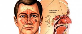
Paranasal sinuses and their functions
The photo shows that in the area of each shell there is an opening through which the sinuses communicate with the nasal cavity. For example, the cephaloid sinus communicates with the nasal cavity in the area of the superior turbinate.
The frontal sinus communicates in the area of the middle concha.
The maxillary sinus, like the frontal sinus, communicates with the nasal cavity at the middle concha.
The frontal sinus is located above the orbit and has an anastomosis in the middle concha.
The sphenoid sinus is located medial (center) to the orbit and has an anastomosis in the superior and inferior turbinates.
Turkish saddle. In its center is the pituitary fossa. In weakened people, the sinuses are often clogged with purulent contents, therefore, to prevent rhinitis, you need to rinse your nose every morning with saline solution at room temperature.
The olfactory zone is represented by special neurosensory cells that contain olfactory receptors. They are contained in the olfactory membrane and in the upper wall of each nasal passage. Olfactory receptors send signals to the first cranial nerve, which transmit them to the brain to the center of smell.
Rhinitis can lead to sinusitis or sinus inflammation. To prevent this complication, you need to start treatment in a timely manner (inhalations, vasoconstrictors, nasal drops).
The anatomical features of the nose are adapted for the best functioning of the body. An incorrectly shaped nose can cause improper drainage of tear fluid, followed by inflammation of the maxillary sinuses and sinuses.
Rhinoplasty is an operation that involves surgically straightening the nasal septum. The incorrect section of bone is removed and a plastic prosthesis is placed in its place.
Anatomical features of the nasal cavity
The nasal cavity is its internal part. Inhaled and exhaled air passes through it. The cavity is divided by the nasal cartilaginous septum into right and left parts. The exit openings of the cavity are called nostrils. Next, the cavity is made up of three pairs of turbinates; they form the nasal passages through which air circulates. Each half of the cavity has four walls:
- The lower wall, also called the bottom, consists of the bones of the hard palate.
- The upper wall is a thin bone plate. Vessels and the olfactory nerve pass through it.
- The inner wall is the nasal septum.
- The lateral wall consists of the nasal conchae. They are divided into three parts. They are interconnected and air moves through them.
Nasal turbinates:
- Inferior turbinate. It is an independent bone, attached to the maxillary bone and the bone of the palate. In its anterior part there is a lacrimal canal. The lower shell is covered with soft tissues that respond to temperature changes.
- The middle turbinate is attached to the palatine and frontal bones. It serves to direct the air flow.
- The superior concha is the smallest and shortest, located next to the olfactory area.
What do you use to pierce your nose in salons?
Nose piercing with a gun is done only by unprofessional and unqualified craftsmen. The thing is that the gun tears the tissue, thereby aggravating the healing process. After a session using a gun, tissue regeneration takes longer, with more frequent cases of infection and suppuration at the puncture site. Therefore, in specialized salons, the nose is pierced only with a sterile needle.
In addition, piercing guns are by no means the safest option in terms of sterility. Even with thorough cleaning, there is a high probability that there will be remnants of foreign epithelium in it, which can lead to diseases such as hepatitis or HIV.
Basic functions of the nasal cavity
The processes occurring in the respiratory system have a decisive influence on the functioning of internal organs and systems. Under favorable conditions, the nose performs basic functions stably and uninterruptedly, ensuring healthy nasal breathing. These tasks include the following.
Respiratory
On its basis, tissues and organs are supplied with oxygen, which is a necessary condition for human life. If there are any factors that interfere with the performance of this function, then the activity of the nervous, cardiovascular, and digestive systems is also disrupted. In addition, if the child’s nasal breathing was impaired for a long time, then disturbances in the development of the facial skeleton and chest organs, slowness of intellectual development and memory loss were observed.
Protective
It involves purifying the air entering the nasal passages from dust and various harmful impurities. The peculiarities of the composition of nasal mucus and the disinfecting abilities of the mucous membrane can significantly reduce the number of harmful microorganisms contained in the air. Manifestations of the protective function of the nose include reflex sneezing and lacrimation, which is a natural protective reaction to the action of an external stimulus;
Function of warming the air entering the nose
This occurs due to the abundant blood supply and filling of the spaces of the nasal concha with blood;
Olfactory
This function is carried out by the olfactory zone of the mucous membrane, which allows one to perceive and interpret various odors that come with the air;
Resonator
It lies in the fact that the nasal cavity, like the cavities of the paranasal sinuses, are air resonators of the voice, capable of giving it tonality and individual sound.
Anatomical atlases and books on the physiology of aging
For those who prefer traditional books:
“Atlas of Human Anatomy” in a gift edition with high-quality detailed illustrations and accurate text explanations can be found
A beautiful, well-organized Russian-Latin reference book on human anatomy for a wide range of readers.
Large atlas of human anatomy in visual tables
The best anatomical tables in good quality.
The book “The Resurrection of the Face or an Ordinary Miracle. Theory and practice of restoring youth” is the most popular book by Natalia Osminina, the author of the Revitonics rejuvenation system, about the physiology of aging of the skin, facial muscles, and restoring the youth of the face.
Natalia Osminina: Resurrection of the face, or an ordinary miracle. Theory and practice of restoring youth
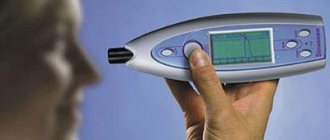
Diagnostic methods
Diagnosis is based on a cytological and histological study of material taken in the initial stages of the disease by scraping or biopsy (cutting out a piece of tissue) from the tumor area. If the basal cell carcinoma has already turned into an ulcerative form, then a smear-imprint is taken from the bottom of the ulcer or erosion for examination.
Dermatoscopy using a special device, a dermatoscope, is highly informative, allowing one to examine the tumor in great detail. It is especially necessary in cases where it is necessary to differentiate basal cell carcinoma from melanoma.
To make a diagnosis of basal cell carcinoma, the doctor may also examine the patient's skin under a Wood's lamp. For this procedure, a special cream is applied to the affected area, then an examination is performed in a dark room.
Photo gallery: devices for diagnosing basal cell carcinomas
Ultrasound is also very important for diagnosis, as it allows one to accurately determine the boundaries, size and depth of tumor growth.
This is very important for choosing a treatment method and deciding on the extent of surgical intervention.
The final diagnosis can be made only after a thorough study of the data obtained from all the studies described above. As a rule, diagnosing basal cell carcinoma of the nasal skin does not cause difficulties for a qualified specialist.
The main difference between basal cell carcinoma and other malignant skin processes is its slow growth and the almost complete absence of distant metastases.
Features of nasal basal cell carcinoma.
The nose is the most prominent part of the face; visible damage (including basal cell carcinoma) on the skin of the nose is often noticed by patients themselves at the very beginning of the disease. Due to its protruding position, it is most exposed to sunlight. For this reason, skin basalioma most often affects the nose. The skin in the area of the cartilaginous part of the nose (tip and wings) is very thin and contains virtually no fatty tissue underneath. As a result, nasal basal cell cartilage penetrates relatively quickly into the cartilage; getting it out of the cartilage is only possible with great difficulty and is associated with disfiguring surgical interventions (see photo below). Basalioma of the nose on its lateral surface at the border with the inner edge of the eye is also very dangerous - here there is a risk of penetration into the periorbital tissue. If you have already had basal cell carcinoma before, the nose requires the greatest protection and attention, because after treatment, 30-40% will develop the disease again, but in a different place. Tanning is contraindicated, and in the summer, in addition to a hat, a good sunscreen is needed.
Why are paranasal sinuses needed?
The evolutionary origin of the sinuses still remains an incompletely clarified issue.
The paranasal sinuses perform the following functions:
- Protective. The air in the cavities helps to absorb the force of impact during skull injuries.
- Baroreceptor. The presence of sinuses allows the body to respond to changes in environmental pressure.
- Resonator. The paranasal sinuses and nasal cavity influence the volume and timbre of spoken speech.
- Thermal insulation. Some sinuses are located on the border with organs that are sensitive to changes in heat and cold, for example, the eyeballs and the roots of the teeth of the upper jaw. The sinuses play the role of an “air cushion” that prevents sudden temperature changes during breathing.
- Moisturizing. Air circulates slowly in the sinuses communicating with the nasal cavity. Due to the fact that it comes into contact with the mucous membrane of the sinuses, the inhaled flow is moistened and warmed. For this reason, if the sinuses are affected, treatment should be immediate.
- Reduction of skull mass. The weight of the bones, despite their relatively large volume, remains small due to the air chambers. The main sinus that plays a role in this is the maxillary sinus.
The structure of the nose. Bones, cartilage, soft tissue
The nose, or rather its visible part, consists of the so-called: root of the nose, back, wings and tip. The internal structure of the nose consists of a hard, bone base, softer cartilage and soft tissue.
Nose bones
The bony skeleton of the nose is formed by the frontal processes of the maxillary bones and the nasal bones. The nasal bones are located in the upper third of the nose and are shaped like a pyramid.
Nasal cartilages
The middle and lower parts of the nose (lower 2/3) consist of cartilage tissue. Cartilage gives shape to the tip of the nose and the lower part of the bridge of the nose.
The cartilaginous skeleton of the nose consists of several symmetrical cartilages and an unpaired cartilage of the nasal septum. The nasal septum cartilage complements the bony nasal septum. It is the anterior edge of this cartilage that largely determines the shape of the dorsum of the nose.
Most people have a deviated nasal septum, but the nose may look symmetrical. A slight curvature of the nasal septum is considered normal and does not require correction.
In the lateral walls of the noses, complementing their bone base, lie the lateral cartilages. In the thickness of the wings there are alar cartilages and small, irregularly shaped accessory and sesamoid cartilages.
Muscles and soft tissues of the nose
On top of the supporting structures is soft tissue, which consists of muscle, fat and skin. The structure, thickness of the skin and fat in the nose varies from person to person, which also affects how the nose looks. As a result, some people have a thin, narrow nose, while others have a fat and convex nose.
The lateral, large pterygoid cartilages of the nose and the frontal process are covered with muscles on top. With the help of these muscles, a person retracts the wings of the nose and compresses the nasal openings.
Muscles are also attached to the legs of the wing cartilage. This is the muscle that lowers the nasal septum and the muscle that elevates the upper lip.
Nasal muscles, training of which can affect the shape of the nose:
Nasal cavity
It is represented by three choanae or nasal conchas, between which the human nasal passages are located. They are localized between the oral cavity and the anterior fossa of the skull - the entrance to the skull.
| Characteristic | Top stroke | Average stroke | Bottom stroke |
| Localization | The space between the middle and superior conchae of the ethmoid bone. | · The space between the inferior and middle conchae of the ethmoid bone; · divided into basal and sagittal parts. | · The lower edge of the ethmoid bone and the bottom of the nasal cavity; · connected to the ridge of the upper jaw and the bone of the palate. |
| Anatomical structures | The olfactory region is the receptor zone of the olfactory tract, exiting into the cranial cavity through the olfactory nerve. The main sinus opens. | Almost all sinuses of the nose open, except for the main sinus. | · Nasolacrimal duct; · The mouth of the Eustachian (auditory) tube. |
| Function | Sensitive – smells. | Air flow direction. | Provides outflow of tears and communication with the inner ear (resonator function). |
Structure of the nasal cavity.
When performing rhinoscopy, the ENT doctor can only see the middle passage; beyond the rhinoscope are the upper and lower ones.
Categories
AllergistAnesthesiologist-resuscitatorVenereologistGastroenterologistHematologistGeneticGynecologistHomeopathDermatologistPediatric gynecologistPediatric neurologistPediatric urologistPediatric surgeonPediatric endocrinologistNutrologistImmunologistInfectious disease specialistCardiologistCosmetologistSpeech therapistElorologistMammologistMedical lawyerNarcologistNeurologistNeurosurgeon NephrologistNutriciologistOncologistOncourologistOrthopedist-traumatologistOphthalmologistPediatricianPlastic surgeonProctologistPsychiatristPsychologistPulmonologistRheumatologistRadiologistSexologist-AndrologistDentistTherapistUrologistPharmacistPhytotherapistPhlebologistSurgeonEndocrinologist
How is surgery performed?
There are 2 main methods of performing surgery to correct the shape and size of the nostrils:
- Removal of skin tissue. It can be used in all cases, regardless of the cause of nostril problems. The surgeon makes neat incisions at the base of the wings of the nose, removes the “loose” part of the skin and applies sutures.
- Contraction of the nasal openings. Used only when necessary to correct too wide nostrils. The doctor makes an internal incision through which the thread is pulled. Then it is tightened to the desired size and fixed.
Most often, rhinoplasty is performed by removing skin tissue. The operation algorithm is as follows:
- The doctor makes incisions at the base of the nostrils - this is done one at a time. The final result depends on the depth of the incision - for example, if the surgeon touches the base with a scalpel, the wings of the nose will become narrow.
- The surgeon removes excess flaps of skin. Here you need to work extremely carefully - excision of too large a fragment of skin can lead to an inharmonious change in appearance. It is impossible to correct such an error.
- The edges of the incision are connected and closed with a cosmetic suture.
If you need to solve the problem of the retraction of the wings of the nose, you will need to restore their support. This type of surgery is performed using the patient’s cartilage tissue, which is obtained from the nasal septum or auricle.
Before the operation, each patient needs to undergo an examination - correction of the shape and size of the wings of the nose is carried out under general anesthesia or local anesthesia (the choice depends on the amount of work to be done). To undergo plastic surgery, the patient must have the results of the following studies:
- general blood and urine tests, biochemistry - results are valid for 2 weeks;
- HIV and ECG tests – data is valid for a maximum of one month;
- blood for hepatitis and syphilis - considered reliable within 2 months;
- fluorography - only a conclusion from a radiologist is required, which has no statute of limitations.
If 6 months before the planned rhinoplasty to reduce the nostrils, plastic surgery on the mammary glands was performed, then the surgeon will need the results of their ultrasound examination.
The recovery period lasts no more than 5 days, during which time the appearance becomes acceptable for going to work. For outpatient treatment, the patient is discharged from the clinic a few hours after the operation - the doctor will make sure that he is feeling normal.
If the rhinoplasty was performed under general anesthesia, you will have to stay in the clinic for 1 - 2 days. In the case of artificial formation of supports for the nostrils, you will need to wear a special plaster cast for a week, which supports the implanted cartilages.
For a month after the operation, you should not visit saunas and steam baths, play sports or expose your face to direct sunlight.
Structure
The nose is the initial part of the respiratory tract where air enters. God not only adorned our face with it, but also endowed it with a vital function for all organs and systems. The structure of the human nose is quite complex. In this article we will look at what the human nose consists of.
The nose is a part of a person’s face located below the bridge of the nose, in the lower region of which there are nostrils that perform respiratory and olfactory functions (see photo).
The structure of the external nose is represented by:
- partition;
- back;
- wings;
- tip.
In a newborn baby, it consists entirely of cartilage. By the age of three, the nose is partially strengthened by bone, like that of an adult. At the age of 14 years, several cartilages occupy 1/5 of its part.
The nostrils are lined with short hairs and trap fine dust, preventing it from entering the lower respiratory tract. In the narrow passages of the nose, cold air has time to heat up so that it can then pass through a number of other organs without causing inflammation of the bronchi and lungs.
The nasal cavity is limited by the palate, which consists of the hard (or bony) palate in front and the soft palate in the back, which does not contain bone. The oral cavity and tongue are also located nearby. The epiglottis is the entrance to the trachea, which in turn leads to the lungs, esophagus and stomach.
Inner parts of the nose:
- cavity;
- paranasal sinuses.
They are connected to each other, have a common muscular wall of the throat and communicate with the inner ear. Therefore, when there is inflammation of any internal ENT organ, there is a risk of developing secondary infection of all three sections and cavities of the throat and ear, for example, purulent otitis media is caused by the leakage of pus from the maxillary sinuses or sinus.
The picture below shows a cross-section of the structure of the nasopharynx: from the inside there is a nasal cavity connected to the throat and the mouth of the auditory tube.
The anatomy of the inside of the nose is very complex. The relief-shaped mucous membrane serves to warm and humidify the air, which then enters the bronchi and lungs. The following types of walls are unified in both cavities:
- Lateral wall - it consists of individual bones, the upper cheekbone, and the hard palate;
- The upper wall is represented by the ethmoid bone. The cranial nerves responsible for smell and touch pass through its openings;
- The lower wall consists of processes of the hard palate and maxillary bones.
The photo shows that in the area of each shell there is an opening through which the sinuses communicate with the nasal cavity. For example, the cephaloid sinus communicates with the nasal cavity in the area of the superior turbinate.
- The frontal sinus communicates in the area of the middle concha.
- The maxillary sinus, like the frontal sinus, communicates with the nasal cavity at the middle concha.
- The frontal sinus is located above the orbit and has an anastomosis in the middle concha.
- The sphenoid sinus is located medial (center) to the orbit and has an anastomosis in the superior and inferior turbinates.
Turkish saddle. In its center is the pituitary fossa. In weakened people, the sinuses are often clogged with purulent contents, therefore, to prevent rhinitis, you need to rinse your nose every morning with saline solution at room temperature.
The olfactory zone is represented by special neurosensory cells that contain olfactory receptors. They are contained in the olfactory membrane and in the upper wall of each nasal passage. Olfactory receptors send signals to the first cranial nerve, which transmit them to the brain to the center of smell.
Rhinitis can lead to sinusitis or sinus inflammation. To prevent this complication, you need to start treatment in a timely manner (inhalations, vasoconstrictors, nasal drops).
The anatomical features of the nose are adapted for the best functioning of the body. An incorrectly shaped nose can cause improper drainage of tear fluid, followed by inflammation of the maxillary sinuses and sinuses.
Rhinoplasty is an operation that involves surgically straightening the nasal septum. The incorrect section of bone is removed and a plastic prosthesis is placed in its place.
The nose performs the following functions:
- olfactory;
- attractive;
- respiratory.
Olfactory function. In the internal cavity there are olfactory receptors, with the help of which we can smell a wide variety of smells. With atrophy of the mucous membrane, we may lose our sense of smell.
Atrophy of the nasal mucosa can appear as a result of steam burns, after taking certain medications, due to a strong infectious process in the ENT organs, and even when inhaling chemicals of various origins.
Respiratory function. The air enters the nose, where it is cleared of pathogenic bacteria and warmed, then goes to the lungs, which ensures the supply of oxygen to the blood and the possibility of life for a person.
Many otolaryngological diseases concern the maxillary sinus. What are the maxillary sinuses? This is a paired organ that is located in the maxillary bone and is located close to the eyes. This part of the body must be treated with special attention, since if the maxillary cavity becomes inflamed, serious complications can occur.
The question of where the maxillary sinuses are located can be answered this way: the location of the organ inside the upper jaw. It is presented in the form of an irregular tetrahedral pyramid. The volume of each sinus is up to 18 cubic centimeters. In humans they can have different sizes.
The inner layer of the organ consists of ciliated columnar epithelium.
The structure of the maxillary sinuses is complex. They consist of:
- Nasal wall. It is also called medial. It contains bones, which gradually turn into mucous membrane. This wall is connected to the nasal passage through a special hole.
- Front or front wall. It is covered by cheek tissue, so its density is higher than the other walls.
- Orbital wall. It is very thin and contains venous vessels and the infraorbital nerve. Therefore, if the disease occurs, there is a risk of complications for the eyes and lining of the brain.
- Rear wall. It is quite dense and is located with the pterygopalatine ganglion, maxillary artery and maxillary nerve.
- Bottom wall. It is located at the level of the nose, but can be located lower. In this case, the roots of the teeth may protrude into the wall of the maxillary sinus.
The purpose of the maxillary sinuses for the human body has not yet been fully studied. All functions that explain what sinuses are needed for were divided into two groups. The first is called external and includes:
- the ability to provide mucus, protect the nasal cavity from pathogenic microorganisms;
- participation in the formation of human speech;
- reflex function;
- participation in the process of smell;
- regulation of pressure inside the nasal cavity.
Due to the voids in the skull, the bones of the upper jaw are not as heavy as the lower jaw.
https://www.youtube.com/watch?v=ytcreatorsru
The second group of functions is called internal. They are responsible for drainage and ventilation. The paranasal sinuses cannot function properly unless there is constant drainage and air exchange. When air flow enters the nasal passage, air exchange occurs within the walls of the maxillary sinus.
The peculiarities of the anatomical shape of the sinuses do not allow air to enter them during inhalation. The unique anatomy of the maxillary sinuses allows them to provide normal nasal breathing. In the maxillary space there is ciliated epithelium, which promotes the movement of mucus, pus and foreign particles into the nasopharynx through the anastomosis.
If there are disturbances in the processes of air exchange and drainage in the paranasal sinuses, then various pathologies develop under their influence. The anastomosis located in the nose can change its size. When the mucous membrane swells, this part of the nose expands. If it is constantly in this state, then air jets will hit the same point and cysts may form.
Narrowing of the anastomosis occurs:
- As a result of severe swelling due to viral diseases.
- If the organ has polyps, tumors and other pathologies.
- Due to the congenital structural features of the organ.
Due to the narrowing of the passage, the mucus begins to stagnate inside, the sinuses become inflamed, fluid and pus begin to accumulate in them, which indicates that sinusitis has begun to develop.
Inflammation of the maxillary sinus is called sinusitis. The pathological process in most cases develops due to infection entering the organ during breathing or through the blood. There are many factors that can trigger the development of the problem.
Inflammation of the maxillary sinuses can occur as a result of:
- Not completely cured runny nose.
- Entry of viruses and pathogenic bacteria into the nasopharynx.
- Acute respiratory viral infection, flu or cold.
- If the jaw bones have been injured.
- Work in hazardous industries.
- Stay in warm and dry air.
- Poor oral hygiene.
- Hypothermia of the body.
- Immune system dysfunctions.
- Deterioration of the secretory glands.
- Allergic reaction.
- Formation of polyps and adenoids in the sinuses.
- Infections of the mucous membrane with fungus, tuberculosis and tumors.
The development of sinusitis in humans can be caused by the use of vasoconstrictor drops for the treatment of rhinitis for a long time.
If the maxillary sinus hurts, then it is necessary to undergo an examination, since it may be sinusitis. The inflammatory process can affect the sinuses on the left, right, and on both sides at once. With the development of disease of the maxillary sinuses, the patient’s well-being worsens. He begins to feel especially bad in the evening. The main symptoms of the disease are:
- The presence of discharge from the nasal passage with impurities of mucus and pus.
- Compression on the face in the area of the bridge of the nose, which is felt more clearly if you tilt your head.
- Feeling of complete nasal congestion, either on the right or left side.
- Memory and sleep disturbances.
- Increase in body temperature to 40 degrees. This symptom develops in an acute form of inflammation.
- Increased fatigue, decreased ability to work, lethargy, apathy.
- Pain. Unpleasant sensations disrupt the quality of life. The temples, nose, gums, eyes hurt, and gradually the unpleasant sensations cover the entire head.
- Breathing disorders.
- The appearance of a nasal voice.
Causes and development factors
It has been proven that there are some causes and factors that predispose to the development of basal cell carcinoma of the nose. These factors are considered:
- Age over 55 years. About 90% of all cases of the disease occur in older people. Moreover, a dependence has been noticed: the older the age group, the higher the incidence of the disease.
- Light skin type and its tendency to freckles. Dark-skinned and dark-skinned people practically do not suffer from nasal basal cell carcinoma.
- Hereditary predisposition. The likelihood of getting sick is higher for those people whose parents or other close relatives suffered from the same pathology.
- Systematic and intense exposure to ultraviolet radiation on exposed areas of the body, one of which is the nose, as well as increased sensitivity to ultraviolet radiation. Proof of this is the fact that in countries with hot climates, basal cell carcinoma on the nose is much more common than in northern latitudes.
- The influence of x-rays and radiation, previous radiation treatment.
- Chronic and long-term exposure to toxic chemicals.
- Frequent mechanical injuries to the nose.
- Inflammatory processes of the skin of the nose, scars, burns and rough scars in this area.
- Reduced immunity of the body due to any long-term chronic disease.
Particular attention should be paid to the main reasons for the development of the most severe, fourth stage of this disease. Typically this is:
- indifference to one’s health and appearance, characteristic of very elderly people;
- lack of attention and care from family members of the sick person;
- living far from medical institutions, inability to see a doctor;
- disorders of consciousness and psyche that often affect older people;
- misdiagnosis, medical errors and incorrect treatment.
Sinuses: diseases
The most common group of diseases affecting the paranasal sinuses is sinusitis (inflammatory damage to the airways).
An oncological process can be observed much less frequently.
Forms of sinusitis:
- Sinusitis. Characterized by inflammation of the maxillary sinuses.
- Frontit. The frontal sinuses are involved in the pathological process.
- Sphenoiditis. The sphenoid sinus, which communicates with the nasal cavity, is affected.
- Ethmoiditis. In this case we are talking about the cells of the ethmoid bone.
Inflammation of the sinuses can occur in acute and chronic forms. Symptoms of the disease directly depend on where the affected sinuses are located.
Common signs of sinusitis:
- Increase in body temperature to 38°C.
- Deterioration in odor recognition.
- Feeling of nasal congestion.
- Marked sensation of pressure on the eyeballs.
- Toothache (when the maxillary sinuses are affected).
- Swelling of the face on the affected side.
If the sinuses are inflamed, then treatment is based on the following principles:
- Drainage. The paranasal sinuses are punctured (pierced) to remove accumulated pus.
- Antibiotic therapy. It is advisable to treat with such drugs if the disease is bacterial in nature.
- Vasoconstrictor drops. They are necessary to relieve swelling of the sinuses surrounding the nasal cavity.
It is very important to know about the peculiarities of the structure and location of the sinuses. This is explained by the fact that any pathology affecting the air cavities can spread to tissues that may be nearby.
Familiarity with the anatomical properties of the sinuses will help to promptly detect the symptoms of a particular disease and, therefore, avoid dangerous complications.
When should you contact an ENT doctor? If, for example, the maxillary sinuses are inflamed, the nose has stopped breathing freely due to swelling and accumulation of mucus - these are serious reasons to visit a doctor.
The presence of even “harmless” symptoms does not tolerate self-medication.
How to determine character by the bridge of the nose
Did you know that the nose has a back? It is the outer area just under the bridge of the nose. It is the junction of both nasal sides. The structure of the back can tell a lot about its owner.
A back with a hump is typical for creative, extraordinary people. They constantly produce new ideas. Erudite. There is a nuance in the localization of the hump.
When it is located close to the tip, its owner is mobile, able to quickly make decisions and act.
When located in the center, its owner is noble and has a sense of justice. Able to support those who need it.
When the nasal curve is located at the bridge of the nose, such a nose speaks of a person as capable of aggression, ready to stand up for himself.
- A convex back characterizes an inveterate individualist. A person works quite successfully alone, not subordinate to anyone. Does not tolerate power over himself. Hardy, ready to work hard, which is what he does.
- The high, wide back is distinguished by the well-known “Greek nose”. First of all, these individuals are strong-willed and purposeful. They clearly understand what they want and achieve it. There is a certain amount of stubbornness, which adorns them more than is a hindrance. Great leaders.
What is the tip of the nose silent about?
An important element in characterizing a person is the tip of the nose.
Curled up characterizes an impressionable, emotional person. Note that if it is raised very high, do not trust your secrets to this person; Pointed upward speaks of wastefulness, a frivolous attitude towards money; The tip directed downwards indicates the owner's suspicion and prudence. It is good to do financial business with such people. If your interlocutor has a sharp point downward, be extremely attentive and careful when communicating. This form often reveals people who are tough and selfish; There are tips like an eagle's beak, which gives a person the image of a “predator”. This is no accident. They are very cunning and capable of taking revenge. You also need to be careful with such people; When the tip looks like it is drooping, this may indicate a person's preoccupation with sexual issues; There are split ends. They belong to individuals who are in constant search, often changing jobs
For such a person it is very important to be emotionally satisfied with his work activity. Modesty and shyness are inherent; Sharp-nosed individuals are characterized by self-control and reflection
They are often musically gifted.
What do the nostrils symbolize?
- Small nostrils distinguish a person who is not wasteful. He is careful with money. For him, that is a sufficient amount of money. The motto is: “There is never too much money”;
- Large nostrils distinguish a generous person with great ambitions. He is characterized by rancor and arrogance. Sometimes he takes on more than he can handle;
- Round nostrils are characteristic of individuals capable of dedication. They are generous without demanding payment for this generosity;
- Nostrils in the form of a rectangle indicate the conservatism of its owner. Everything is fine with his money;
- Triangular-shaped nostrils are characteristic of stingy and extremely thrifty people;
- Narrow and long are characteristic of people who are able to support emotionally. They are characterized by empathy. They are good friends.
So, now you understand a little and can determine the character of a person by the shape of his nose. Apply this knowledge in practice. Observe people from a physiognomy perspective. You'll see, this is very interesting.
Tags: physiognomy
PARNASAAL SINUSES
The human skull has one feature that distinguishes it favorably from the skulls of apes that are anatomically very similar to us. It's light. According to evolutionary teaching, this was an important condition for the transition to upright walking and was achieved in several ways. Firstly, the bones of the facial skull became smaller and more graceful, the lower jaw lost its massiveness, and secondly, the bones of the brain became thinner. Finally, air cavities turned out to be a surprisingly simple and convenient device, like caves of different sizes in the thickness of a row of bones. They are called sinuses. Their names specify their location, that is, in which bone they are located. But where can we get air to fill the sinuses, because once it is introduced, it is gradually absorbed by the blood vessels?! Therefore, a constant influx of new portions is needed, otherwise the very meaning of such an option for lightening the skull would disappear. That is why all sinuses located close to the nasal cavity have an obligatory connection with it, or anastomosis, and are often referred to as paranasal sinuses . Next, we will take a closer look at the structure of the human nasal sinuses .
TYPES AND STRUCTURE OF THE NASAL SINUSES
Paired maxillary sinus , also known as the maxillary sinus, is the largest (5-30 cm3).
Located in the upper jaw, it has a very representative neighborhood. So, with one wall it connects to the nasal cavity, and it is this wall in its upper part that has a relatively wide anastomosis connecting the sinus cavity with the middle nasal passage. The bottom of the maxillary sinus reaches almost to the roots of the teeth, which even in some cases extend into its cavity. The thinnest upper wall is also the lower wall of the orbit. Stepping 1 cm from the root of the nose to the side along the cheek, you can place your finger on the area of the front wall of this sinus. The frontal sinus is, like the previous one, a steam room. The Old Believer position of the Holy Cross began with touching the pads of the index and middle fingers to the forehead, which exactly corresponds to the projection of both (right and left) sinuses. The size of the sinus can vary significantly from a pea to a small medallion. It is extremely rare that it may be absent. The posterior wall of this usually flat (like the frontal bone itself) sinus limits it from the cranial cavity, that is, directly behind the posterior wall is the frontal lobe of our brain. The lower wall has an opening leading into the frontonasal canal, connecting the frontal sinus with the anterior section of the same middle nasal meatus.
The sinuses of the ethmoid bone are divided by thin bony septa into 5-12 cells, conventionally designated as anterior, middle and posterior, with the anterior and middle cells opening into the middle nasal meatus, and the anastomosis with the posterior ones - in the upper nasal meatus. The sinuses of the ethmoid bone also contact the orbit (from the side), the cranial cavity (from above), and border on other sinuses.
The sphenoid sinus is located in the sphenoid bone and is a pair. Through its walls it is adjacent to the pituitary gland (top), carotid artery (side), cranial cavity (top and side), nasal cavity and nasopharynx cavity (bottom). It has an anastomosis with the nasal cavity in the posterior sections of the upper nasal passage.
So, in order not to get confused about what opens where, let’s summarize. The nasal cavity contains three paired turbinates and nasal passages. The paranasal sinuses communicate with the nasal cavity through their anastomoses, opening into a certain nasal passage: into the upper one - the posterior cells of the ethmoid bone and the sphenoid sinus; in the middle - the anterior and middle cells of the ethmoid bone, the maxillary and frontal sinuses; into the lower - nasolacrimal canal.
INFLAMMATION AND BLOOD SUPPLY OF THE PARANASAL SINUSES
Microorganisms can penetrate the sinuses through the anastomosis, causing acute inflammation in them.
This is manifested by local and general headache, fever, and weakness. With sinusitis, the cause of which, in addition to nasal diseases, can also be dental pathology, there is a feeling of heaviness, tension in the cheek area, severe nasal congestion, pain when chewing, and purulent discharge from the nose. This sinus is quite deep, and the outlet, as already mentioned, is located high relative to its bottom. Therefore, the outflow of pus is greatly hampered. It is often necessary to resort to puncture (puncture) of the sinus to evacuate its contents and administer medications. This manipulation is not pleasant, but in some cases only it can effectively help the patient. Inflammation of the sinuses is very dangerous due to its location; their close proximity to important organs - the eyeball, brain, pituitary gland - can cause irreparable harm. If you start the process of inflammation, the pus can destroy and melt the bone walls of the sinuses.
Let us mention that the nasal cavity is connected, in addition to the paranasal sinuses, also with the orbit by a special bone nasolacrimal canal. The duct of the same name passes through it. Therefore, when we cry, the liquid released from the nose at this time is a tear. The nasolacrimal duct opens into the lower nasal passage.
Blood supply and venous outflow from the sinuses is carried out through arteries and veins that supply blood to the corresponding bones. The outflow of lymph occurs into regional nodes for the bone.
What to do to prevent skin from peeling
Peeling can be eliminated with comprehensive preventive measures. Be sure to review your diet and include dishes rich in B vitamins, tocopherol and retinol. Eat more:
- fresh carrots;
- green vegetables;
- sea fish;
- dairy products;
- nuts;
- whole grain porridge.
Drink at least 2 liters of clean water daily to prevent lack of moisture in your cells. During the off-season, take vitamin complexes to increase the body's defenses.
A red, scaly nose does not tolerate hot or ice water. To wash your face, use mineral water at room temperature. Avoid alkaline soap. Cleanse with cosmetic milk or oatmeal.
- Grind the flakes with a coffee grinder and place in a plastic container.
- Before washing, pour a little powder into your palm and dilute with warm water until it becomes sour cream. Apply to face, massage the sides of the nose and rinse.
- Do not rub your skin with a towel. Gently blot the moisture with a napkin, wipe the dermis with tonic and apply moisturizer.
Do not use alcohol-based products for daily care. They dry out and increase flaking. When going outside in the summer, protect your face from ultraviolet radiation with special means. In winter, use a thick foundation that protects from frost and wind.
If the area around your nose is covered with white scales, under which red spots have formed, do not rush to clean them with your fingers. First, soak the peeling with water and lubricate with Vaseline. Take a piece of gauze, folded several times, and rub the affected areas, removing the scales. After completing the manipulation, rinse the epidermis with chamomile infusion, dry and lubricate with cream.
Causes of pain in the nasal passages
Nasal pain is quite common. Experts say there are several reasons for this.
- Presence of rhinitis.
This reason is familiar to everyone. A runny nose occurs due to a viral or bacterial infection entering the body. With a cold, swelling of the mucous membrane, congestion of the nasal passages and the formation of mucus often occur. But it is worth noting that with a viral infection, the nose does not always hurt from a runny nose. This phenomenon occurs due to the fact that a person tries to blow out all the mucus. He does this often and forcefully. Therefore, the blood vessels and tissues in the nose are stressed. - The presence of more dangerous diseases such as syphilis or tuberculosis. These diseases lead to the destruction of bone tissue. Often in the initial stages this process is painless. But when the disease is advanced, the patient may experience severe pain.
- Incorrect treatment of a runny nose. This reason is that the patient simply refuses to visit a doctor and carries out treatment on his own. If the drugs are taken incorrectly or for a long time, an adverse reaction occurs, which leads to the development of pain in the nasal passages.
- The occurrence of sinusitis. This disease occurs when a person begins to suffer from a viral infection and allows the disease to take its course. The patient often suffers from severe pain in the sinus area. Also accompanying symptoms are pain in the head, nasal congestion, weakness and malaise.
- The appearance of herpes.
This infection can occur not only in the lips, but also in the nasal cavity. The disease refers to a viral infection. It is characterized by the appearance of small formations on the inner layers of the mucous membrane. Often, patients are unaware of the presence of herpes, and painful sensations appear as a result of blowing their nose. There is no need to treat the disease, as it goes away on its own after a few days. - Trauma to the nasal cavity. This phenomenon occurs when injuries occur in the facial area. To avoid further problems, it is worth visiting a doctor for a thorough examination.
- The occurrence of furunculosis and streptoderma. Most often, the disease occurs in preschool children. The reasons for this phenomenon are dirty hands, which children take into their mouths and shove into their noses. With this disease, there is no runny nose, but a rash in the form of boils appears on the wings of the nose, tip or septum. In addition to all this, other signs are observed in the form of increased temperature, breathing problems and general weakness.
- Allergic reaction. This disease is not uncommon nowadays. It occurs in preschool children, pregnant women, and people between the ages of twenty and twenty-five. The patient may suffer from rhinitis, itching, sneezing and watery eyes. Pain in the nose occurs due to severe swelling of the mucous membrane. To eliminate the unpleasant symptom, you need to find out the cause of the pain.
Diseases of the nasal cavity and treatment
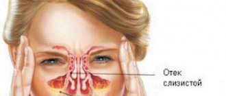
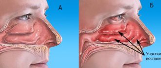
Acute rhinitis
Acute rhinitis is an acute inflammation of the nasal mucosa, which can occur as a consequence of other infectious diseases or as an independent disease. In acute rhinitis, hyperemic and swollen mucous membrane of the nasal cavity. There is a feeling of heat and, accompanied by headaches, impaired nasal breathing, increased secretion, and lack of smell.
At the first signs of acute rhinitis, aspirin is prescribed. Warming, hot tea, and effects on reflexogenic zones are indicated. Drug treatment consists of prescribing vasoconstrictors and antihistamines. indicated for severe swelling of the mucous membrane. For inflammatory processes in the mucous membrane, antibacterial agents are prescribed.
Chronic rhinitis
Disease of the nasal mucosa. Clinically, chronic rhinitis is manifested by nasal congestion, difficulty breathing through the nose, mucus secretion, etc. Chronic rhinitis can cause tonsillitis, etc.
There are several types of chronic rhinitis: vasomotor, allergic, hypertrophic, medicinal. Vasomotor rhinitis occurs due to decreased vascular tone in the nasal cavity. The individual reaction of the body to irritants becomes the cause of allergic rhinitis. When the connective tissue of the nasal cavity grows, hypertrophic rhinitis develops. Long-term use of vasoconstrictor drugs causes drug-induced rhinitis.
Ozena
The cause of ozena is atrophy of the nasal mucosa. Clinical manifestations of ozena: thick, foul-smelling discharge from the nasal cavity, impaired nasal breathing, lack of sense of smell, and the formation of dry crusts.
Treatment is carried out with medication. Immunity enhancing drugs, antibiotics, and vitamins are prescribed. Local treatment is aimed at softening and removing crusts from the nasal cavity. In severe cases, surgery is performed.
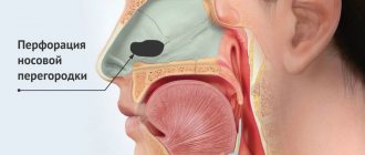
Deviated nasal septum
The causes of a deviated septum are:
- Uncoordinated development of facial skeletal structures
- Polyps
- Hypertrophied nasal concha
- Injuries
- Tumors
A deviated nasal septum makes nasal breathing difficult and causes congestion, mucous or purulent discharge, and headaches. Treatment is usually done surgically.
Adhesions in the nasal cavity
Fusions of the nasal septum and the lateral wall of the nasal cavity are called synechiae. Obstruction of the nasal passages (congenital or acquired) is called atresia.
The narrowing of the nasal passages due to fusion causes disruption of nasal breathing. In some cases, adhesions cause sinusitis. Treatment of adhesions is carried out surgically.
Hematomas of the nasal cavity
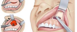
Hematomas are formed as a result of the accumulation of blood between the periosteum and the bone of the nasal septum. A hematoma can cause narrowing of the nasal passage, impaired nasal breathing, pain, and swelling. Sometimes the hematoma festeres and turns into an abscess, which is dangerous for intracranial complications (brain abscess, meningitis, etc.). An abscess of the nasal septum is manifested by severe swelling and pain.
Treatment of a fresh hematoma is limited to puncture and suction of blood. For an abscess, surgery is performed.
How to care for your piercing
Anyone who has dared to get a puncture should understand that nothing ends immediately after visiting the salon. Any piercing requires equally careful and long-term care, be it traditional, such as an ear piercing, or unusual, such as a piercing of the bridge of the nose.
Care after a puncture should be as follows:
Before starting the care procedure, wash your hands thoroughly and disinfect them.
This is extremely important, otherwise you risk introducing an infection into the wound. It is necessary to rinse the frame twice a day with saline solution to keep it clean and not accumulate discharge.
In case of dust or dirt, it is necessary to apply an antiseptic. Therefore, carry it with you for the first 10 days after the puncture. Minimize contact of the puncture with clothing, hair and any other foreign objects. Eat right, maintain your immune system, drink plenty of water and get enough sleep. This will help your body heal. After the first 10 days after the piercing, you can reduce the care to once a day. When the primary healing stage has passed, you can replace the medical earring with your own, but if you feel severe discomfort, you need to return the medical earring and wait some more. Sometimes it is not so easy to insert a medical earring, so we recommend that you contact the artist who had your piercing done.
What not to do:
- You should not constantly disturb the puncture: fiddle with the jewelry, scroll it, touch it.
- If you want your puncture to remain with you and please you for many years, then you should not take out the earring during the healing period (3-4 months) - the hole will simply heal.
- Do not use hydrogen peroxide or products containing alcohol. They can complicate the healing process, even leading to a chemical burn.
- If possible, do not smoke or drink alcohol for several days after.
- It is not recommended to visit the solarium and swimming pool for two weeks, and baths and saunas for a month.
It is also necessary to care for the septum, however, in addition, it is recommended to drip drops containing sea salt, treat the puncture with a cotton swab soaked in an antiseptic, and it is strictly forbidden to tear off the resulting crusts, in order to avoid infections.