Causes
In relation to congenital benign tumors of the nasal cavity, the causative factors are various exo- and endogenous teratogenic effects on a woman during pregnancy. Trigger factors for the appearance of benign tumors of the nasal cavity in adults are long-term adverse effects on the nasal mucosa. They may be associated with the presence of a chronic disease of the nasopharynx of infectious (chronic rhinitis, sinusitis, sinusitis, nasopharyngitis, adenoids) or allergic (allergic rhinitis, hay fever) origin; dust or smoke in the work area; inhalation of various irritating substances (for example, among workers in the chemical or pharmaceutical industries); frequent injury to the nose and its mucous membrane.
Symptoms of benign tumors of the nasal cavity
Benign tumors of the nasal cavity at the beginning of their development occur without any clinical manifestations. Symptoms occur when the tumor reaches a significant size and begins to interfere with the normal flow of air into the nasopharynx. In this case, the patient experiences difficulty in nasal breathing, which usually serves as a reason for him to contact an otolaryngologist. There is also a decrease in the susceptibility of odors (hyposmia), a sensation of a foreign body in the nasal cavity and nosebleeds, especially intense if the tumor is vascular.
As a result of impaired ventilation of the nasal cavity, a secondary infection often occurs with the development of rhinitis or rhinosinusitis. In such cases, patients with benign tumors of the nasal cavity complain of mucous or mucopurulent discharge from the nose, headache and pain in the inflamed sinus area.
Some benign tumors of the nasal cavity (angioma, chondroma, osteoma) have infiltrative growth and can spread to the paranasal sinuses, pharynx, orbital cavity, and brain. The growth of such tumors affecting the pharynx has a clinical picture similar to benign tumors of the pharynx and is manifested by impaired swallowing (dysphagia) and breathing. Tumor growth into the orbit is characterized by exophthalmos, diplopia, narrowing of visual fields, limited mobility of the eyeball, and decreased visual acuity. The spread of a benign tumor of the nasal cavity to the structures of the brain can be manifested by increased headaches, unilateral flattening of the nasolabial fold, epileptic seizures, disorders of the cranial nerves and other symptoms.
Osteomas and chondromas often grow into the bone structures that form the nasal cavity and the walls of the paranasal sinuses, causing their destruction. As a result, the clinical picture of these benign tumors of the nasal cavity is characterized by a curvature of the nasal septum and various facial deformities.
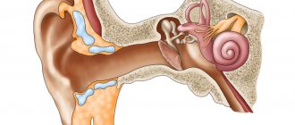
Diagnosis of benign tumors of the nasal cavity
Benign tumors of the nasal cavity are diagnosed by an otolaryngologist. Rhinoscopy is performed, which allows the doctor to examine the formation, differentiate it from scleroma and a foreign body, and determine by its appearance what type of tumor it belongs to. Asymptomatic benign tumors of the nasal cavity at the initial stage can be discovered accidentally during rhinoscopy for another disease. Nasal cavity formations that are difficult to diagnose are an indication for consultation with an oncologist and an endoscopic biopsy.
Impaired sense of smell in benign tumors of the nasal cavity is detected during olfactometry. In order to study the extent of tumor invasion into structures adjacent to the nasal cavity, radiography of the paranasal sinuses, radiography and CT of the skull, pharyngoscopy, CT and MRI of the brain are performed; consultation with an ophthalmologist with testing of visual acuity, exophthalmometry, determination of visual fields and ophthalmoscopy (examination of the fundus of the eye). To identify pathogenic microflora in the presence of an infectious process, a swab is taken from the throat and nasal cavity.
Treatment of benign tumors of the nasal cavity
Due to the disruption of normal respiratory function, the risk of malignancy and proliferation, benign tumors of the nasal cavity are an indication for surgical treatment. A limitation to surgical intervention may be the patient’s advanced age and the presence of chronic decompensated diseases (heart failure, coronary artery disease, severe hypertension, respiratory failure, bronchial asthma, diabetes mellitus, renal failure, cirrhosis of the liver, etc.).
The method of removing a tumor of the nasal cavity depends on its type, size and nature of its growth. Small fibromas, adenomas and papillomas are removed endoscopically under local anesthesia using an electrocoagulating loop. A bleeding nasal polyp is excised along with a section of the nasal septum at its attachment site. To prevent relapses, the base of the tumor is cauterized by cryotreatment or electrocoagulation. Large benign tumors of the nasal cavity are excised with a scalpel, radio wave knife or using a laser. To clearly differentiate tumor cells from surrounding tissues during surgery, a surgical microscope is used.
Small vascular benign tumors of the nasal cavity are removed by laser, electrocoagulation or cryodestruction. Removing large angiomas is associated with the risk of massive bleeding, so they are removed after preliminary ligation of the carotid arteries. When an angioma spreads deep into the surrounding tissue, occlusion of the vessels feeding it or sclerosis of the tumor is used.
Osteomas and chondromas that grow into the bone walls and structures adjacent to the nose often have to be removed in parts, using not only endonasal, but also external surgical approaches. The operation may be accompanied by resection of bone structures and a significant amount of facial tissue with the formation of defects requiring reconstruction using plastic surgery methods.
Source: www.krasotaimedicina.ru
Treatment
It is very important that cancer treatment is not only effective in eliminating the tumor, but as safe as possible in terms of side effects. The most effective method is surgery to remove the nasal tumor, but radiation therapy and chemotherapy are also possible.
We recommend reading Cancer of the endometrium (body) of the uterus - causes, symptoms, signs, treatment
The operation is performed within healthy tissue, during which the tumor can be removed using a scalpel, electrocoagulator, laser or cryodestruction. The area of operation is determined by the extent of the lesion. Along with the cancer cells, the affected structures, which can become the walls of the orbit and nose, are also eliminated. If necessary, they can be replaced with modern cosmetic prostheses made of titanium.
Conservative treatment of cancer through radiation therapy is also possible. In some cases, irradiation is carried out in combination with surgery. Damage to cancer cells by radioactive rays can be carried out as an independent method of therapy only for small tumors or if surgical intervention is impossible. The disadvantage of this method is that using radiation it is not always possible to completely get rid of all cancer cells.
Chemotherapy may often be prescribed preoperatively to stop the growth and shrink the cancerous tumor, and after surgery to eliminate residual cancer cells. The use of strong chemicals is fraught with side effects, and therefore this method of treatment cannot be called completely safe.
Nasal hemangioma and other benign tumors of the nose
Benign nasal tumors are formed from various tissues present in the nose and paranasal sinuses. Hemangioma is a benign tumor that most often develops in the first days of a child’s life and actively grows in the first six months of his life. After the child is one year old, the tumor begins to involute; most of the hemangiomas disappear by the age of seven, the rest by the age of twelve. More often, hemangiomas form in girls. The cause of the disease is a disturbance in the development of blood vessels in the embryonic period. Nasal hemangioma can grow deep into the skin, tissues of the organ and to the sides, destroy tissues, provoke bleeding, and disrupt the functions of the organ.
Nasal fibroma is a benign formation that can spread to the orbit. Nasal fibroma is rare, characterized by active growth and quickly grows into the orbit and paranasal sinuses. The tumor causes nosebleeds and nasal congestion. Fibroma that has grown into the orbit and nasal sinuses causes various complications: constant lacrimation, decreased visual acuity, exophthalmos, and can cause deformation of the skull bones. Fibroids rarely develop in the paranasal sinuses, most often developing in the maxillary sinus.
Adenomas in the nose can be true adenomas, a papilloma-like tumor, or a malignant destructive adenoma. Adenomas do not produce secretions, despite the fact that the tumor contains glandular epithelium. Adenomas in the nose are rare and are characterized by nosebleeds, nasal congestion, and can lead to the development of exophthalmos, purulent dacryocystitis and other complications. Benign and malignant adenomas grow slowly, and the prognosis for an expanded malignant adenoma is unfavorable. Treatment of the disease is radical surgery; radiation therapy and radiation therapy are not used due to low efficiency.
Benign neoplasms of the nose are osteoma, chondroma, angioma, papilloma, chordoma, lipoma, myxoma, bleeding polyp, ganglioneuroma, dermoid cyst and other neoplasms. Tumors are formed during intrauterine development of the fetus with a negative impact on the woman’s body during pregnancy. They can appear in chronic diseases of the nasopharynx; the appearance of tumors is influenced by poor environmental conditions, injuries, and infections.
Forms
The majority of patients with nasal cavity tumors are diagnosed with benign tumors. Such formations do not harm neighboring structures and systems. Benign forms of the disease include:
- papillomas;
- vascular formations;
- angiogranulomas;
- osteomas;
- chondromas.
After the appearance of the first signs indicating the presence of a benign neoplasm, it must be eliminated without delay, since there is a high probability of its malignant degeneration.
Malignant tumors of the nose include:
- sarcomas - characterized by rapid development with penetration into nearby structures;
- squamous epithelial cancer - unlike squamous cell cancer of the nose, it is slow in progression with scant symptoms;
- neurogenic neoplasms - formed from nerve tissue.
In each individual case, the prognosis depends on a number of factors, such as the type of neoplasm, the stage of development at the time of detection and how adequate the therapy was.
Most often, patients encounter cancer of the nasal mucosa. If there is a suspicion of cancer of the nasal sinuses or other parts of the ENT organ, the sooner a full examination is carried out, the greater the patient’s chances of a full recovery.
We recommend reading Kidney cancer - symptoms in women and men, treatment, signs
Cancer of the nose and paranasal sinuses: symptoms
A tumor in the nose develops in the maxillary sinus (maxillary sinus cancer), and cancer also affects the nasal cavity. Nasal cancer, the symptoms and signs of which become pronounced at a late stage, accounts for 1.5% of all cancers; men are more often affected. The reasons for the development of the disease are not fully known; most often, nasal cancer develops in workers of nickel production enterprises, woodworking and leather industries, in people who smoke and who often suffer from nasal infections. Symptoms and signs of nasal and sinus cancer vary. At the initial stage of development, the symptoms are similar to those of various diseases - rhinitis, sinusitis and other disorders.
Then the patient begins to experience headaches, discomfort, pain in the sinuses, and a change in sense of smell. The nose is constantly stuffy, mucous discharge from the nose bothers me. The pain can radiate to the upper jaw, temple, and a constant severe headache - such symptoms develop with cancer of the maxillary sinuses. Cancer of the posterior external part of the maxillary sinus is manifested by difficulty while eating, difficulty opening the mouth - the cancer grows into the masticatory muscles. Cancer of the anterior lower part of the maxillary sinus often affects the hard palate and upper jaw, leading to tooth loss and the appearance of wounds on the gums. Cancer of the nose and paranasal sinuses can lead to displacement and loss of the eye, and facial deformation.
Squamous cell carcinoma is the most common type of cancer; adenoid cystic carcinoma, adenocarcinoma, poorly differentiated transitional cell carcinoma, olfactory esthesioneuroblastoma and melanoma are less common. Carcinoma of the nose is not as dangerous as cancer of the paranasal sinuses, which has a less favorable prognosis. If early stage cancer is detected on the nose, then the five-year survival rate of patients is more than 50%, for cancer of the paranasal sinuses it is no more than 25%. Sinosanal poorly differentiated carcinoma has an aggressive course - the tumor affects the paranasal sinuses and nose, and is characterized by the appearance of ulcers and a tendency to necrosis.
Cancer of the nose and paranasal sinuses has clinical manifestations depending on the location of the tumor. Often a tumor in the thickness of the alveolar process is mistaken for an inflammatory process. It manifests itself in growth after tooth extraction. It is very difficult to differentiate tumors from the upper internal sinus. A tumor of the upper outer part of the maxillary sinus can remain unrecognized for a long time and manifest clinical symptoms in the form of pain. A tumor that has grown into the orbital area is manifested by swelling of the lower eyelid. A tumor that affects the infratemporal fossa, pterygopalatine fossa leads to swelling of the eyelid, the development of chemosis with exophthalmos.
Main stages of the tumor process
After confirming the diagnosis, the patient is prescribed appropriate treatment tactics, depending on the stage of the disease. Oncologists distinguish several stages of the malignant process: