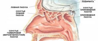
The maxillary sinuses are small cavities that are located on the sides of the nasal passages. Their main function is to warm and purify the inhaled air. They are also resonators when pronouncing sounds. The maxillary sinuses are connected to the nasal cavity by small passages, the size of which is no more than 3 mm.
Puncture of the maxillary sinuses is most often prescribed in the absence of effect from conservative therapy
An inflammatory process in the maxillary sinuses can occur as a result of pathogenic microorganisms entering this area, as well as due to injuries to the nose or a deviated nasal septum. Inflammation is accompanied by the accumulation of pus. In this case, a disease such as sinusitis develops.
Indications for diagnostics
The following indications for X-ray examination of the PPN are distinguished:
- head injuries;
- headache in the forehead, eyes, bridge of the nose;
- headache when bending the head forward, which can radiate to the upper jaw;
- nosebleeds;
- frequent or severe nasal congestion;
- discomfort in the nasal area after ARVI;
- a combination of symptoms such as rhinorrhea, lacrimation, fear of light;
- long-term temperature above 37.2;
- suspicion of inflammation, cavity formation or tumor;
- adenoid hypertrophy in children.
Diagnosis of the paranasal sinuses of the face is carried out before surgery and to monitor the treatment of pathologies over time. In the first case, a contrast research method is used.
Survey area
The picture is taken in black and white, which clearly shows the bone skeleton of the nose, the size of the nasopharynx, the structure and contours of the air cavities (aka sinuses). Using the image, the doctor analyzes the integrity of the bones and cartilage tissue, the condition of the hollow formations adjacent to the nose:
- frontal (frontal) sinuses, located in the frontal bone of the skull;
- lattice labyrinth;
- the main sphenoid sinus, divided by a septum;
- paired maxillary (maxillary) cavities;
- the lower part of the temporal bones with a cellular structure (mastoid process).
According to the norm, in the absence of pathologies, the outline of the bones should be smooth, the pneumatization (airiness) of the sinuses should not be impaired, that is, there should be no neoplasms or fluid or foreign objects
Abnormalities and diseases diagnosed from the image
One of the main diseases of the nasal cavities, sinusitis (inflammation of the mucous membranes), has several different forms. X-rays of the paranasal sinuses can identify sinusitis. Sinus hyperplasia or hyperplastic sinusitis is characterized by increased growth of mucosal cells. This area will appear thicker in the image. Catarrhal, edematous-catarrhal or exudative (with accumulation of fluid) form of sinusitis is displayed on an x-ray as a sharp darkening of the inflamed sinus. Allergic sinusitis is manifested by severe swelling.
X-ray of the paranasal sinuses differentiates the disease by location:
- inflammation of the maxillary sinuses – sinusitis;
- inflammation of the frontal sinus - frontal sinusitis;
- pathology of the mastoid processes – ethmoiditis.
The extent of the spread of the inflammatory process is also determined. With monosinusitis, the disease affects only one paranasal cavity; polysinusitis is diagnosed when inflammation is localized in several sinuses. If all right or all left sinuses are involved in the process, a diagnosis of hemisinusitis is made; damage to all cavities means the presence of pansinusitis.
Use in pediatrics
An X-ray of the nasopharynx in a child, in addition to the history and rhinoscopy data, and the results of laboratory tests, is a good help for diagnosing and distinguishing various pathological processes of the paranasal sinuses. In early childhood (up to 3 years), the use of radiation methods is undesirable, so x-rays are prescribed in extreme cases.
How harmful is x-ray for a baby?
Radiation exposure when using x-rays is extremely small. There is an opinion that the harm caused to the body when undergoing modern radiation diagnostic methods is less, but this is not so. Unlike a computed tomography scan, which takes many slices and produces many images, x-rays deliver much less radiation. Therefore, the risk to human health is minimal.
Prevention
The main preventive recommendation concerns the treatment of colds. Any infections must be treated correctly and completely. Otherwise, they can go into a chronic stage and cause tissue proliferation.
Do not try to cure adenoma on your own; consult your doctor for prescriptions.
If you work in hazardous work and are constantly in contact with aggressive compounds, try to protect yourself from their exposure. Use respirators and other protective equipment. In addition, it is important to strengthen the immune system, eat right and lead an active lifestyle without bad habits. Smoking is one of the main risk factors.
If you have difficulty breathing, you should immediately consult a doctor. This may be a consequence of a cold, but sometimes it signals an adenoma. The only effective treatment is surgery. Removing the capsule restores respiratory function and avoids the development of serious complications.
results
An x-ray of the paranasal sinuses is assessed by such an indicator as pneumatization, that is, the airiness of the sinus cavities. Normal pneumatization occurs in the absence of pathologies in the area under study, and the color of the paranasal sinuses is similar to the color of the orbit. Thus, in the presence of a pathological process, the sinuses darken, that is, their color on the x-ray will be lighter than that of the orbit.
An X-ray of the nasal bones is assessed by the integrity of the bone skeleton of the skull; if it does not correspond to the norm, it shows fractures, cracks or displacements.
What the study shows
X-ray of the SNP helps determine:
- sinusitis in acute or chronic form;
- cyst;
- benign or malignant tumor;
- injury to the nose or facial bones;
- deviated septum;
- osteomyelitis (purulent-necrotic pathology of bone marrow and bones);
- osteoporosis.
The video talks about how and what radiography of the paranasal sinuses helps to identify. Filmed by the Moscow Doctor channel.
Symptoms of sinus inflammation
Characteristic signs of inflammation of the nasal sinuses are:
Acute inflammation usually goes away on its own within a couple of weeks, but the chronic form can torment for a long time, when periods of recovery turn into relapses of the disease.
Signs in babies
For children, inflammation of the sinuses is a common disease, almost always of a bacterial nature.
Certain diagnostic difficulties lie in the wide range of manifestations of such pathology.
It is especially difficult to diagnose in newborns.
Older children with inflammation complain to their parents that they have:
- pain appears around the eyes;
- mucus and pus are discharged from the nose;
- stuffy nose;
- breathing is difficult.
The acute phase is characterized by a significant increase in temperature. In children, this inflammation lasts a long time, mouth breathing appears, coughing, nasal passages are clogged, and there is an unpleasant odor from the mouth.
In babies, the disease is accompanied by lack of appetite, poor sleep, tearfulness, weight loss, and moodiness. Eyelids often swell.
A CT scan may be performed to confirm the presence of sinus inflammation.
Are x-rays of the sinuses harmful and how often can they be done?
In case of inflammation of the SNP, images are taken several times during one course of treatment. To observe cysts and benign formations over time, it is necessary to take an x-ray once a year. X-rays performed twice within 12 months are considered optimal. In any case, the procedure should be performed as many times as prescribed by the attending physician.
It does not cause harm due to the low radiation dose, equal to 0.02 with a digital x-ray and 0.04 millisievert with a film x-ray. The acceptable annual dose for humans is 150 millisieverts.
Contraindications and restrictions
Contraindications to X-ray examination include pregnancy and children under 7 years of age. The influence of gamma rays on the fetus can provoke the development of pathologies. The procedure can be performed if it is impossible to do an ultrasound or the woman’s health justifies the possible risk to the fetus.
Are radiography prescribed for children?
It is not advisable to send children for x-rays due to the negative effects of radiation on the skeletal system. However, if there is no alternative, if the doctor considers it necessary to take an x-ray, you should not refuse. Some experienced ENT doctors and radiologists believe that the diagnostic method is suitable for children from 5 years of age.
Education for children
Adenoma in the nose in children appears due to frequent colds. When the immune system does not have time to cope with pathogens, a large amount of decay products from waste products of viruses and bacteria enters the blood. This further weakens the defense, causing numerous complications.
When the tonsil gets infected, its size increases. This is a kind of protective mechanism to prevent the pathogen from penetrating deep into the tissue. If the child has constant illnesses, then the tonsil does not have time to take a normal shape, and cells grow deeper into the nose.
Young children can rarely say what exactly is bothering them. If the child does not speak yet, then you can understand what is happening by constantly opening his mouth. Since nasal breathing is difficult, the baby will breathe through the mouth and act restless at night. If he is still breastfed, then difficulties will arise during breastfeeding.
In most cases, children develop a bleeding polyp. In this situation, nosebleeds constantly occur. Osteoma occurs infrequently, mainly due to previous diseases. Chondromas appear as a consequence of disturbances in embryonic development during the period of intrauterine formation.
Preparing for an X-ray
Preparation for the procedure comes down to two rules:
- remove jewelry and other metal objects located in the head area (chain, earrings, hairpins, hoop);
- wear a special apron (lead), which is issued immediately before the x-ray.
There are no restrictions related to food or liquid intake. It is necessary to clarify when and how to take an x-ray when observing the disease over time. For example, if a patient is having a cuckoo sinus irrigate, x-rays are taken before the sinuses are cleared of fluid.
How does the procedure work and how long does it last?
The duration of the study is several minutes.
X-rays are taken depending on the indications as follows:
- The patient enters a special room. Children are accompanied by adults.
- In a standing or sitting position, the radiologist fixes the head in the position required to take the image. This can be a straight or lateral plane, a projection of the chin, the occipito-frontal part or the occipito-mental part. One of the parents holds the child's head.
- You need to take a deep breath and hold your breath for a few seconds.
- The doctor will inform you that the procedure is complete.
- The picture develops within 15-20 minutes.
Is X-ray dangerous? This is explained in the video from the Live Healthy channel.
Nose piercing options
There are several options for how to properly pierce your nose:
- Piercing one of the wings of the nose (this is the most common method, as it is considered one of the most painless);
- Piercing the nasal septum, which separates the nostrils from each other (this procedure is very painful, since there are many receptors in this area of the nose);
- Piercing the bridge of the nose (this is a very complex and dangerous method of piercing the nose, so it is not particularly common).
Interpretation of results
The interpretation of the results of an x-ray examination may be as follows:
- normal (taking into account the patient’s age);
- the presence of dark spots in the paranasal sinuses;
- thickening;
- visible trauma or the presence of a foreign body.
Norm
An X-ray of the sinuses of a healthy person looks like this:
- The nasal septum divides the nasal cavity into symmetrical sides of a triangle.
- The white stripes running to the right and left of the divided area are the nasal passages.
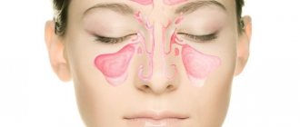
- The triangular cavities on the sides of the nose are the maxillary sinuses.
- Between the eye sockets there is an ethmoid sinus with thin walls, the cells of which should be clearly visible.
- Above the orbits, the frontal sinuses are defined, which can have different shapes. Their separation by bone partitions is allowed.
- There must be air in the sinuses. Their edges, like the contours of the bones, should be clear and even.
Darkening, cavities and thickening in the image
We can talk about diagnosing a pathology if the images show the following:
- darkening the area;
- thickening;
- the presence of a cavity of different shapes;
- deformation of bone tissue.
Acute inflammation is characterized by thickening of the mucosa and deformation of its borders (with purulent contents). The presence and level of fluid are determined by horizontal demarcation in the sinuses. An x-ray will not tell you about the composition of the contents; for this you will need to make a puncture (puncture). If the inflammation is chronic, the mucous membrane thickens and the lumen in the sinus becomes smaller.
Sinusitis
Sinusitis on an x-ray looks like a darkening, which has characteristic differences in various pathologies.
The pictures should be decrypted like this:
- With hyperplastic sinusitis, there is thickening of the mucosa directly next to the bone. In this case, the internal contour becomes wavy and blurry.
- With catarrhal sinusitis, thickening of the mucosal walls with complete or partial darkening is noted. The presence of a light cavity in the center of the sinus indicates a chronic process.
- With exudative sinusitis, darkening of the paranasal sinuses occurs with a horizontal delimitation, reflecting the level of fluid filling.
- With vasomotor and allergic sinusitis, pronounced swelling of the mucous membrane is noted.
Sinusitis
Sinusitis is an inflammation of the lining of the maxillary sinuses.
The following types of sinusitis are diagnosed:
- Exudative. The presence of fluid in the upper sinuses on one or both sides.
- Parietal. Localization of inflammation near the bone walls. The edges of the mucous membrane are deformed and directed inside the sinus.
- Polypous. There is bulging of areas of the mucous membrane, which can be either single or multiple.
Neoplasms and cysts in the sinuses
If photographs of the paranasal sinuses reveal a cavity with dense contents, this indicates the presence of a benign or malignant formation. In most cases, neoplasms are found by chance during the diagnosis of other pathologies. A sinus cyst is defined as a light, rounded area located outside the sinus mucosa. Its edges are clear and even.
Bone injuries
If a bone is broken or displaced, it will be visible on an x-ray. Bone injuries may appear on x-rays as dense fragments in the sinuses. The image helps to establish the location of the fracture and the displacement of bone fragments, if present. Severe fractures are accompanied by bleeding, which will appear as fluid in the sinuses. The doctor may also detect an old injury that appears as a callus on the image.
Foreign bodies
The foreign body in the image has contours in accordance with what exactly got into the nasal cavity. Typically, these are beads or other round objects that children place in their noses. On x-rays they appear as areas of varying darkness.
Possible consequences of x-ray
By imaging the paranasal sinuses with an x-ray machine, you may experience some side effects. Possible consequences of X-ray radiation, especially frequent exposure, include irreversible skin damage (for example, cancer). In people prone to blood diseases, changes in the composition of the plasma are possible, and leukemia is likely to develop. X-rays have a detrimental effect on the fetus, since it is tens of times more susceptible than an adult organism. The fetus may develop various tumors and congenital developmental anomalies; a woman may have a miscarriage.
SOURCES: https://idiagnost.ru/issledovaniya/rentgen/kogda-provoditsya-rentgen-pazuh-nosa https://apkhleb.ru/rentgen/nosa-pridatochnyh-pazuh https://hromosoma.com/rentgen/pazuh- nosa-10071/
Causes
In most cases, the proliferation of cells in the nasopharyngeal tonsil is experienced by children who are often and seriously ill. However, pathology also develops in adults. This is due to the following reasons:
- infectious processes in the nasal sinuses. The pathogen provokes strong tissue proliferation, which often occurs with weak immunity;
- If the body's defenses cannot cope with the load, then the activity of the immune system is disrupted. Then any disease can cause the development of a tumor;
- long-term use of tobacco and alcohol negatively affects a person’s condition, increasing the likelihood of neoplasms;
Tobacco and alcohol use. - allergies are one of the risk factors;
- work in hazardous production (in which there is constant exposure to chemicals, dust or smoke);
- mechanical injuries of the nasal cavity and mucous membranes;
- constant presence in rooms with low air humidity and a lot of dust;
Frequent exposure to dusty rooms with low humidity. - hereditary predisposition or genetic factor;
- difficult pregnancy (the pregnant woman had infectious diseases, used potentially dangerous drugs or was under the influence of toxic substances). Difficult childbirth (hypoxia, trauma) also increases the risk;
- improper care of the baby in the first months of life (nutrition, features of breastfeeding, ignoring diseases).