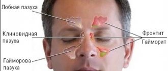
The largest of the paranasal sinuses (sinuses) is the maxillary, or maxillary. Its volume is determined by the age and individual characteristics of people. The functions of this paranasal sinus are to warm and humidify the inhaled air. The mucous membrane of the maxillary sinus is lined with a large number of glands that produce mucus. If their excretory ducts become blocked, a cyst may form. It is dangerous because it contains pus. Treatment of the pathology can be carried out conservatively or surgically.
What is a maxillary sinus cyst?
According to ICD-10, this pathology is called a cyst or mucocele of the nasal sinuses. With this disease, a benign cystic neoplasm is formed in the maxillary sinus, resembling a bladder in appearance. Its internal cavity is filled with liquid - purulent or sterile, which depends on the severity and duration of the disease. The walls of the formation are thin and elastic, lined with epithelial cells. In most patients it is localized at the bottom of the maxillary sinus. The tumor is dangerous because if it is large, it can completely block the access of air.
Causes
The general reason for the appearance of this neoplasm is a disruption of the normal outflow of secretions or complete blockage of the glands on the mucous membrane of the maxillary sinus. Even when the excretory ducts are blocked, mucus continues to be produced. It accumulates in the maxillary sinus, where it has nowhere to go. As a result, the gland stretches and takes the shape of a ball, which is a cyst.
If the size of the tumor does not exceed 1 cm, then the patient does not notice any particular discomfort. Otherwise, it completely fills the sinus cavity, which is why treatment is carried out surgically. Risk factors for the development of this pathology are:
- chronic sinusitis, rhinitis and other diseases in which the functioning of the maxillary sinuses is impaired;
- violation of the structure of the anastomosis - the outlet of the maxillary sinus;
- caries, periodontal disease and other foci of infection in the oral cavity;
- frequent allergic reactions;
- congenital anatomical features, for example, facial asymmetry;
- nasal injuries;
- prolapse of the hard palate;
- deviated nasal septum;
- immunodeficiency states.
Classification
Depending on the location, cysts of the right and left maxillary sinuses are distinguished. In another classification of this pathology, the criterion is the type of separated contents. It may be the following:
- mucous discharge – mucocele;
- serous fluid – hydrocele;
- purulent discharge – pyocele.
Least of all, experts have studied the origin of false cysts, which are cyst-like formations. They are typical for male patients. The causes of false cysts are pathologies of the upper teeth, the action of allergens or infections. The difference between such neoplasms is the absence of an epithelial lining inside the cystic bladder. Taking into account their origin, two more types of cysts are distinguished:
- Odontogenic. They are formed as a result of infection from foci of inflammation at the roots of the teeth and adjacent tissues. Odontogenic cyst of the maxillary sinus is of two types: follicular (appears in children 10-13 years old due to insufficient development of the impacted tooth base or inflammation of milk teeth) and radicular (its cause is caries).
- Retention (true cysts). They are formed due to obstruction of the glands that produce mucus. The inside of the retention cyst of the maxillary sinus is lined with a layer of epithelial cells.
Additional questions
► ICD-10 code
Designated as K09 or J33.8. Depends on the cause of formation and type of maxillary cyst.
► Why is it dangerous?
The danger arises in a situation of suppuration or inflammation of the cyst. In the best case, frontal sinusitis or sinusitis will occur, in the worst case, inflammation will spread to the soft outer tissue. It is fraught with the following complications: in the nasal cavity - inflammatory processes, sinusitis; in the orbit - abscesses, phlegmons; intracranial diseases - encephalitis, meningitis, thrombosis, etc.
We invite you to familiarize yourself with What is an abutment in dental implantation?
► Do they take you into the army?
If a conscript has cysts of the maxillary sinus, he will not be accepted into the army. They are given a deferment from service for the duration of the operation and subsequent rehabilitation. The commission at the military registration and enlistment office will refer the future defender for treatment to the ENT department.
Symptoms of a maxillary sinus cyst
The danger of this pathology is that in most patients it does not manifest itself in any way. It is diagnosed incidentally as a result of an X-ray, computed tomography, or magnetic resonance imaging scan performed for another disease. The cyst only causes discomfort if it is located in a certain location or is large in size. In such cases, the patient may experience the following symptoms:
- distension and pain at the site of the tumor;
- breathing problems, congestion from the neoplasm;
- pressure in the eyes, feeling of fullness;
- frequent nasal discharge;
- swelling of the cheek;
- headache, worse when tilting the head down;
- severe discomfort and intense pain in the bridge of the nose and forehead when diving under water.
Article on the topic: What are the main reasons for the appearance of a left kidney cyst and methods of its treatment?
The cyst of the right maxillary sinus does not differ in symptoms from the tumor in the left. When a cystic bladder ruptures, yellow or orange fluid begins to flow from one nostril. This process is not always harmful to health. What is dangerous is suppuration of the contents of the cystic bladder, which is indicated by the following symptoms:
- pain in the eyes, cheeks, teeth;
- heat;
- purulent runny nose;
- general signs of intoxication of the body.
Preparation and organization of the operation
In a situation where removal of a maxillary sinus cyst is required, you need to thoroughly prepare for surgery. Surgery is indicated if the formation causes discomfort in the nose. Small cysts usually cannot be removed surgically.
The dimensions for removal are determined by the doctor. A benign formation can be quite large and cause a lot of trouble. Surgeon intervention is required if the cyst is larger than 1 centimeter. First of all, the formation blocks the access of oxygen and contributes to difficulty breathing. In such cases, surgery is definitely required.
Cyst removal methods:
- Endoscopic removal of a tumor is a modern, safe method of treatment. This type of operation is the most gentle. Fiber optic technology is used for this purpose. The bone is not subjected to mechanical damage, the operation is carried out under control provided by special equipment. Removal of the formation using endoscopic equipment is a method indicated for most patients. The operation takes a short time and does not cause complications. The cyst is removed through the mouth of the nose.
The surgical intervention lasts 25-40 minutes. Nowadays, this technique is the safest and least invasive. The operation does not involve mechanical damage to the sinus. After surgery, no complications develop, and the recovery period does not take more than a week. The patient must spend this time in the hospital. After just 7 days, the patient can return to work and normal lifestyle. You should continue to follow your doctor's recommendations.
- Another option for cyst removal is Denker surgery. The anterior wall of the sinus is opened and the necessary incision is made above the upper lip. The operation is performed under anesthesia. The surgeon removes the cyst using special medical instruments. This surgical method is used to provide access to a tumor on the anterior wall of the nasal sinus.
Treatment of maxillary sinus cysts requires strict adherence to all medical recommendations. The doctor may prescribe the use of certain medications that suppress pathogenic cells and restore the structure of the nasal mucosa. Medicines will promote rapid recovery and reduce symptoms of the disease. In therapy, it is important to constantly maintain the functioning of the nasal passages. This can be achieved with medications prescribed by a doctor.
If there are indications for surgery to remove a cyst, careful preparation is carried out.
Surgery will be required if there is discomfort in the nose. Typically, small cysts are not removed surgically. Surgical manipulations will be required when the diameter of the neoplasm is more than 1 cm, it blocks oxygen and complicates breathing. Then an operation must be performed, since breathing during sleep can suddenly stop. Dangerous complications of the absence of surgery for a large cyst are: meningitis, encephalitis, thrombosis. Complications may affect the visual analyzers and the orbit.
Endoscopy
A safe modern method of surgical intervention. This option is the most gentle; it requires fiber-optic equipment. The bone is not mechanically damaged, the doctor controls the situation during the operation using equipment. This method is prescribed to most patients with a cyst in the maxillary sinus.
The manipulations do not last long and do not provoke complications. The cyst of the right maxillary sinus or left is removed through the mouth of the nose, this takes up to 40 minutes. The sinus is not damaged, the total recovery time is no longer than 1 week. Depending on the patient’s condition, rehabilitation is carried out in a hospital or at home. After 7 days, the patient is allowed to return to his work and normal lifestyle. It is important to continue to follow the rules set by your doctor.
Operation according to Denker
During manipulation, the wall of the sinus is opened and an incision is made above the upper lip. It is necessary to undergo anesthesia first. The surgeon performs the removal using special instruments. This method is implemented when the cyst is located on the anterior wall of the sinus. After the operation is completed, the doctor prescribes medications to suppress pathogenic microorganisms and restore the correct structure of the nasal mucosa. Taking medications will help you recover faster and maintain the functioning of your nasal passages.
Complications
The main danger is not the cyst itself, but its contents, which can become infected at any time. For this reason, the cystic cavity is considered a potential source of chronic infection. When the cyst of the maxillary sinuses becomes inflamed, pus begins to accumulate in them, which often leads to rupture of the capsule. This is indicated by the secretion flowing from the nose that is yellow in color and has an unpleasant odor.
Some doctors believe that such a process is good, but the leaked pus can get into the ear, which will lead to otitis media. In addition to suppuration, complications of the cyst include other pathologies:
- osteomyelitis;
- impaired visual acuity, diplopia due to compression of the optic nerve;
- changes and deformations of the skull bones;
- exacerbation of chronic sinusitis;
- episodes of apnea;
- constant migraine;
- lack of oxygen in the body.
How dangerous is this pathology?
As the disease progresses, swelling of the turbinates, the formation of polyps, and changes in the shape of the jaw occur. Acute respiratory infections aggravate the course of the pathology and provoke dangerous decay. Complications of the cyst are deformation of the cranial bones, suppuration, atrophy of bone structures and their rejection, impaired visual function (diplopia). This can be avoided if the cyst in the maxillary sinus is treated in a timely manner.
Exacerbations of chronic purulent sinusitis with a fever and the occurrence of progressive pain in the face or head are likely. Permanent hypoxia leads to problematic functioning of the cardiovascular system. Brain function may deteriorate.
Diagnostics
To detect a maxillary sinus cyst, a complex of laboratory and instrumental studies is used. The following procedures reflect the exact clinical picture:
- X-ray. To obtain an image of the sinuses, a contrast agent is injected into them, which helps to identify formations of any size.
- CT scan. It is necessary to determine the location and structure of the tumor. The technique reveals the thickness of the membrane and the internal structure of the cystic bladder and gives indications for surgery.
- Puncture of the maxillary sinus. The cyst is punctured with a thin needle. When yellow contents flow out of the nose, the diagnosis is confirmed. The technique does not give accurate results, since it helps to identify only large tumors. The procedure belongs to the diagnostic category.
- Sinusoscopy. Another diagnostic procedure is performed using an endoscope, which is inserted through the anastomosis of the maxillary sinus. This is necessary to identify and study the neoplasm itself and its localization. This method also helps to detect polyps of the maxillary sinus, i.e. hyperplasia of its mucosa.
Features of therapy
This type of formation can also be called a maxillary sinus cyst. Being a pathological condition, it is considered dangerous due to the proximity of the sinus to the membranes of the human brain.
Sinuses are cavities, the inner surface is lined with mucous tissue with a large number of glands. The mucus that is produced in this space performs a protective function for the body.
The ducts of the glands are very narrow and can overlap, as a result of which the gland becomes overfilled with its own secretion. Under internal pressure, it becomes deformed and stretched, turning into a spherical cyst. The formation can occupy the entire area of the sinus.
The main method of treating a cyst of the right maxillary sinus is surgery.
The presence of a cyst in the sinus may not be felt by the patient, and is revealed only by an x-ray. In the early stages of cyst development, it may not be visible to X-ray examination if the condition is accompanied by inflammation or swelling of the sinus membranes. The cyst grows slowly, so that a person can live with it for quite a long time without experiencing any problems.
When a cyst is visible on an x-ray, it appears as a shadow in the sinus cavity, shaped like a ball.
Here are the classic symptoms that accompany the presence of a cyst:
- Nasal breathing becomes difficult when the cyst reaches a large size.
- A discharge resembling purulent discharge begins from the nasal passage - yellow in color, with a characteristic odor. This symptom signals a cyst rupture.
- A headache occurs and is severe, radiating to the frontal area, temple or back of the head. The sensations can be permanent, or they can occur in certain situations - when the body position changes, during sudden movements, when the weather changes.
- Often pain occurs spontaneously in the area of the maxillary sinuses. When pressing on the area of the affected sinus, the patient feels pain.
- The patient may experience a feeling of pressure and fullness.
- Problems with nasal breathing, discharge and headaches are localized on the side on which the cyst is located.
- Patients often suffer from irritability, decreased performance, and dizziness.
- In some cases, the active growth of the cyst is accompanied by deterioration of memory and sleep, and loss of appetite.
What these symptoms mean, you should check with your doctor.
If at least one symptom from this list occurs, you need to contact a specialist for help, the cost of delay in the event of pain is too high. After conducting the necessary examination, further steps to solve the problem will be known.
Here are the situations in which a cyst of the right maxillary sinus or left sinus most often occurs:
- inflammatory diseases of the nasopharynx;
- diseases of the maxillary sinuses, which can cause blockage of the gland duct;
- an allergic reaction, especially long-term exposure to an allergen on the patient’s body;
- if the nasal septum is curved or there are changes in the shape of the cartilage or bone formations of the facial part of the skull;
- dental problems of the upper jaw, advanced caries, pulpitis.
Cysts growing in the cavity of the maxillary sinus can be of several types.
| Cause of occurrence | Type of cyst |
| Inflammatory process in the sinus | A true cyst occurs due to blockage of the gland duct. When a formation occurs, internal mucous cysts continue to produce secretions, and the cyst increases in size. |
| A false cyst does not have a mucous membrane that produces mucus. | |
| Dental problems | Odontogenic formations grow at the apex of the root of the diseased tooth, increasing in size and reaching the sinus cavity. Since the plate between the roots of the teeth and the sinus is very thin, this is a fairly common phenomenon in people who do not treat their teeth during treatment. |
| The follicular form of cysts develops due to a disturbance in the development of tooth follicles. |
Right maxillary sinus cyst often grows due to dental problems
Before prescribing treatment, a diagnostic examination is required.
The following methods are used:
- X-ray examination. An image of the object will be obtained, from which one can judge its size.
- X-ray examination with the introduction of a contrast agent. To do this, the sinus is punctured and the substance is injected, then an image is taken.
- Computed tomography makes it possible to obtain more accurate information about the location of the formation and its size.
- Endoscopy, performed with fiber optic instruments, makes it possible to examine the cyst, assess its size and prescribe the correct treatment tactics for the disease.
In combination with complaints, one option for managing the situation can be chosen:
- monitoring the cyst if it is not of significant size;
- removal of the formation if it is large and causes discomfort to the patient.
So, we have discussed the symptoms, types and causes of maxillary sinus cysts. Treatment will only be carried out surgically. In this case, such manipulation cannot be avoided. Neither physiotherapeutic measures, nor heating, nor any medications can eliminate the cyst of the left maxillary sinus or the right. In addition, physiotherapy is contraindicated, as the disease can develop into extensive sinusitis.
There is no specific size of a maxillary sinus cyst that suggests its removal. Indications for surgical intervention are the presence of complaints and complications in the patient. Surgery for maxillary sinus cysts is carried out in any clinic specializing in this. In public clinics this is done absolutely free.
In private medical institutions, removal of a retention cyst of the maxillary sinus, as well as its other types, will depend on certain conditions. As a rule, the cost of such an operation is about 40 thousand rubles. However, before this, the patient must undergo some diagnostic measures described above.
Treatment of maxillary sinus cyst
If the pathology does not bother the patient in any way, then emergency measures and specific treatment are not required . The doctor simply advises to monitor the cyst and fight the disease that caused its formation. In general, the decision on the treatment method depends on the specialist. When choosing a treatment regimen, the doctor takes into account the following factors:
- complaints from the patient himself;
- degree of neglect;
- the presence of concomitant diseases.
Conservative
This type of treatment is aimed at slowing down the growth rate of the cystic formation, therefore it is used only for small sizes. Many experts express the opinion that conservative therapy is not highly effective. No medicine can completely get rid of a cyst. The drugs only reduce the symptoms of the pathology, but the tumor itself remains until surgical removal. If the doctor nevertheless makes a choice in favor of conservative therapy, he may prescribe the following drugs:
- Salt solutions: Aquamaris, Humer, Physiomer, Marimer. Adults and children over 2 years old are prescribed 1-2 injections into each nostril up to 4 times a day. The drugs can be used for a long time.
- Normalizing the outflow of cyst contents: Sinuforte. One press should be made into each nasal passage. The course of treatment lasts 6-8 days. When used every other day, Sinuforte can be used for 12-16 days.
- Local antibiotics: Isofra, Polydexa, Bioparox. Use intranasally: one injection into each nostril up to 4-6 times a day. You should not use the drugs for more than 1 week.
- Systemic antibiotics: Lincomycin, Amoxicillin, Azithromycin. These are serious drugs that should only be prescribed by a doctor. The dosage and course of treatment is determined by the disease and individual characteristics of the patient.
- Local corticosteroids: Nasonex, Beconase. Dosage for adults and children over 12 years of age – 2 inhalations into each nasal passage once (200 mcg of the drug per day).
- Vasoconstrictor sprays: Otrivin, Xylen, Tizin, Sanorin, Rinazolin, Nazol, Nazivin. Apply 1-2 drops in each nostril up to 3 times throughout the day. You should not use vasoconstrictors for more than 5 days, as they are addictive.
Article on the topic: Amorolfine for nail fungus - release form, composition, instructions for use, analogues and price
Cyst removal
If the size of the formation is too large, the doctor prescribes surgery. The main indication for surgery is a deterioration in the patient’s quality of life. Removal of a maxillary sinus cyst is carried out using different methods. A certain type of operation is chosen taking into account the size and location of the formation. There are 3 options for surgical removal:
- Classic maxillary sinusotomy according to Denker. During this operation, the maxillary sinus is opened through an opening in the upper jaw. Next, using a curette, the cavity is cleaned, removing not all pathological contents. Disadvantages of maxillary sinusotomy: it is performed under anesthesia, the patient remains in the hospital for another week. The advantage is the ability to remove hard-to-reach tumors. In addition, such an operation is the only method for removing a cyst on the posterior wall of the maxillary sinus.
- Caldwell-Luke operation. Consists of trepanation of the maxillary sinus. The cystic bladder is removed through the hole. This procedure is rarely used today because the risk of injury to the anterior wall of the sinus is high.
- Puncture. This is a temporary measure in which its contents are pumped out through a puncture of the maxillary sinus. Disadvantages of puncture: removal does not always end with recovery; there is a risk of complications (fistulas, large abscesses). The advantage is temporary relief.
- Endoscopy. This is a more gentle method. An endoscope with video equipment is inserted through the anastomosis of the maxillary sinus to clean out the sinus cavity. Advantages: no incisions, duration 20-60 minutes, low risk of complications and damage to the maxillary sinus. There are no downsides to the operation.
Folk recipes
If a retention or odontogenic cyst of the maxillary sinus does not bother the patient, then the doctor may prescribe treatment with medications and folk remedies . Alternative medicine offers the following recipes:
- Take 5-6 drops of fresh aloe juice per tablespoon of vegetable oil. Drop a whole pipette into each nostril up to 2-3 times a day.
- Take a couple of forest cyclamen tubers, grate them, and then squeeze out the juice through cheesecloth. Every day in the morning, drop 2 drops into each nasal passage. After a week of treatment, take a break for 2 months, then repeat another treatment cycle.
Types of jaw cysts
For reasons, an upper jaw cyst occurs:
- Retention - occurs when the excretory ducts of the glands in the mucous membrane are blocked.
- Odontogenic – develops due to the pathological influence of dental problems.
According to the mechanism of formation and location of odontogenic and retention formations, they are classified into 2 types:
- True - its shell consists of epithelial cells that produce mucus. This disorder occurs due to anatomical problems in the nasal sinuses or due to chronic lesions of the nasopharynx.
- False or pseudocyst - it does not contain epithelial cells. Such formations are associated with dental disorders, incorrect anatomical structure of the jaw, dental diseases, or errors in treatment by the dentist.
Also, the cyst can be single or multiple; the left and right side of the location does not affect the symptoms of the tumor. According to ICD 10, the disease refers to diseases of the nose and sinuses and has code J34.1.
In the field of medicine, there is a separate classification of formations on the maxillary sinuses. A cyst of the left or right maxillary sinus can be of the following types: mucocele with mucous contents, hydrocele with serous fluid, and pyocele with purulent contents.
In addition, based on their origin, these neoplasms are divided into three types. Let's look at them separately.
There are various ways to remove maxillary sinus cysts. The classic version looks like this: the surgeon makes an incision under the patient’s upper lip, opens the wall of the bone cavity with a chisel and excises the cyst. The disadvantages of such an intervention are that it is quite traumatic and requires a long period of rehabilitation.
In our clinic, operations on maxillary sinus cysts are performed using endoscopic technologies. Under visual control, the doctor makes a small (several millimeters in size) puncture in the gum and through it removes the pathological formation. No “puncture” manipulations are performed in this case. The entire intervention lasts no more than 20 minutes.
Recovery of the body after endoscopy occurs very quickly, so we do not hospitalize patients. You are allowed to leave the clinic on the same day.
- A follicular cyst develops at the site of an unerupted tooth. It is formed instead of a tooth and is located in the alveolar edge of the jaw bone.
- A retention cyst is most often diagnosed on the upper jaw and is the most common. The formation develops due to a violation of the patency of the gland ducts. The airway becomes blocked, and cicatricial changes lead to the formation of a cyst.
- An odontogenic cyst often forms in the maxillary sinus. This formation is localized due to the penetration of infection from the roots of the tooth into the upper jaw. Odontogenic cyst can be perihilar and follicular. The latter develops from root granulomas.
After the formation of a cyst, the iron continues to produce secretions. The cyst grows and its walls gradually stretch. With strong growth, the cyst can reach large sizes. Symptoms of a cystic formation can easily be confused with a common cold. But such an acute respiratory viral infection cannot go away for a long time. A sick person needs to pay special attention to this. If the cyst continues to develop, surgical intervention cannot be avoided.
If a cyst is detected, immediate treatment must be started. Otherwise, the pathology will lead to negative consequences. In some cases, surgery is performed.
A maxillary sinus cyst can close the entire cavity and cut off oxygen supply. The maxillary sinuses have a large number of glands that produce a special protective secretion. It prevents pathogenic microbes from entering the body. When the excretory ducts are blocked, the glands will begin to overflow, and over time a spherical cyst may form.
Prevention
An important condition for the prevention of maxillary sinus cysts is maintaining oral hygiene. It is recommended to treat caries and periodontal disease in a timely manner and regularly visit the dentist. Additionally, to prevent the formation of cysts in the maxillary sinuses, the following must be done:
- promptly treat runny nose, rhinitis, sinusitis and other respiratory diseases;
- avoid long-term allergies and eliminate them by taking antihistamines;
- Be sure to seek help from a doctor if you have a deviated nasal septum.
Postoperative period
After removal of a cyst or mucocele of the nasal sinuses, the patient should be monitored in the hospital for several more days. Depending on the intervention methods, the patient may experience swelling, pain and discomfort. If necessary, drainage is installed and painkillers are prescribed. In some cases, patients develop a fever.
Classic surgery is very traumatic, which is why specialists have recently given preference to endoscopic techniques and microsinlusrotomy.