Price
Diagnostic methods
| Name of service | Price |
| Ultrasound of the paranasal sinuses using the Sinuscan device | 800 ₽ |
Advantages
- The latest, constantly updated equipment
- Interest-free installments for all services
- Online consultations with an ENT doctor
- Visit of an ENT doctor to your home
- Friendly and qualified staff
- 24/7 ENT assistance
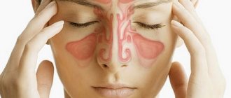
Ultrasound examination (ultrasound) of the paranasal sinuses is an instrumental diagnostic method that the doctor uses to assess the condition of the paranasal sinuses and identify the presence of a pathological process in them. To conduct the study, a sinusoscope is used - a type of ultrasound sensor that is designed specifically for examining the paranasal sinuses.
The study is one of the highly informative methods for diagnosing the condition of the sinuses located around the nasal cavity. Ultrasound of the sinuses in Moscow at the Ear, Nose and Throat Clinic is performed by experienced doctors using a modern sinusoscope. This allows you to identify diseases in the early stages and prescribe adequate therapy.
Diagnostics
Ultrasound examination of the paranasal sinuses is a method used in diagnosing various diseases of the corresponding structures. The procedure is necessary to identify the following pathologies:
- inflammation of the nasal mucosa (rhinitis);
- adenoiditis - an inflammatory process in the nasopharyngeal tonsil;
- inflammation of the frontal, maxillary sinuses - frontal sinusitis and sinusitis;
- cysts, tumors, polyps and other neoplasms in the paranasal sinuses.
The affordable price and safety of the procedure for the patient make the study an indispensable part of the mandatory diagnostic program for patients with ENT pathologies.
Methods for examining the nose and paranasal sinuses.
1Anatomy and physiology of ENT organs.
Anatomy of the nose.
The nose is the beginning of the respiratory tract.
It is divided into 3 sections: the external nose, the nasal cavity, and the paranasal sinuses.
External nose
resembles an osteochondral pyramid covered with skin. The following elements of the external nose are distinguished: root, dorsum, slopes, wings and tip. The walls of the external nose are formed by tissues: bone, cartilage and skin.
The bony part consists of the paired nasal bones, the frontal processes of the maxilla and the nasal process of the frontal bone.
Cartilages of the external nose: triangular, alar and accessory.
The skin has the following features:
1. many sebaceous glands
2. a large number of hairs in the vestibule of the nose, which perform a protective function
3. many blood vessels
The blood supply to the nose is carried out from the system of the external and internal carotid arteries, venous outflow through the facial and orbital veins, then into the cavernous sinus and into the internal jugular vein. This structure of the venous system can contribute to the development of orbital and intracranial complications.
Nasal cavity
divided by the nasal septum into two halves, has anterior openings - the nostrils and posterior openings - the choanae, leading to the nasopharynx.
Each half of the nose has 4 walls.
· The medial wall, or septum of the nose, is formed by a quadrangular cartilage in the anterior section, a perpendicular plate of the ethmoid bone at the top and in the mid-posterior section a bone called the vomer.
· The upper wall consists of a perforated plate of the ethmoid bone, through which branches of the olfactory nerve and vessels pass.
· The lower wall, or floor of the nasal cavity, is formed by the processes of the upper jaw, which at birth may not fuse together and form a cleft called the “cleft palate.”
· The lateral wall has a complex structure. On the inner surface there are three bony protrusions - the turbinates.
: top, middle, bottom.
The shells are covered with cavernous tissue, which, under the influence of physical, chemical and psychogenic stimuli, promotes rapid swelling of the mucous membrane and narrowing of the nasal passages. Under the shells there are nasal passages - upper, middle and lower.
The space between the edges of the turbinates and the septum is called
the common nasal passage
. In young children, the inferior turbinate fits tightly to the bottom of the nose and with minor inflammation of the mucous membrane, nasal breathing is completely disrupted.
The opening of the nasolacrimal canal opens into the lower nasal passage; a delay in its opening in a newborn leads to impaired outflow of tears, cystic dilation and narrowing of the nasal passages.
The paranasal sinuses - the maxillary, frontal, anterior and middle cells of the ethmoidal labyrinth - open into the middle meatus.
The sphenoid sinus and the posterior cells of the ethmoidal labyrinth open into the upper nasal passage.
The nasal cavity is divided into three zones:
- vestibule (entrance),
- respiratory - from the bottom of the nose to the middle turbinate, it is lined with ciliated epithelium and contains many cells that secrete mucus and serous secretions
-olfactory - the upper part of the nasal cavity, lined with olfactory epithelium.
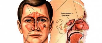
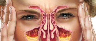
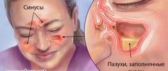
Paranasal sinuses
- These are the air cavities around the nasal cavity, which communicate with it through the outlet openings in the nasal passages. There are four pairs of sinuses:
· The maxillary sinus is located in the body of the upper jaw and borders at the top with the orbit, at the bottom with the oral cavity (the tops of the roots of the teeth can protrude into the lumen of the sinus), on the front wall there is a depression - the canine fossa (the sinus is usually opened here).
· The frontal sinus is located in the thickness of the frontal bone and borders the orbit below, the anterior cranial fossa at the back. In 10-15% of cases this sinus may be absent.
· The ethmoid labyrinth is located between the orbit and the nasal cavity and consists of 5-20 air cells, which are divided into three groups - anterior, middle, posterior.
· The sphenoid (main) sinus is located in the body of the sphenoid bone. Next to it are located: the carotid artery, cavernous sinus, optic chiasm, pituitary gland.
Physiology of the nose.
The nose performs the following functions:
1. Respiratory function - the nasal cavity and sinuses are involved. If nasal breathing (breathing through the mouth) is impaired, the body receives 78% of normal oxygen, headaches, fatigue, increased intracranial pressure, etc. appear. in children, this leads to improper teething, curvature of the nasal septum, deformation of the facial skeleton, bronchial asthma, bedwetting and other pathologies.
2. Protective function – the air is cleaned, warmed and moistened.
3. Olfactory function, decreased sense of smell is called hyposmia, complete absence is called anosmia, perversion of smell is called cacosmia.
4. The resonator function is to enhance the tones of the voice and give it an individual timbre. Impaired passage of air in the nasal cavity and sinuses causes a closed nasal sound, and
with free breathing through the nose, but a violation of the movement of the soft palate (cleft soft palate, paralysis), an
open nasal sound is observed.
Methods for examining the nose and paranasal sinuses.
Patients are examined in a specially equipped room, protected from bright sunlight. The patient is positioned on a chair next to the instrument table to the right of the light source. The examiner puts a frontal reflector on his head and illuminates the nose area with a beam of reflected light.
Stages of patient examination:
1. History
2. Examination of the external nose – shape, skin color, palpation: soft tissue swelling, bone crepitus
3. Anterior rhinoscopy - performed using a nasal speculum. Attention is paid to the shape of the septum, the condition of the nasal turbinates, the color of the mucous membrane, the presence of mucus, pus, and crusts.
4. Posterior rhinoscopy – a nasopharyngeal speculum and a spatula are required. The nasopharynx, choanae, orifices of the auditory tubes, and vomer are examined.
Respiratory function is examined using the Vojacek test - a piece of fluffy cotton wool is brought to one nostril, closing the other, and its movement is observed.
Olfactory function is determined using four standard solutions. These can be: 0.5% acetic acid solution (weak odor); pure wine alcohol (medium smell); valerian tincture (strong); ammonia (ultra-strong).
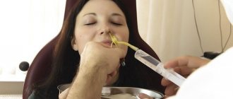
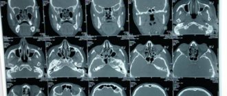
The paranasal sinuses are examined using radiography, diaphanoscopy (examination in a dark room using a light bulb - the method has historical value), puncture of the sinuses using a Kulikovsky needle, as well as trephine puncture of the sinuses (frontal).
General methods of treatment.
Treatment is divided into two groups - conservative and surgical.
Conservative treatment includes: toileting the nose with cotton wicks (or washing with soda-saline solution, infusions of medicinal herbs), infusion of medicines into the nose with drops (for adults 3 - 5 drops, for children - 1 - 3), administration of ointments (cotton wool is wound on a probe , also medicinal substances are administered using turundas), insufflation of powders (using a special powder blower), inhalations, warming thermal procedures.
Surgical treatment methods include: trimming the turbinates (conchotomy), resection of deviated nasal septum, ultrasound of the inferior turbinates, galvanocaustics (cauterization of the mucous membrane with electric current), cryotherapy (cauterization of the mucous membrane with liquid nitrogen), cauterization of the mucous membrane with chemicals
“Anatomy, physiology, methods of ear research»
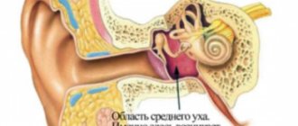
Anatomy of the ear. The ear is the organ of hearing and balance. It is located in the temporal bone and is divided into three sections: external, middle, internal.
The outer ear is the pinna, the external auditory canal and the eardrum, which is the boundary between the outer and middle ear.
Auricle
formed by cartilage, covered with perichondrium, skin and fatty tissue, which is located at the bottom of the auricle, forming the lobe.
External auditory canal
consists of a membranous-cartilaginous section and a bone section. The transition from one tissue to another has a narrowing. The skin of the cartilaginous section contains hair follicles, sebaceous and sulfur glands. The external auditory canal borders in front with the joint of the lower jaw (sharp pain when chewing during inflammatory processes), at the top with the middle cranial fossa (in case of fractures of the base of the skull, cerebrospinal fluid may leak from the ear).
Eardrum
represents a thin membrane of pearl-gray color. It consists of three layers: outer - skin, inner - mucous, middle - connective tissue, which has two types of fibers (radial and circular), which ensures the taut position of the membrane. It has two sections: a loose (front - upper) part and a tense (large) part.
The middle ear consists of the tympanic cavity with the auditory ossicles, the auditory tube, and the mastoid process.
Tympanic cavity
– irregular cube, about 1 cm cube, located in the temporal bone. It contains three auditory ossicles: the malleus (connected to the eardrum), the incus, and the stapes (bordering the inner ear). The bones are connected to each other by joints and held by muscles and perform the function of transmitting sound vibrations.
Eustachian tube
connects the tympanic cavity with the nasopharynx and is located at an angle. It consists of a short bony section (1/3 of the length) and a long membranous-cartilaginous section, which is in a closed state and opens when swallowing and yawning. At this moment, a portion of air enters the tympanic cavity and balances the atmospheric pressure with the pressure in the cavity. The mucous membrane has ciliated epithelium with cilia. The auditory tube performs a protective, drainage and ventilation function. If the tube is obstructed, hearing may be impaired.
In children, the auditory tube is short, wide and located horizontally. This facilitates easy penetration of infection from the nasopharynx.
Mastoid
represents air cavities that communicate with each other. An infection from the tympanic cavity can cause inflammation in the mastoid process.
The inner ear is represented by a bony and membranous labyrinth and is located in the temporal bone. The space between the bony and membranous labyrinth is filled with perilymph (modified cerebrospinal fluid), the membranous labyrinth is filled with endolymph. The labyrinth consists of three sections - the vestibule, the cochlea, and three semicircular canals.
vestibule
the middle part of the labyrinth and connects to the tympanic membrane through the round and oval windows. The oval window is covered by the stapes plate. In the vestibule is the otolithic apparatus, which performs a vestibular function.
The cochlea is a spiral canal in which the organ of Corti is located - this is the peripheral part of the auditory analyzer.
The semicircular canals are located in three mutually perpendicular planes: horizontal, frontal, sagittal. In the expanded part of the canals (ampulla) there are nerve cells, which, together with the otolithic apparatus, represent the peripheral section of the vestibular analyzer.
Physiology of the ear
There are two important analyzers in the ear - auditory and vestibular. Each analyzer consists of 3 parts: a peripheral part (these are receptors that perceive certain types of irritation), nerve conductors and a central part (located in the cerebral cortex and analyzes irritation).
The auditory analyzer starts from the auricle and ends in the temporal lobe of the hemisphere. The peripheral part is divided into two sections - sound transmission and sound perception.
The sound conducting department - air - is:
auricle - picks up sounds
external auditory canal – obstructions reduce hearing
· eardrum – vibrations
chain of auditory ossicles, stapes plate inserted into the window of the vestibule
· perilymph - vibrations of the stapes cause vibrations of the perilymph and, moving along the curls of the cochlea, it transmits vibrations to the organ of Corti.
There is also bone conduction
, which occurs due to the mastoid process and bones of the skull, bypassing the middle ear.
Sound-receiving department
These are the nerve cells of the organ of Corti. Sound perception is a complex process of converting the energy of sound vibrations into a nerve impulse and conducting it to the centers of the cerebral cortex, where the received impulses are analyzed and comprehended.
The vestibular analyzer provides coordination of movements, body balance and muscle tone. Rectilinear movement causes displacement of the otolithic apparatus in the vestibule, rotational and angular movement causes movement of the endolymph in the semicircular canals and irritation of the nerve receptors located here. Next, the impulses enter the cerebellum and are transmitted to the spinal cord and to the musculoskeletal system. The peripheral part of the vestibular analyzer is located in the semicircular canals.
Methods for studying the auditory analyzer.
· History taking
External examination and palpation
· Otoscopy – determines the condition of the external auditory canal and the condition of the eardrum. It is carried out using an ear funnel.
· Functional studies of the ear. Includes examination of auditory and vestibular functions.
Auditory function is examined using:
1. Whispering and colloquial speech. Conditions: soundproofed room, complete silence, room length at least 6 meters. (norm: whispered speech – 6m, spoken – 20m)
2. Tuning forks determine air conductivity - they are brought to the external auditory canal, bone conductivity - tuning forks are placed on the mastoid process or on the parietal region.
3. Using an audiometer, sounds entering the headphones are recorded in the form of a curve called an audiogram.
Methods for studying vestibular function.
Rotational test is carried out using a Barany chair
Caloric test - warm water (43g) is injected into the external auditory canal using a Janet syringe, and then cold water (18g)
· Pressor or fistula test - air is pumped into the external auditory canal using a rubber balloon.
These tests make it possible to identify autonomic reactions (pulse, blood pressure, sweating, etc.), sensory reactions (dizziness) and nystagmus.
The human ear perceives sound pitches from 16 to 20,000 hertz. Sounds below 16 hertz are infrasound, above 20,000 hertz are ultrasound. Low sounds cause vibrations of the endolymph, reaching the very top of the cochlea, high sounds - at the base of the cochlea. With age, hearing deteriorates and shifts towards low frequencies.
Approximate limit of sound volume location:
· Whispered speech – 30db
· Conversational speech – 60db
· Street noise – 70db
· Loud speech – 80db
· Scream at the ear – up to 110db
· Jet engine – 120db. In humans, such a sound causes pain.
Anatomy of the pharynx.
This is a hollow muscular organ located at the level of 1-6 cervical vertebrae. The wall consists of 3 layers - muscle, fibrous layer and mucous layer. In the submucosal layer there is a lot of lymphoid tissue in the form of individual nodules and large clusters that form the tonsils.
The pharynx is divided into 3 sections:
· nasopharynx (upper part) – from the vault to the hard palate
Oropharynx (middle part) - at the level of the oral cavity
laryngopharynx (lower part) - begins at the level of the root of the tongue and passes into the esophagus.
There are 7 openings in the throat:
· in the nasopharynx there are 2 choanae connecting the pharynx with the nasal cavity; 2 mouths of the auditory (Eustachian) tubes, located on the side walls of the nasopharynx.
· In the oropharynx there is a pharynx through which the pharynx communicates with the oral cavity. The pharynx is bounded above by the soft palate, the uvula, on the sides by the anterior and posterior palatine arches, and below by the root of the tongue.
· The laryngopharynx is the entrance to the larynx and esophagus.
In the pharynx there is Pirogov’s lymphoid ring, consisting of 6 tonsils:
two tubal tonsils (surrounding the entrance to the auditory tubes),
· one nasopharyngeal tonsil (located on the roof of the nasopharynx), which is called adenoids,
two palatine tonsils located between the palatine arches in a triangular niche,
· one lingual tonsil, located at the root of the tongue.
The lymphoid tissue of the pharynx contains lymphocytes, which play an important role in the formation of local and general immunity. Antibodies are formed in the tonsils that destroy bacteria and viruses.
The paired palatine tonsils are of greatest importance in clinical practice due to their structural features.
The structure of the palatine tonsils:
On top of the tonsils are covered with a fibrous membrane - a capsule, from which connective tissue fibers extend deeper. As a result, cells filled with lymphocytes, mast and plasma cells are formed - these are follicles. On the free surface of the tonsils there are cracks or lacunae that go deep into the tonsils and branch there. In the lacunae, desquamated epithelium, lymphocytes, microbes, food debris accumulate and form plugs that contribute to the development of the inflammatory process in the tonsils. Shallow lacunae clear themselves when swallowing, while in deep ones the plugs persist and lead to the development of a chronic process.
The intersection of the respiratory and digestive tracts occurs in the pharynx. The pharynx performs 4 functions - breathing, swallowing, speech production and protective.
Research methods.
1. History
2. External examination - the submandibular lymph nodes are palpated.
3. Examination of the middle part of the pharynx - pharyngoscopy. This is done using a spatula. The oral mucosa, soft palate and uvula, anterior and posterior arches, the surface of the tonsils, and the presence of lacunae contents are examined.
4. Examination of the hypopharynx - hypopharyngoscopy. It is carried out using a laryngeal mirror.
5. Digital examination of the nasopharynx is carried out in children to determine the size of the adenoids
1
Treatment methods
Physiotherapeutic. Prescribed for diseases with a chronic course. With the help of ultrasound, a magnetic field, and electric current of different frequencies, it is possible to reduce the severity of the pathological process and stimulate its transition into the remission phase.
Operational. The method is effective for detecting neoplasms in the paranasal sinuses. After identifying tumors, cysts or polyps, the doctor, using miniature instruments, removes the pathological tissue. Due to this, a radical improvement of the patient occurs with the elimination of the cause of unpleasant symptoms.
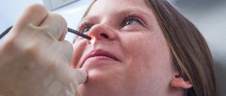
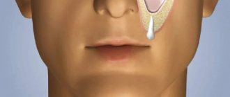
Ultrasound of the nasal turbinates - what is it?
In the case when the nasal mucosa is involved and drug treatment is powerless, there is only one option left - surgery on the inferior turbinates. In such a situation, the best solution would be ultrasound vasotomy.
A — condition of the inferior turbinates after surgery. B - before surgery
What is it and how effective is it? This is the most important and frequently asked question from patients. Ultrasonic disintegration is a surgical procedure on the inferior turbinates of the nose.
Ultrasound affects the mucous surface and the blood vessels passing there. Under the influence of ultrasound, they are destroyed, during which they are replaced with new ones.
The procedure helps reduce the mucous layer and restores nasal breathing. Ultrasonic cauterization is a simple and short procedure. Its duration is approximately 5-20 minutes. It is performed under anesthesia; depending on the severity of the patient’s condition, local or general anesthesia may be used. The first one is most often used.
Along with this method, there are Laser vasotomy and Radio wave disintegration of the inferior turbinates. Vasotomy in ENT offices can be called by different names; you can fully understand what vasotomy is HERE.
Ultrasonic disintegration can be right-left-sided, as well as mixed. The latter is the most common, since a runny nose involves both nasal passages. This surgical intervention is considered quite effective, but the disadvantage is the short-lived result of therapy. It can last differently for each person, depending on individual characteristics, as well as the severity of the pathology.
Features of the study
The study is performed in A-mode. Unlike a traditional ultrasound, where the outline of organs is visible on the screen, in this case the doctor receives a diagram with a straight line in the center. The paranasal sinuses normally contain air, which does not reflect sound waves. This causes only a straight line to be visualized.
If there is fluid inside the sinuses (exudate, pus, blood) or neoplasms (cysts, tumors, polyps), the line deviates. This is due to the reflection of sound waves from soft tissues or pathological contents of the sinus. Based on the data obtained, the doctor forms a conclusion and proposes a targeted treatment method.
Seeing a doctor in a timely manner will help maintain your health.
Don't delay treatment, call right now. We work around the clock. tel. (24 hours a day)