Subtotal darkening of the maxillary sinuses on radiography
As a rule, subtotal darkening of the maxillary sinuses is a consequence of chronic sinusitis in the acute stage.
Very often, a common cold can be fraught with a long-term persistent runny nose, which is difficult to get rid of.
Over time, patients develop headaches, especially with a sharp turn of the head, impaired sense of smell and taste, weakness and swelling of the eyelids, and runny nose.
With untimely or inadequate treatment, the latter symptom can develop into a very dangerous disease called sinusitis and subsequently lead to subtotal darkening of the maxillary sinuses.
X-ray: where to start
During the examination, the specialist will make a preliminary diagnosis of “sinusitis” and send you for an x-ray to clarify suspicions.
An x-ray is a collection of images of the anatomical structures of the body obtained by x-ray irradiation.
An X-ray image, like any negative image, consists exclusively of black and white shades.
This is due to the fact that each tissue in the human body has its own degree of absorption of gamma radiation. The more tissue absorbs, the brighter and more intense it is visible in the image.
To diagnose certain diseases, it is extremely necessary to perform fluoroscopy in the optimally correct position, since only in this projection the areas required for inspection become visible. For example, there are several sinuses in the skull:
- frontal (frontal) sinus,
- wedge-shaped (main),
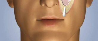
- maxillary (maxillary),
- lattice labyrinth.
In order for the nasal sinuses to be as visible as possible in the image, it is necessary to shoot in the following projections:
- axial – mainly used to assess the condition of the skull base, rocky part of the temporal bone and visualization of the main sinuses,
- lateral – important for examining the frontal, determining the size of the sphenoid and maxillary sinuses, the condition of the anterior parts of the facial bones and the base of the skull,
- nasofrontal – optimal for studying the frontal sinuses, cells of the ethmoidal labyrinth and orbits,
- The most clear way to trace the pneumatization of the maxillary sinus is to position the patient with the chin and nose resting on the X-ray machine stand (nasochin position).
For correct diagnosis, it is very important that the image has good contrast and sharpness and does not have any extraneous shadows or artifacts. All this together will make it much easier for the doctor to make the correct diagnosis.
As a rule, complete darkening of the maxillary sinuses indicates that a large amount of pus has accumulated inside them due to an extensive inflammatory process caused by harmful microorganisms.
X-ray of the maxillary sinus in normal and pathological conditions
Before moving on to diagnostic criteria, it is necessary to evaluate the quality of the image. To do this, you need to pay attention to the clarity of the ethmoid bone structure and the pneumatization of the nasal sinuses.
The sinuses are hollow structures that have the same pneumatization as the orbits of the eyes, due to which they have the same dark color on an X-ray image. Therefore, during the initial assessment of the condition of the maxillary sinuses, it is necessary to simply compare them with the orbits.
In the initial stage of sinusitis, X-rays can reveal thickening of the affected contours, which may indicate inflammation of the mucous membrane. Thickening of the maxillary sinus mucosa can be observed in several cases:
- catarrhal (acute) inflammation,
- chronic process (exacerbation),
- swelling after treated sinusitis or allergies.
Also, do not forget that the ethmoid labyrinth is the first structure in which inflammation develops during sinusitis.
In the absence of timely treatment, the process spreads to other sinuses, including the maxillary sinus, which is determined by subtotal darkening on the radiograph.
Therefore, if there are no changes in this sinus, it is worth taking a close look at the structure of the ethmoid bone.
Symptom of "milk in a glass"
As acute sinusitis progresses, liquid contents accumulate in the maxillary sinus - an infiltrate, which is detected on an x-ray as a level of liquid against a background of subtotal darkening - the “milk in a glass” symptom. Subtotal darkening can be unilateral or bilateral, with different levels of fluid. As a rule, the darkening is homogeneous, homogeneous, with signs of thickening of the mucous membrane.
With sinusitis, an x-ray may not reveal the “milk in a glass” symptom, but there may simply be a homogeneous darkening in the entire maxillary sinus - a total darkening. This indicates that the entire sinus is clogged with pus and requires immediate surgical intervention.
Puncture of the sinus for the purpose of pumping out the contents significantly improves the general condition of the patient - the temperature decreases, headaches decrease. Also, pumping out the liquid allows you to study its nature more closely, so the puncture can also be carried out for diagnostic purposes, among other things.
Despite the fact that emptying the cavity brings great relief to the patient, the treatment is not considered complete and must be supplemented with a course of antibiotic and hormonal therapy, which can be combined with the use of vasoconstrictors and rinsing the sinuses with various antiseptic solutions.
Subtotal and total darkening of the maxillary sinuses may also indicate the presence of dense neoplasms in the nose - sarcoma, osteoma, chondroma, etc. The formation can be homogeneous or have a dense membrane with liquid contents.
If the neoplasm is small in size and does not contribute to the accumulation of infiltrate, then the patient is not characterized by intoxication and the presence of pain. If the cyst is large, secondary sinusitis develops, the treatment of which may not give the desired result. Therefore, you must first differentiate the nature of the formation and take measures to remove it.
How else are the sinuses examined?
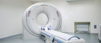
In addition to radiography, the most complete and voluminous picture of all pathological processes will be provided by studies such as magnetic resonance and computed tomography.
They will provide more accurate information about the pathological process in the affected sinus, inside and outside the process. The method is expensive, but informative.
There is also a method of ultrasound scanning of the maxillary sinuses, which does not provide much information, but is able to distinguish swelling from infiltration.
In any case, the attending physician must select a diagnostic method and study the conclusions, since it is he who is able to select adequate therapy depending on the type of pathological process in the affected sinus.
Source: https://za-rozhdenie.ru/lor/gajmorit/subtotalnoe-zatemnenie-gajmorovyh-pazuh
Treatment of sinusitis
Treatment of the disease includes a special set of measures that are aimed at suppressing the source of infection and restoring drainage in the maxillary sinus. Typically, acute sinusitis is treated with conservative methods, which primarily ensure a good outflow of sinus contents. Chronic sinusitis is treated using pharmacotherapy, which can only be selected and prescribed by a doctor. If there is excessive discharge of mucus and pus from the nasal sinuses, as well as severe pain, they are washed.
What is subtotal darkening of the maxillary sinuses?
Subtotal darkening of the maxillary sinuses - what is it and how dangerous? This conclusion can be heard in various diseases of the ENT organs. In simple terms, it is an inflammation of the mucous membrane of the maxillary sinus with accumulated secretions. An X-ray is often recommended for patients with sinusitis, sinusitis or sinusitis.
Subtotal darkening of the maxillary sinuses - causes
If the doctor sent the patient for an X-ray of the nose, then he suspects inflammation of the mucous membrane with a possible accumulation of discharge or pus. The image shows the maxillary sinuses, frontal and ethmoidal labyrinth. Subtotal darkening of the maxillary sinuses makes it possible to determine the advanced state of the disease. The more shadows there are in the picture, the more purulent masses have already accumulated.
The reason for the accumulation of such secretions is the active reproduction of pathogenic microorganisms. The causative agents of sinusitis or sinusitis are streptococci or pneumococci.
They can become active after prolonged rhinitis, when treatment was ineffective or incorrect. With the accumulation of secretions and inflammation of the mucous membrane, severe swelling appears, which does not allow the mass to leave the sinus.
Mucus begins to accumulate, which is an ideal environment for the proliferation of pathogenic microorganisms.
Pus accumulates in the maxillary sinuses not only due to a bacterial infection. If the picture clearly shows thickening of the mucous membrane, then this condition could have formed due to:
- Acute inflammation;
- Allergy progression;
- Swelling after sinusitis;
- Long-term chronic inflammation.
Do not forget that sinusitis or frontal sinusitis can develop due to a cyst, which can also be seen in the image. The causes of darkening are adenoids and polyps. Such formations can provoke frequent runny nose, which over time transforms into sinusitis.
X-ray of PPN for pathology of the maxillary sinuses
X-ray of the paranasal sinuses (PSN) helps to assess the stage of development of sinusitis and sinusitis already at the primary stage of the disease. This diagnostic method is prescribed if:
- There is nasal discharge after a respiratory infection;
- The discharge does not go away within 7-10 days;
- Nosebleeds appear;
- The body temperature remains high, but there is no nasal discharge;
- Noticed hyperemia or redness of the skin in the paranasal sinuses;
- When tilting the head, a pressing pain appears.
Total darkening of the maxillary sinuses indicates severe neglect of the disease, this is especially evident when the frontal sinuses are darkened. At the initial stages of development of sinusitis, discharge accumulates only in the PPN.
If the doctor diagnoses nasal pathologies caused by adenoids, cysts or polyps, then treatment will include surgery. The PPNs themselves in a healthy person are semi-oval in shape, their color matches the color of the eye sockets. At the slightest accumulation of pus or inflammation of the mucous membrane, the doctor notes a parietal darkening.
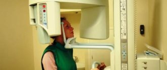
The X-ray procedure of the PPN is painless. To do this, just position yourself correctly on the device stand. The patient's position depends on the type of projection indicated by the doctor.
The radiologist must indicate the correct position. Often, patients, either in a vertical or horizontal position, press their nose and chin tightly against the apparatus stand.
It is enough to fixate for a few seconds to get a clear image.
X-rays during this procedure can range from 0.12 to 1.18 mSv, the amount of mSv depends on the type and power of the equipment used.
Relatively small doses of x-ray radiation are unacceptable for pregnant women and preschool children.
For women in this position, rays can affect the development of the fetus, and in a child under 6 years of age they cause changes in the bone tissue of the skeleton, so doctors prescribe such a study as a last resort.
What can be seen on an x-ray?
The healthy nasal cavity in the image has a clear border and an average thickness of the walls of the nasal cavity. When X-raying the maxillary sinus with suspicion of the initial stage of sinusitis, radiologists note thickening of the affected contours. This condition is characterized by the appearance of inflammation of the mucous membrane.
The ethmoid labyrinth is also visible in the image, because in this structure the accumulation of secretions occurs first. If darkening is not visible, then it is important to carefully examine the structure of the ethmoid bone tissue.
Infiltration or pus often causes the so-called “milk in a glass” symptom. This name comes from the property of the discharge to always be located in a horizontal position, no matter what position the patient occupies. Darkening with this symptom is either unilateral or bilateral.
If the disease is advanced, then the accumulation of pus becomes total. This condition is fraught with the spread of purulent masses into neighboring tissues and vessels. There is an increased risk of infiltrate penetrating into the brain, which causes meningitis.
When pus enters the bloodstream, it causes sepsis and inflammation of internal organs.
Accumulation of exudate with subtotal and total darkening also develops with the appearance of dense neoplasms in the nasal cavity, for example, chondroma, osteoma or sarcoma. Such cells may have a dense outer shell, but inside they will have a liquid consistency.
Source: https://nasmork.net/chto-takoe-subtotalnoe-zatemnenie-gajmorovyh-pazuh/
Causes of the disease
Very often, the cause of sinusitis is infections, which include: streptococci, Haemophilus influenzae, staphylococci, mycoplasmas, fungi, viruses and chlamydia.
In addition, the development of parietal sinusitis can be provoked by the following diseases and conditions:
- ARVI and respiratory tract diseases,
- chronic tonsillitis and pharyngitis,
- inflammation of the roots of the eighth teeth,
- sudden and frequent changes in pressure when diving and flying,
- measles and scarlet fever,
- prolonged allergic runny nose;
- deviated septum,
- narrowness of the nasal passages.
The disease becomes chronic due to untimely, incomplete or incorrect treatment of the above diseases. A particular insidiousness of sinusitis lies in the delayed appearance of symptoms, which can manifest themselves only after some time - 2-4 weeks after the end of the primary disease. The likelihood of developing sinusitis increases several times during the off-season, when there is a natural lack of vitamins and the body’s protective functions decrease.
Children under seven years of age do not suffer from sinusitis due to the structure of the nasal passages.
Upon reaching this age, the cause of sinusitis in children is most often inflamed adenoids, which prevent full nasal breathing and represent a constant source of infection.
Subtotal darkening of the maxillary sinuses
What is subtotal darkening of the maxillary sinuses? If an x-ray shows a decrease in transparency in the lower and middle parts of the maxillary sinus, this may indicate the presence of infectious inflammation or a benign tumor - a cyst, a polyp. X-rays are prescribed to patients if sinusitis, polypous sinusitis, cancer, etc. are suspected.
X-ray examination of the paranasal sinuses (SNS) allows us to assess the level of pneumatization (presence of air) in the mentioned anatomical structures.
The image assesses the degree of inflammation of the maxillary sinuses, i.e. maxillary sinuses, and prescribe adequate treatment.
The article discusses the interpretation of radiographs, as well as pathologies that are detected during subtotal and parietal eclipse of bone cavities.
X-ray PPN – what is it?
X-ray PPN is an instrumental method for examining the paranasal sinuses, which makes it possible to identify pathologies in the early stages of development. An X-ray image gives a clear idea of what pathological processes occur in the paranasal sinuses. Transillumination of intranasal structures allows you to determine:
- degree of inflammation of the mucous membranes;
- the presence of serous or purulent exudate in the nasal cavity;
- formation of benign or malignant tumors.
During the examination of the patient, the radiologist takes two pictures - in a supine and standing position. The absence of any pathologies in the maxillary sinuses looks like this on x-ray:
- the bone walls of the paranasal sinuses have a clear outline;
- the contours of the ethmoid cells located at the level of the nasal septum are not blurred;
- Pneumatization of the PPN is no different from the reference one, which is located at the level of the eye orbits.
If, after an instrumental examination, darkened areas are detected in the image, the patient is referred for a computed tomography scan. After an accurate diagnosis, he prepares a suitable drug treatment aimed at eliminating the inflammatory reactions in the maxillary sinuses.
Why is a PPN x-ray prescribed?
In the bones of the skull there are several pairs of air cavities, which are called paranasal or paranasal sinuses (sinuses). Their inner surface is covered with soft tissues, in particular ciliated epithelium. Its inflammation often leads to the development of sinusitis.
Sinusitis is a collective term that refers to a group of respiratory diseases characterized by inflammation of the PPN. Radiologists pay attention to the fact that not every sinusitis is sinusitis. Only when the maxillary (maxillary) sinuses are affected is a diagnosis of “maxillitis” or “sinusitis” made.
X-ray visualization of the maxillary sinus allows us to identify several forms of sinusitis:
- polyposis;
- parietal-hyperplastic;
- purulent;
- catarrhal
An otolaryngologist cannot diagnose “sinusitis” only on the basis of the patient’s medical history and complaints.
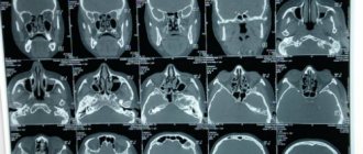
When receiving an unclear image, it is impossible to say with certainty that the patient suffers from one or another type of pathology. In this case, the diagnostic examination is supplemented by computed tomography.
Interpretation of the radiograph
Even with an X-ray examination of the maxillary sinuses, it is not always possible to say that the cause of pathological symptoms is sinusitis. Deciphering the image requires analysis of a number of anatomical structures:
- eye orbits;
- oral cavity;
- frontal sinuses;
- lattice labyrinth.
Deciphering one x-ray takes an experienced specialist no more than 10 minutes of time. If there are obvious dark spots in the bone cavity, the doctor may accidentally diagnose cancer. What does the specialist’s medical report indicate and how is the image interpreted?
- exudative maxillitis - light spots with a clear horizontal border in the upper part of the maxillary sinuses;
- parietal-hyperplastic maxillitis - parietal darkening in the area of the bone walls, associated with swelling of the mucous membrane; the wavy contour of the bone cavity faces the inside of the sinus;
- exudative maxillitis - total darkening of the maxillary sinuses associated with the accumulation of fluid in the anatomical structures;
- polypous sinusitis - protrusion of the parietal part of the sinus into the bone cavity;
- purulent sinusitis – subtotal (almost complete) darkening of one or both maxillary sinuses.
The above transcripts are provided for informational purposes only and therefore cannot be used for independent diagnosis and drawing up a treatment regimen.
Only a doctor can assess the degree of impairment of pneumatization of the maxillary sinus using an x-ray. When making a diagnosis, they take into account structural changes in the ethmoidal labyrinth, as well as the clarity of the contours of a single cell.
What pathologies are detected using X-ray of PPN?
X-ray of the PPN reveals characteristic changes in the intranasal structures. In a healthy person, the paranasal sinuses look like semi-oval dark formations. To identify pathology, you need to compare the degree of their staining with the shade of the eye orbits. Parietal darkening of bone cavities indicates the development of a whole range of respiratory diseases.
Hypertrophic maxillitis
Hypertrophic maxillitis is a chronic inflammation of the maxillary sinus, accompanied by thickening of the mucous membrane. In the image, the pathology does not appear as darkening, but as a wall layer with a ragged, curved edge. The interpretation of the radiographic image is described as follows: parietal darkening of the maxillary sinus due to hypertrophy (thickening) of the mucosa.
If the diagnosis contains the phrase “total darkening,” this indicates a complete violation of the pneumatization of the bone cavity.
In other words, the patient is found to have complete obstruction (airlessness) of the maxillary sinus. Treatment of the disease involves the use of antibiotics and nasal corticosteroids.
If the pathology has developed against the background of allergic rhinitis, antihistamines are included in the treatment regimen.
Oncological diseases
Transillumination of the maxillary sinuses allows us to determine the presence of benign or malignant tumors in the intranasal structures. By analyzing the x-ray, a specialist can identify:
- osteoma is a benign neoplasm that occurs in bone tissue; the tumor grows very slowly and almost never becomes malignant, so its course is considered favorable;
- sarcoma is an actively growing malignant tumor, which consists mainly of connective tissue; the rapid development of pathology leads to the destruction of bone structures in the paranasal sinuses;
- chondroma – a cartilaginous tumor prone to malignancy (malignancy); It is diagnosed mainly in young children and adolescents.
Benign and malignant tumors are subject to surgical removal and subsequent drug treatment.
Cyst in the paranasal sinuses
As a rule, a cyst on an x-ray is a surprise to a specialist. The process of tumor formation does not cause any discomfort to the patient, so in most cases it is diagnosed accidentally during an endoscopic or radiographic examination.
Spontaneous opening and evacuation of purulent secretion from the cyst into the surrounding tissues can lead to complications. A cyst is a hollow neoplasm containing fluid inside. In the picture, the cyst looks like a rounded small shadow located in the middle or lower part of the maxillary sinus. Treatment of cystic tumors requires surgery.
Exudative sinusitis
If the maxillary sinuses are filled with serous effusions, a horizontal level of fluid will be visible on the x-ray. In most cases, this indicates the development of exudative sinusitis - a respiratory disease in which a lot of fluid accumulates in the maxillary sinuses.
When carrying out differential diagnosis, a puncture is taken from the affected sinus to accurately determine the type of pathology. In otolaryngology, the following forms of sinusitis are distinguished:
- catarrhal - filling of the paranasal sinuses with serous effusions, accompanied by redness (hyperemia) and swelling of the mucous membrane;
- hemorrhagic - a dangerous and quite rare form of the disease in which blood accumulates in the maxillary sinuses;
- purulent - bacterial inflammation of the accessory sinuses, characterized by the accumulation of purulent exudate in the bone cavities.
Hemorrhagic sinusitis is dangerous due to increased vascular permeability, in which infection can easily penetrate the systemic bloodstream.
To treat the catarrhal form of the disease, vasoconstrictor drops (decongestants), mucolytics, antiallergic and antiviral agents are used. Purulent inflammation can be eliminated only by taking systemic antibiotics and local antimicrobial agents.