MSCT of the paranasal sinuses
– an improved technology of computed tomography, the essence of which is layer-by-layer scanning of the area under study with a stream of X-rays and recording the radiation passed through the tissue with several rows of detectors. By obtaining many thin sections (layer-by-layer images), a detailed image of the maxillary, frontal, ethmoidal and sphenoid sinuses is achieved, which makes MSCT a popular diagnostic method in otolaryngology. MSCT of the paranasal sinuses is prescribed to identify inflammatory lesions of the sinuses (sinusitis, sinusitis, ethmoiditis, sphenoiditis), tumor processes, injuries and foreign bodies, as well as control of conservative and surgical treatment. The use of contrast agents improves the visualization of neoplasms of the paranasal sinuses and makes it possible to assess their prevalence.
MSCT of the paranasal sinuses is a relatively young technique that is gradually gaining popularity due to a number of advantages over other research methods. Its main advantages are high information content and non-invasiveness. During the procedure, the skin is not damaged, and the patient experiences minimal inconvenience. The study is performed in a relatively short period of time, especially in comparison with magnetic resonance imaging. MSCT of the paranasal sinuses allows you to display images in real time, which makes it an excellent assistant for monitoring the implementation of minimally invasive procedures. This study is not as sensitive to the patient’s movements as MRI, and can be prescribed by doctors even if the patient has electronic devices and metal implants in the body.
MSCT of the paranasal sinuses is an improved version of classical computed tomography. It is successfully prescribed by otolaryngologists, oncologists and maxillofacial surgeons to clarify the diagnosis and determine further treatment tactics. The distinctive features of the technique are a high degree of image detail and the use of several scanning planes. This makes it possible to create a three-dimensional model of the study area in 3D mode for a more thorough study. Compared to conventional computed tomography, MSCT of the paranasal sinuses is characterized by the use of lower doses of X-ray radiation, higher research speed and improved image quality. The procedure can be performed routinely or on an emergency basis.
In what cases is a study prescribed?
When is a computed tomography scan of the nose indicated? Indications for use are the following conditions:
- suspicion of a violation of the integrity of the bones of the nose and sinuses;
- risk of hemorrhage;
- traumatic brain injuries;
- frequent headaches of unknown origin;
- frequent nosebleeds accompanied by increased blood pressure;
- suspicion of the presence of a foreign object in the nasal area;
- chronic nasal diseases;
- malignant tumor, presence of purulent exudate;
- a complication of infectious pathology of neighboring organs, which has spread to the nasal sinuses.
Contraindications
These types of diagnostics are also similar in terms of contraindications. MRI and CT are not performed:
- people with a weight over 150 kg and a waist size over 150 cm;
- if there are foreign bodies in the body, as this can cause severe bleeding;
- children under 5 years of age (only in extreme cases is MRI allowed for children under anesthesia);
- in the first trimester of pregnancy, as this carries a risk for the development of the fetus.
What can a CT scan show?
Computed tomography of the sinuses allows you to see images of layer-by-layer sections of the scanned space. The study shows the contours, density, structure, volume, mineralization of PPN.
A CT scan of the nose and sinuses will help evaluate the effectiveness of the therapy if the diagnosis was established a long time ago.
Pathology diagnosed in a timely manner using CT scanning will help to avoid serious consequences and complications.
So, what does a sinus CT scan show? Using this method, the following diseases can be identified:
- sinusitis at the initial stage and during chronic process;
- neoplasms of the nasal mucosa. The study will also answer the question of whether the tumor is malignant or not;
- sinusitis;
- the cause of regular migraines;
- sphenoiditis, frontal sinusitis, ethmoiditis;
- polyps;
- the cause of the inflammatory process, the treatment of which does not bring effect;
- will help visualize deformation and damage to the nasal bones;
- will show the location of foreign bodies in the sinuses.
Computed tomography of the anatomy of the nose and paranasal sinuses
09.04.2014
Due to the fact that almost all imaging for chronic sinusitis is performed using CT, this article focuses on CT anatomy and also discusses the most common anatomical variations. Before reading the article, readers are advised to review the anatomy of this area.
ANATOMY
Understanding the anatomy of the nasal cavity and its anomalies is important as it leads to an understanding of the radiographic anatomy necessary to plan the surgical approach.
Nasal cavity
The nasal cavity warms and humidifies the inhaled air and filters out small foreign bodies it contains before the air enters the lungs. It is lined by the same cylindrical epithelium that lines the airways of the lungs - the trachea and bronchi. This epithelium secretes mucus that coats the walls of the nasal cavity, which participates in the process of mucociliary clearance, in which small foreign particles are eliminated.
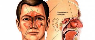
The paranasal sinuses also drain into the nasal cavity, in addition, it ensures the flow of air to the olfactory epithelium in the olfactory recesses, thereby participating in the act of recognizing odors. The olfactory recesses (sinuses) are located in the superior nasal passages, medial to the paired superior turbinates. The nasal cavity is surrounded by air-bearing, mucous-lined cavities called the paranasal sinuses, which include the paired maxillary (maxillary), frontal, ethmoid and sphenoid sinuses. The sinuses communicate directly with the nasal cavity, into which their secretions, secreted by the mucous membrane of the sinus walls, are removed. The cilia of the epithelium flicker towards the natural drainage holes, allowing secretions to be removed from the sinus cavities into the nose. The bottom of the nasal cavity is the hard palate. On the lateral walls there are spiral folds of mucosa covering the nasal turbinates and draining the paranasal sinuses. The curved surfaces of the turbinates increase the contact of inhaled air with the mucous membrane. The roof of the nasal cavity consists of a cribriform plate in the center and on the sides - the lower walls of the cells of the ethmoid bone. In the middle, the nasal cavity is divided into two halves by a septum, which is partly bone and partly cartilage, and is lined, unlike the lateral walls, with flat epithelium.
Paranasal sinuses
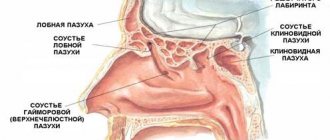
The paired frontal, maxillary, ethmoid and sphenoid sinuses surround the nasal cavity. The ethmoid sinuses form the roof, and the maxillary sinuses form the lateral walls. The frontal ones are located in front and above, and the wedge-shaped ones are located behind and above the ethmoid ones. The main drainage openings are the anterior ostiomeatal complexes (AMCs) (see Fig. 1), which drain the frontal and maxillary sinuses and the anterior and middle thirds of the ethmoid sinuses.
Figure 1. CT scan of the nasal cavity. Normal anterior ostiomeatal complex The anatomy of the ethmoidal infundibulum and the uncinate process is clearly visible.
The uncinate process (CP) and the lateral wall of the nasal cavity form the ethmoidal infundibulum (FV). The above sinuses are drained into the RV through different openings. The opening of the maxillary sinus (MS) and the recess (or duct) of the frontal sinus (FS) (see Fig. 2) open into the most anterior part of the FMS and are clearly and consistently visible on CT scans.
Figure 2. CT scan of the nasal cavity. The frontal sinus opens into the middle meatus just lateral to the anterior portion of the middle turbinate. The anterior-most ethmoid cells are called “nasal agar cells.”
Other openings are formed from the anterior and middle groups of ethmoid cells and also open in the RV, but are not visible even on 2 mm frontal CT scans. When the frontal, maxillary sinuses, and anterior and middle cells of the ethmoidal labyrinths are involved, this pattern is called an infundibular pattern. This pattern is most common in chronic sinusitis.
Processes involving the anterior ethmoidal cells influence the drainage of the ICP and LA into the RV. The most common cause of frontal sinusitis and sinusitis is disease of the anterior ethmoid cells in combination with rhinitis, both viral and bacterial in nature. Inflammation and swelling of the mucous drainage holes leads to disruption of secretion and mucociliary clearance, which leads to inflammation in the upper jaw and left atrium. Cleansing and curing the process in the anterior ethmoid cells leads to the cleansing of the upper jaw and left atrium. The indicated outflow pathways from the ICP and LA were documented using stained droplets. For a more detailed introduction to the physiology of the paranasal sinuses, readers are referred to manuals on otorhinolaryngology, for example, edited by Stammberger and Messerklinger.
The posterior OMK (Fig. 3) is located in the sphenoethmoidal recess and drains the posterior ethmoidal cells and sphenoid sinuses.
Figure 3. CT scan of the nasal cavity. The vomer separates the two openings of the sphenoid sinuses, which are located in the sphenoethmoidal recess. The opener is pneumatic (standard).
The vomer separates the two openings of the sphenoid sinuses. In chronic sinusitis, this complex is less frequently involved because the anatomical variants are less common. A sagittal reconstruction of the OMK showing the curved edge of the lunar duct with an inferior fold of mucosa covering the uncinate process is shown in Figure 4. The frontal recess is also visible.
Figure 4. CT scan of the nasal cavity. The reconstructed sagittal image of the OMK demonstrates a curved edge of the lunar duct with an inferior fold of mucosa covering the uncinate process. The frontal recess/duct is also visible.
Nasal septum
The anterior upper part of the nasal septum consists of cartilage. Its posterior upper part consists of the perpendicular plate of the ethmoid bone, and the lower part consists of the vomer and nasal crest of the upper jaw and palatine bones. The bony part of the nasal septum is often pneumatized. When the pneumatized part inflates, it can disrupt the air flow into the wedge-shaped holes (which are adjacent to both sides of the opener on frontal CT scans) in the wedge-ethmoid pockets. Spikes and curvatures of the nasal septum can disrupt air flow, especially if they compress the mucous membrane of the nasal turbinates or the lateral wall of the nasal cavity. Severe deviations of the nasal septum can deform the turbinates and completely stop the flow of air.
Lateral wall
The anatomy of the lateral wall of the nasal cavity is quite complex and most anatomical anomalies occur there. Typically, a person has 3 nasal conchas: superior, middle and inferior. Sometimes there is a 4th nasal concha, the uppermost one. The spaces between the turbinates along their outer edges and the side wall of the nasal cavity are called the nasal passages. The inferior turbinate is attached to the lateral wall of the nasal cavity and only the nasolacrimal duct opens into the inferior nasal meatus. Most of the inhaled air passes through the middle, rather than the lower, nasal passage. Functionally, the middle nasal meatus is the most important, since the upper nasal passage, the left nasal passage, and the anterior and middle cells of the ethmoidal labyrinths open into it. Most anatomical variants occur in the middle meatus and turbinate region. The posterior cells of the ethmoidal labyrinths and the sphenoid sinuses open into the superior nasal passage.
Uncinate process
The uncinate process (CP), which forms the anterior part of the OMC, is one of the most important bone structures. The CO acts as an air valve, preventing direct entry of air into the ICP and ensuring normal mucociliary clearance. Simple allergic or even viral rhinitis, as well as the anatomical variant of CO, can lead to a narrowing of the rhinitis, which results in a disruption of the normal drainage of the paranasal sinuses. This bony structure is central to functional endoscopic sinus surgery (FESS), as it is the first structure that is identified and removed during FESS. CO begins from the lateral wall of the nasal cavity, sometimes from the inferior turbinate, and protrudes into the area of the air flow. Its upper edge is attached to the floor of the anterior ethmoidal cells and then decreases in height towards the nasopharynx, with its upper free edge forming the semilunar meatus, which opens directly into the middle meatus. The orbital floor or medial wall is located lateral to the CO, and the RV lies between them, opening directly into the ICP through its openings in the anterior, middle and upper corner. Several ethmoidal cells, as well as the frontal sinuses, also drain into the RV. The frontal recess, or duct, opens into the anterior ethmoidal part of the middle meatus. An increase in the so-called agger nasi cells, the most anteriorly located ethmoid cells, can cause a narrowing of this drainage channel. Thus, both the frontal and HF cells, as well as the anterior and middle ethmoid cells, open into the RV.
See continuation here.
Author: Charles Lee , MD, Chief of Diagnostic Radiology, University of Kentucky.
Posted in Articles | Tags: anatomy, research, CBCT, computed tomography, , ENT organs, nasal cavity, PPN, tomography
Advantages and disadvantages
CT scan of the paranasal sinuses is a highly accurate method for diagnosing pathologies of the nose and its sinuses. A detailed scan helps to understand what stage the pathological process is at and prescribe the most appropriate treatment in this situation.
What are the advantages of CT scanning of the nose and paranasal sinuses? The following advantages of this diagnostic method can be highlighted:
- high-quality images of the sinuses;
- low radiation exposure;
- high research speed;
- absence of negative effects on the body of the subject;
- clear visualization of all nasal formations;
- absence of discomfort and pain during the procedure;
- complexity - based on the scan results, you can assess the condition of bone formations, blood vessels and tear ducts.
But computed tomography of the nose and paranasal sinuses also has disadvantages. They are hidden in contraindications. The study cannot be carried out under the following conditions:
- pregnancy, since CT scans can cause fetal malformations;
- childhood - the child cannot stop moving for a long time. In addition, belts make them afraid;
- allergic reactions to iodine used as a contrast agent. But contrast may not be used in the study;
- lactation period;
- thyroid diseases;
- failure of renal, hepatic, cardiac functions;
- diabetes;
- multiple myeloma.
Contraindications to CT scan of the sinuses
CT is an effective diagnosis, but has some limitations to the examination that you need to know!
Like any other procedure, a computed tomography examination has some contraindications. This is largely due to the specifics of this method, since during the CT scan there is direct irradiation, albeit not strong.
Contraindications to CT scan of the sinuses are:
- pregnancy
- small age - up to 3 years
- when using contrast, computed tomography is excluded for people who have an allergic reaction to iodine, since all contrast agents are made on its basis
- diabetes
- heart diseases
- renal or liver failure
- lactation period in women
Important! Of course, some exceptions to contraindications can be made, but this is the privilege of only the treating doctor.
How is diagnostics carried out?
CT scan of the paranasal sinuses is performed as follows:
- Specialists assess the condition of the subject.
- The essence of the research method and the purpose of its implementation are explained to the patient.
- The patient is put on a suit that will protect the body from exposure to x-ray radiation.
- Next, the patient is placed on his back on the couch. Arms should be extended along the body. The head is fixed in the headrest. You may need to tuck your chin to your chest and look up.
- Contrast is administered if necessary. 50 ml of contrast is usually injected into the cubital vein through a catheter. For a short time, the patient may feel warmth and a specific taste. These sensations pass on their own and quickly.
- The table together with the subject is pushed into the tomograph. Next, the doctor goes into the room where the equipment for recording and processing the device’s signals is located. The specialist has the opportunity to communicate with the patient during the examination using a sound device.
- During a CT scan of the nose, the table moves up or down. This is necessary to register images in different projections.
- The entire examination takes 5-10 minutes. Upon completion, the patient is sent home or to the ward.
Methodology
When performing MSCT of the paranasal sinuses, the patient lies with his back on a movable table, which slides into the tomograph in such a way that the head is located in the scanning area, fixed using belts and rollers. Staying still during the examination is extremely important because... The clarity of the image and the reliability of the information received depend on this. If MSCT of the paranasal sinuses with contrast is prescribed, an intravenous injection of a dye is given before the procedure. During operation of the device, the patient hears noise; this type of diagnosis does not imply the occurrence of other unpleasant or painful sensations. The duration of the procedure is several minutes. With contrast, the scanning duration increases to a quarter of an hour. The data obtained during the examination is provided in the form of a written report and printed photographs or on electronic media.
Cost of MSCT of the paranasal sinuses in Moscow
The price of this diagnostic method can vary widely. Fluctuations primarily depend on the location of MSCT. A multislice computed tomography scan in a private medical clinic will cost more than in a government organization. The cost of scanning the paranasal sinuses is influenced by the urgency of the procedure (after preliminary registration or urgently), as well as the technical parameters of the tomograph used. Some centers operate around the clock and provide discounts for performing the procedure in the dark. If contrast is used during the study, the price of MSCT of the paranasal sinuses in Moscow varies depending on the amount of the drug administered, which increases in direct proportion to the patient’s weight.
Decoding the results
Based on the results of a computed tomography scan of the nose, the doctor evaluates the following parameters:
- how the nasal septum and bones that form the sinuses are located;
- along what paths does the sinuses drain?
- symmetry of the right and left sides of the nose, sinuses. This parameter will help to determine the anatomical location of the bones and, if necessary, carry out surgical correction in the future;
- pneumatization of the sinuses, its degree.
The interpretation of the results of a CT scan of the nose and paranasal sinuses can be presented in the table:
| Pathology | Signs on CT | Features of the study | What research methods can be combined with? |
| Sinusitis | Fluid in the axillary lumens, thickened soft tissues | CT scanning for sinusitis is not one of the main diagnostic methods. Prescribed only in the absence of effect from treatment and to differentiate diseases | X-ray examination of PPN as an additional method |
| Polyps | In the case of a single polyp, a formation is visualized on a stalk, which comes from the membrane of the axillary wall. If the polyps are multiple, the shape of the sinus will be changed | It is difficult to detect polyps located in the alveolar bay (formation in the maxillary sinus) | Initially, an X-ray of the PPN is taken. CT scan is prescribed to clarify the data |
| Sinus neoplasms | The bone tissue is destroyed, a formation is visualized in the soft tissues | Difficulties in differentiating benign and malignant formations | If a CT scan fails to differentiate a benign and a malignant tumor, then biopsy material is taken from the altered tissue |
| Odontogenic cysts of the maxillary sinuses | Homogeneous darkening of an intense nature, the upper contour is rounded and clear. The mucous membrane above the cyst may be thickened | There may be a need for differential diagnosis of a polyp in the alveolar bay | This pathology is usually diagnosed using radiographic examination. CT helps to eliminate the shadow of the bones and clarify the size of the cyst. Magnetic resonance imaging may be required to accurately determine the boundaries of the cyst. |
| Rhinogenic cysts of the maxillary sinus | The darkening is homogeneous, round. It is adjacent to the axillary wall. The upper contour is clearly visible | The mucous membrane is not thickened. |
Which is better: CT or MRI
What is better to do: CT or MRI of the sinuses? Each method has specific indications. This or that study is prescribed based on the suspected disease.
Thus, with CT, bone structures are well visualized, and with MRI, soft tissues are clearly visible. For example, CT is useful for detecting tumors that have penetrated the sinus walls. CT is also suitable for diagnosing sinusitis and sinusitis, but only MRI of the sinuses can determine their type and detect the cause. Magnetic resonance imaging will also help diagnose mucosal diseases, polyps and labyrinthitis. These methods can complement each other. Read more about the signs of sinusitis and methods of treating it→
CT scan of the sinuses is a safe and informative study that allows you to diagnose many pathologies due to the high accuracy of the resulting image. Using this method, you can determine the indications for surgery and monitor the effectiveness of the treatment.
Author: Vladislava Spiridonova, specialist, especially for Moylor.ru
Indications
CT and MRI of the sinuses are prescribed in the following cases:
- severe trauma to the nasal passages;
- headaches of unknown localization;
- injury to the facial bones;
- unreasonable discharge of mucus from the nasal passage;
- allergic rhinitis;
- pain in the nose, radiating to the dentition;
- constant bleeding;
- suspicion of cancer;
- pain when touching the face.
Only a doctor can decide which is better or an MRI of the sinuses. When it is necessary to examine a neoplasm that has “migrated” from another anatomical area through the bony nasal septum, a CT scan is prescribed. If we are talking about vascular pathologies or mucosal atrophy, you need to do an MRI.