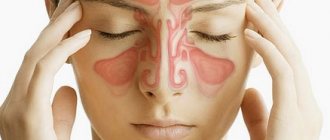
A common pathology of the respiratory system is inflammatory damage to the maxillary sinuses, which is dangerous due to its complications.
In order to recognize and effectively combat the disease in a timely manner, it is necessary to become more familiar with its causes, the location of the fluid during the inflammatory process, the main symptoms and treatment methods.
Inflammation of the maxillary sinuses (sinusitis) - what is this disease?
Sinusitis is an inflammation of the maxillary sinuses, accompanied by the secretion of mucus and pus, making breathing difficult.
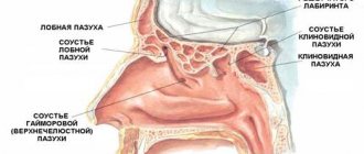
The maxillary sinuses are located in the nasal cavity, they are also called maxillary or maxillary. For a better understanding, you need to imagine a container, a bottle with a volume of 10 ml filled with air, where the base is the outer wall of the nasal cavity, and the upper part is the zygomatic process of the jaw.
What is sinusitis
This will be the maxillary sinus, which, through a small hole located at the base of the orbit, connects to the middle nasal passage. It is not the closedness of the space that becomes the main reason for the development of inflammatory processes, given the thinness of the lower wall of the maxillary sinus.
Attention! Since the maxillary cavities border both the orbit and the dental arch, if they are damaged, meningitis, eye and dental diseases can occur.
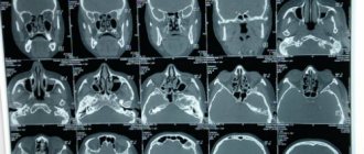
So, sinusitis is an inflammatory disease of the maxillary sinuses, accompanied by the release of mucopurulent secretion from the nasal passages, difficulty breathing, increased body temperature, headaches, and photophobia. On an x-ray, the liquid (pathological secretion) is visible, like milk in a glass with a clearly visible level.
What does sinusitis look like in the picture?
Functions of the paranasal sinuses
Each person in the area of the facial bones has several groups of accessory air cavities: the maxillary, frontal, sphenoid and ethmoid labyrinth. Their main function is to humidify and warm the inhaled air, thereby protecting the upper respiratory tract from hypothermia. The cilia lining the inner lining of the paranasal sinuses trap particles of dust and pathogenic bacteria.
The pneumatization of each cavity is different. With infectious or allergic inflammation of the mucous membranes, the walls of the sinuses begin to actively thicken, viscous exudate accumulates, which is produced by the epithelium and displaces air. The patency of the drainage paths is blocked, the outflow of secretions is hampered, an acute inflammatory process develops, and when a secondary infection occurs, the mucus turns into purulent masses with a fetid odor.
Patients complain of an increase in the amount of green nasal discharge, a deterioration in general health, an increase in body temperature, and a nagging headache that gets worse when bending over. Breathing becomes difficult, there is constant nasal congestion due to swelling of the mucous membranes, lacrimation, the sense of smell is impaired, and taste sensations are distorted.
Causes
The inflammatory process in the maxillary sinuses occurs as a consequence of:
The appearance of growths (adenoids, polyps) that do not allow normal breathing. Mucus accumulated in the sinuses creates a favorable environment for the activity of pathogenic microorganisms, which leads to inflammatory processes.
In most cases, children with reduced immunity are prescribed removal of such growths. Here it is important to distinguish adenoids from tonsils, which, on the contrary, are one of the components of the entire immune system and are necessary to protect the body from pathogenic microorganisms. Adults are less likely to suffer from adenoids than children.
Developed complications or side effects in respiratory diseases (flu, tonsillitis, tonsillitis, rhinitis, etc.).
Dental problems . Often, damage to the maxillary sinuses is observed when the upper molars are destroyed, provoking the appearance of inflammatory foci in the soft tissues. In such situations, the otolaryngologist refers the patient for a consultation with a dentist to confirm or refute the diagnosis.
If you do not take any measures to eliminate inflammation of the maxillary sinuses, then with the slightest weakening of the immune forces, tissue decay develops, which poses a danger to the body.
Deviation of the nasal septum , which can be either acquired in case of injury or congenital. As a result, frequent inflammation of the maxillary sinuses occurs and using a medicinal method of treatment does not make any sense. The best option is surgical intervention to help clear the nasal passages and normalize the body's respiratory function.
Excessive use of vasoconstrictor drugs . Most people who often experience nasal congestion do not turn to qualified specialists, but try to solve the problem on their own. These medications are only a temporary solution to the problem of the maxillary sinuses; breathing is restored only for a couple of hours. After which, drying out, swelling of the mucous membrane and inflammatory damage to the maxillary sinuses occurs.
Video about the causes of sinusitis:
The paranasal sinuses are pneumatized, what does this mean?
Normally, pneumatization is preserved, which means that the adnexal cavities function naturally, and the person does not suffer from sinusitis or sinusitis.
Diagnostics
X-ray examination is used to examine the paranasal sinuses. This is a traditional diagnostic method, which is prescribed if the patient complains of:
- pain in the paranasal sinuses;
- fever;
- chills;
- purulent nasal discharge and some other symptoms.
In addition to collecting anamnesis (questioning the patient) and X-ray examination, the method of rhinoscopy is actively used. All this allows you to determine the condition of the paranasal sinuses, whether you have sinusitis, what its shape and severity are.
To increase the accuracy of diagnosis, the pneumatization criterion is used. It, in turn, can be normal (preserved), reduced or increased.
In the process of assessing the level of pneumatization, the inflamed sinus is compared with a healthy one. In addition, during the examination, it is necessary to analyze the current state of the bone walls of the sinuses, the presence/absence of pathological neoplasms (cysts, polyps). In what cases can the level of pneumatization change? What reasons influence this important indicator and evaluation criterion?
The paranasal sinuses may be poorly pneumatized after the onset of sinusitis, which is caused by pathogenic microorganisms.
Diagnostics
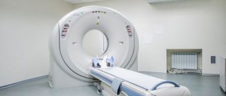
To determine the degree of filling of the sinuses with air, radiography, computed tomography or endoscopic examination (rhinoscopy) is performed. The picture is taken in several projections so that the condition of the cavities can be accurately assessed.
Normally, pneumatization is preserved, which means that the adnexal cavities function naturally, and the person does not suffer from sinusitis or sinusitis. When the diagnostic parameter decreases, the image reveals darkening or asymmetry of the inflamed sinuses, narrowing of the nasal passages, thickening of the walls of the mucous membranes, and increased fluid levels.
When assessing the degree of pneumatization, the patient’s age is taken into account, since the anatomical development of the paranasal sinuses ends only during puberty. In children under 2 years of age, only the maxillary sinuses function; by the age of 12–14 years, the rest are formed.
deviated nasal septum.
How to relieve swelling of the mucous membrane of the maxillary sinuses?
To quickly relieve swelling, the doctor usually prescribes rinsing the paranasal sinuses with medicinal solutions and systemic antimicrobial therapy, if the bacterial or mycotic nature of the disease is detected. It is impossible to purchase antibiotics at a pharmacy on your own today, so at the first symptoms of sinusitis you should consult a doctor. To relieve swelling, a specialist may prescribe:
- local drugs;
- systemic anti-inflammatory drugs;
- physiotherapy in the form of laser therapy.
Modern hardware methods for the treatment of sinusitis make it possible to promptly stop the further development of a dangerous disease with the help of laser therapy, which will avoid suppuration and surgical intervention.
The use of topical medications in the form of drops, sprays or ointments also requires a professional approach. Different in their composition and method of action, such medications should also be used only as prescribed by a doctor. They may contain hormones, with the help of which the swelling of the mucous membrane of the maxillary sinuses and soft tissues of the face will be quickly eliminated. Correct use of local drugs, as part of complex therapy, will help avoid acute purulent inflammation.
Prescribing a course of physiotherapy with the combined use of anti-inflammatory, vasoconstrictor and antimicrobial drugs prescribed by a doctor will help to quickly improve the patient’s general condition. Treatment of inflammation of the maxillary sinuses should always be comprehensive. Some drugs relieve the symptoms of the disease, while others eliminate the cause. Undergoing physical procedures prescribed by an otolaryngologist helps improve the local immune response and speed up the recovery process after acute inflammation.
Undergoing physical procedures prescribed by an otolaryngologist helps improve the local immune response and speed up the recovery process after acute inflammation.
Types of pneumatization of the nasal sinuses
What is it, pneumatization of the paranasal sinuses? This is the filling of the sinuses with air, which is possible due to direct communication with the nose. This process is involved in respiration.
Symptoms
Signs of the disease can manifest differently in each person, it all depends on the characteristics of the body. And an important factor is the form of the disease; if it is an acute inflammation of the maxillary sinuses, then the symptoms will be pronounced, but if it is chronic, then there may be no visible manifestations.
External manifestations
Among the main signs of inflammatory damage to the maxillary sinuses , which give grounds to assert its presence, there are:
- labored breathing;
- feeling of nasal congestion;
- headache;
- greenish-yellow discharge;
- rapid fatigue;
- general malaise;
- high body temperature;
- tearfulness;
- pain in the eyes;
- complete loss of smell;
- swelling of the face;
- pain when pressing on the area around the eyes.
Forms
| Form name | Description |
| Acute | It is characterized by the duration of the inflammatory process, the release of purulent secretions from the nasal passages, the presence of an unpleasant odor, swelling of the cheeks and high temperature. The cause of its appearance is a viral disease such as rhinitis, flu, whooping cough, measles, as well as an allergic reaction, injury, and surgical procedures. The symptoms are very pronounced. Treatment takes 3-4 weeks. |
| Chronic | The disease is characterized by a rather long and sluggish course, in which the phases of exacerbation and remission alternately change. Associated symptoms include frequent headaches, ailments, stuffy nose, and other manifestations, taking into account the advanced state of the pathological process. The maxillary sinus is affected by inflammation. |
| Catarrhal | The pathological condition develops against the background of acute infectious diseases during the cold season, allergies. It is accompanied by swelling of the mucous membrane, but without the release of purulent secretion from the nose. This condition is also called edematous or edematous-catarrhal. |
| Purulent | It is produced by acute or chronic bacterial inflammation of the mucous membrane of the maxillary nasal cavities. In the absence of proper treatment, nearby vital organs are at risk, in particular the eyes, brain, and vestibular apparatus. |
| Polyposis | This form is also called proliferating sinusitis of the maxillary sinuses. The pathology occurs in the paranasal cavities and is of an inflammatory-infectious or allergic nature. As a result, an abnormal proliferation of the connective tissue of the maxillary sinuses occurs, which activates the growth of growths on the mucous membrane - polyps. |
| Left-sided, right-sided and two-sided | Sinusitis can affect both sides (bilateral inflammation), which is very rare, the right or left side, then we are talking about the left-sided or right-sided form of the disease. |
ENT about acute and chronic sinusitis:
Types of pneumatization of the nasal sinuses
There are unfilled spaces in the facial bones of every person's skull. These are the sinuses of the nose, which in medicine are called paranasal or paranasal.
Air passes through them, so they are connected to the nose. Depending on the location of the sinuses, they form groups:
- the maxillary cavities are paired and are located in the upper jaw;
- there is a frontal cavity in the frontal bone;
- in the main bone of the skull there is a wedge-shaped cavity;
- The ethmoid cavity is formed from parts of the ethmoid bone.
The paranasal sinuses perform the following functions:
- breathing, because they are the ones who moisturize, warm and purify the inhaled air before it enters the lungs. This makes it clear why impaired nasal breathing generally worsens the well-being of the entire body;
- protection, since they are the ones who hold back coarse particles. So, when such particles get on the mucous membrane, it becomes irritated and the person sneezes, thereby clearing the nasal passages;
- sense of smell, special epithelial tissue helps detect odors;
- The paranasal sinuses take part in the formation of the timbre of the voice.
The accessory nasal cavities are formed inside the womb and complete their development during puberty. They have an epithelium that produces mucus. So, the largest sinus is the maxillary sinus. Pneumatization of the cavity can affect the formation of depressions other than the existing four, as well as deform the cavity.
The frontal sinus is divided into 2 parts, and its sections are pneumatized in different ways. The same thing happens with the sphenoid sinus, which also has 2 parts and their pneumatization is different.
The paranasal sinuses are examined externally by a doctor, when the nasal and oral cavity and their swelling are visible. Upon palpation, you can notice pain in the patient. However, a deep examination can only be carried out using rhinoscopy.
A more complex diagnostic technique is probing, which is carried out either through the frontal or through the sphenoid cavity.
If the doctor suspects the presence of inflammation in the sinuses, he may send you for an X-ray examination. Pneumatization of the maxillary cavities as a result of such a study may be as follows:
- Reduced.
- Saved.
- Promoted.
It should be noted that pneumatization is not a diagnosis, but merely terminology to characterize an x-ray image that describes the paranasal sinuses. If the result says that it is preserved, this is considered normal, which means there is no disease.
The sinuses are pneumatized to a reduced degree as a result of:
- underdeveloped internal spaces of the nose;
- the appearance of inflammation in them;
- accumulations of exudate;
- cysts.
Increased pneumatization occurs most often with diseases of the endocrine system, usually such as gigantism and acromegaly.
Reduced pneumatization appears with inflammation of the maxillary sinus, when its lower septum is clearly thin. This could be due to a dental problem. In some cases, during the treatment of caries or dental filling, the healing material from the filling gets into the air-accumulating cavities, which provokes the problem.
In addition, with inflammation of the mucous membrane, sinusitis occurs and pneumatization also decreases. It is caused by bacteria or viruses, and also occurs as a result of complications after an untreated runny nose.
Healthy paranasal cavities are always filled with air (pneumatized), which moves along the passages of the nose.
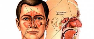
When pus appears in the sinuses, the diseases are differentiated depending on which sinus is affected by the inflammation of the cavities:
- sinusitis occurs due to inflammation of the upper;
- frontal sinusitis appears with frontal disease;
- ethmoiditis is an inflammation of the ethmoid;
- Sphenoiditis is a lesion of the main sinus.
Among other things, simultaneous inflammation of all cavities, which is called pansinusitis, often occurs. Most often, the maxillary and frontal cavities become inflamed due to their anatomical location. Be that as it may, any pain in the area of the accessory cavities is a signal to consult a doctor.
An x-ray of the paranasal cavities immediately shows a decrease in air filling of the affected sinus.
In addition, quite often a horizontal location of the fluid is revealed (if the x-ray procedure was performed while standing), and darkening of the cavities along the edges also occurs, which indicates thickening of the mucous membrane.
This disease cannot be delayed, so the doctor prescribes complex therapy consisting of medications and physiotherapeutic procedures. Due to the fact that the disease is severe, often with purulent, stagnant discharge, treatment cannot be done without antibacterial agents.
Late diagnosis and delayed treatment can lead to serious consequences due to the proximity of the nasal cavities to other vital organs. The ears, eye sockets and even the brain are at risk, since infection from the maxillary cavities can spread to its membrane.
Therefore, if you notice such symptoms, you should immediately seek advice from a specialist:
- painful sensations in the forehead, eyes, nose. They can especially intensify when the head is tilted forward;
- a bursting sensation in the area of the nose and eyes in the absence of discharge;
- constant long-term congestion;
- copious purulent discharge;
- profuse lacrimation, fear of light;
- high body temperature;
- general weakness, loss of appetite, apathy;
- swelling of the eyelid or cheek, redness of the eye from the inflamed cavity may be observed;
- dry cough at night;
- an unpleasant odor from the nasal cavity, often not felt by patients due to a decrease or complete loss of smell.
We invite you to familiarize yourself with Types of forest felling, fines for illegal logging
Share on social networks! by HyperComments (7 votes: 3.86 out of 5)Loading…
What is it, pneumatization of the paranasal sinuses? This is the filling of the sinuses with air, which is possible due to direct communication with the nose. This process is involved in respiration.
- maxillary sinuses,
- frontal sinuses,
- sphenoid sinus,
- ethmoid cavity.
The nasal cavities play an important role in the functioning of the human body. They are involved in breathing, effectively moisturize, warm and clean the inhaled air, and protect against large particles from entering the mucous membrane using the protective mechanism of sneezing. They provide a good sense of smell and allow you to distinguish odors. Participate in the formation of the voice and its timbre.
The degree of air filling of the sinuses is assessed in comparison with the healthy side (maxillary sinuses) or the transparency of the orbit. It is the last indicator that is best taken as a basis when assessing the survey results, since it is unchanged. If the description of the image indicates that pneumatization, or the filling of air in the sinuses, is not impaired, there is no need to worry.
- Diagnostics
- Causes
- When to see a doctor?
- on this topic
https://www.youtube.com/watch?v=YyTix-jNYFI
Paranasal sinuses are air cavities located in the bony joints of the skull and communicating with the nasal cavity.
There are 4 groups of paranasal sinuses: maxillary or maxillary, frontal, ethmoidal labyrinth, sphenoid or main.
In addition, the frontal and sphenoid sinuses are further divided into two parts.
The sinuses begin to form during prenatal development and are fully formed only after puberty.
The inside of the sinuses is lined with ciliated epithelium with goblet cells that synthesize mucus.
This helps warm and humidify the inhaled air. In addition, the resonance increases.
What it is? This is a medical term that refers to the presence of air spaces in the bones.
Pneumatization of the nasal sinuses is the process of filling the nasal cavities with air throughout their entire volume.
Only in this case do normal conditions for the functioning of the sinuses occur. When the paranasal sinuses are not properly filled with air, various types of diseases occur. Therefore, it is one of the diagnostic criteria for nasal pathologies.
If the process of filling with air in the paranasal cavities is disrupted, an inflammatory process begins, due to which respiratory function is impaired, the sense of smell is reduced and discomfort occurs.
Diagnostics
In the case of diseases associated with paranasal sinusitis, instrumental types of diagnostics are resorted to, namely radiography. When describing radiography, the type of pneumatization must be indicated.
Pneumatization of the sinuses is visible on x-ray
If this condition has any pathologies, then depending on where the disorders occur, the following diseases can be diagnosed: sinusitis, frontal sinusitis, sphenoiditis, ethmoiditis. All these diseases are inflammatory in nature and require timely treatment. Otherwise, complications associated with hearing and vision impairment may occur.
If the X-ray results indicate that the nasal sinuses are pneumatized, this indicates that the air in the cavities circulates normally and no pathologies are observed. Also, as a result of a study with such indicators, they can write that pneumatization of the sinuses is preserved.
This may be due to infectious processes occurring in the oral cavity, for example, caries.
Another reason for a decrease in air filling level may be the penetration of a broken piece of tooth or filling into the sinuses.
An increase in filling level occurs in various endocrine diseases.
Treatment of pathologies occurs using conservative methods. If no positive dynamics are observed, then they resort to radical methods of treatment. If there is frequent occurrence of exudate in the sinuses, a puncture may be prescribed.
Diagnostics
At the first signs of sinusitis, you should contact an otolaryngologist - a practicing specialist who diagnoses and treats disorders of the ENT organs.
To establish a diagnosis for purulent and catarrhal sinusitis, the doctor questions the patient about all possible manifestations of the disease, examines the nasal cavity, and refers him for an X-ray examination of the maxillary cavities. Accumulated pus in the frontal and maxillary sinuses is visible in the image as a fluid level. In this case, an accurate diagnosis is very easy to establish.
Difficulties in determining sinusitis arise in its chronic form, when a computed tomography scan of the paranasal sinuses is required to identify a foreign body, polypous lesion and other changes.
Reference! If there is no positive result after applying therapeutic treatment, then surgery is inevitable.
Video about the diagnosis and treatment of sinusitis:
When does decreased or increased pneumatization occur?
This condition is observed with the development of an inflammatory process in the sinus, accumulation of fluid (exudate, pus), the appearance of a cyst or their congenital underdevelopment.
Depending on the cavity in which the inflammatory process is localized, the following can be diagnosed:
Pansinusitis can also be diagnosed, a condition in which all paranasal sinuses are involved in the inflammatory process. As a rule, it is in this case that there is the greatest risk of complications.
The most common inflammation of the frontal cavities or maxillary cavities. This is due to the peculiarities of their placement. An X-ray examination is required to make a diagnosis. A decrease in pneumatization of the maxillary sinuses makes it possible to finally confirm this diagnosis. The main cause of the disease is the penetration of various viruses or bacteria into the cavity, and hypothermia.
If the inflammatory process is accompanied by an accumulation of exudate or pus, an x-ray can show its level relative to the nasal cavity. In this case, there will be no air in the lower part of the sinus. This can only be noticed when the patient is in an upright position during the examination. Both paranasal cavities or one may be involved in the inflammatory process.
There are other causes for this condition. A disorder such as decreased pneumatization of the maxillary sinuses can develop as a result of dental diseases. This is due to the thin partition between them. During dental treatment, particles of medications and filling material can penetrate into the cavity. These foreign substances provoke the occurrence of an inflammatory process.
The frontal nasal sinus and other paranasal sinuses are also characterized by a decrease in pneumatization when an inflammatory process occurs in them. Therefore, the X-ray method of examination is one of the main ways to establish a diagnosis. In this case, not only its results should be taken into account, but also the patient’s complaints about deteriorating health.
If the maxillary sinuses are pneumatized beyond normal, this may indicate that the patient has serious diseases of the endocrine system. Therefore, further treatment should be carried out not by an ENT doctor, but by a specialized specialist. Most often, this condition is observed when a person has pathologies such as gigantism and acromegaly.
For some patients, after an examination, the ENT makes a final diagnosis, the formulation of which includes “impaired pneumatization of the maxillary sinuses.” It comes as a real shock to many. In fact, there is nothing terrible - most often this is only one of the symptoms of sinusitis, and with proper treatment it can be eliminated.
Read more Order on internal investigation in the organization
The content of the article
Treatment
To relieve the inflammatory process in the paranasal sinuses and significantly improve the patient’s condition, various methods are used, the choice of which is based on the severity of the disease and the individual characteristics of the body.
Surgical
Puncture
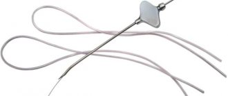
This method of treatment involves puncture of the maxillary sinuses. But given the frequent occurrence of complications after this procedure, today doctors use a more modern method of treatment - the use of the Yamik device, which allows fluid to be removed from the sinuses without a puncture. The procedure is performed under anesthesia and the patient does not feel any painful symptoms.
The doctor inserts a soft catheter into the nasopharynx through the nasal passage and uses a balloon to inject air. By creating a seal in the cavity, the accumulated pus and mucus are sucked out using a syringe. The sinuses are pierced most often in patients with chronic sinusitis.
Video about the technique of the YAMIK procedure:
Medication
Ointments . To deliver the active substance to the nasal cavities, turundas are used. Oxalic ointment, Fleming's, and Levomekol are highly effective. A cotton swab with ointment is inserted into the nose and held for 15 minutes.
Antibiotics . They are used exclusively for bacterial diseases. In most cases, Flemoclav, Amoxiclav, Cefotaxime, Cefazolin, Summed, Clarithromycin, Polydexa, Isofra, Ofloxacin, etc. are prescribed.
Antihistamines – Loratadine, Clarisens.
Drugs with an analgesic effect - Aspirin, Ibuprofen.
Decongestants , for example Phenylephrine.
Phenylephrine
Physiotherapeutic
Thanks to physiotherapeutic procedures, it is used as an additional treatment to stimulate blood circulation, reduce swelling and relieve pain during inflammation of the maxillary sinuses.
Pneumatization is reduced
The reason for insufficient filling of air voids may be:
- inflammation of the mucous membranes;
- cysts, polyps;
- congenital anomalies of the structure of the sinuses;
- deviated nasal septum;
- incorrect dental treatment;
- cystic fibrosis;
- underdevelopment of the sinuses;
- sinusitis, frontal sinusitis, ethmoiditis, sphenoiditis;
- allergic sinusitis;
- consequences of unsuccessful rhinoplasty;
- entry of a foreign body into the sinus cavity;
- malignant tumors;
- mechanical injuries of the skull;
- accumulation of purulent exudate;
- dental caries, periodontal disease.
Most often, a decrease in pneumatization is observed with inflammation of the maxillary sinuses and the development of acute sinusitis. A large amount of pus accumulates in the cavities, which displaces air. When polyps, cysts and other neoplasms grow in the sinuses, there is little free space left, so the air is not contained in sufficient volume. Advanced polyposis with spread into the drainage passages and nasal cavity leads to a lack of pneumatization.
Inflammation of the mucous membranes can spread to one or several voids at once, and occur in acute or chronic form. Provoking factors leading to a decrease in pneumatization include smoking, chemical burns, atrophy, epithelial necrosis, allergic irritation of the soft tissues of the nose and a depressed state of the immune system.
Improper dental treatment can cause perforation of the bottom of the maxillary sinus, a chronic inflammatory process, and the entry of filling material and pieces of food into its cavity.
What to do if your sinus hurts
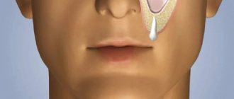
To reduce swelling, relieve inflammation and pain in the sinuses, it is necessary to rinse the nasal passages with a solution of sea salt or herbal infusion of heather, calendula, and sage. Procedures are carried out 3-4 times a day.
To avoid overdrying and the development of swelling of the mucous membrane, oil instillations are used in a lying position. The mixture is prepared from olive and corn oil, taken in equal quantities, infused with St. John's wort for three hours.
Then, on the projections of the maxillary and frontal sinuses, you need to apply a warming ointment , for example, take 1/1 tsp on one match head of Zvezdochka balm. warm ghee.
The treatment course is 7-10 days.
Features of treatment for adults and children
Inflammation of the maxillary sinuses in adults and children is treated for 3-14 days, depending on the stage of the disease. The means used are:
Antibacterial drugs that suppress the activity of pathogenic bacteria (Amoxicillin).
Amoxicillin
Analgesics that help relieve pain (Apetaminophen, Ibuprofen, Aspirin).
Mucolytic agents to reduce nasal discharge (Guaphenesin).
Guafenesin
Corticosteroids that relieve inflammation (Beclamethasone dipropionate, Prednisolone).
Medicines used to relieve swelling of the mucous membrane (Pseudoephedrine hydrochloride).
Pseudoephedrine hydrochloride
Inhaled antibiotics acting directly on the mucous membrane (Fluimucil).
Fluimucil
Video about the treatment of sinusitis in a child:
What does pneumatization of the sinuses indicate?
Pneumatization of the sinuses is a characteristic of the results of an x-ray examination.
This indicator can reveal the presence of an inflammatory process in the paranasal cavities, swelling of the mucous membrane, accumulation of exudate or pus, the occurrence of a cyst and other pathological conditions.
What is it - pneumatization of the paranasal sinuses? This is the filling of the sinuses with air, which is possible due to direct communication with the nose. This process is involved in respiration.
The cavities are located on the front part of the skull, depending on their location they are divided into the following types:
- maxillary sinuses;
- frontal sinuses;
- sphenoid sinus;
- ethmoid cavity.
The nasal cavities play an important role in the functioning of the human body.
They are involved in breathing, effectively moisturize, warm and clean the inhaled air, and protect against large particles entering the mucous membrane using a protective mechanism - sneezing. They provide a good sense of smell and allow you to distinguish odors. Participate in the formation of the voice and its timbre.
Based on the results of an x-ray examination, pneumatization of the sinuses can be determined as increased or decreased. If it is saved, this means that there are no violations.
We invite you to familiarize yourself with what does dismissal by agreement of the parties mean and how to receive the correct payment?
The degree of air filling of the sinuses is assessed in comparison with the healthy side (maxillary sinuses) or the transparency of the orbit.
If the description of the image indicates that pneumatization, or the filling of air in the sinuses, is not impaired, there is no need to worry.
In addition, the age of the patient must be taken into account. After all, the degree of air filling of the sinuses in adults and children can differ significantly, which is associated with anatomical features.
This condition is observed with the development of an inflammatory process in the sinus, accumulation of fluid (exudate, pus), the appearance of a cyst or their congenital underdevelopment.
Preventive measures
In order not to provoke the development of inflammation of the maxillary sinuses, the following recommendations must be followed :
- Get a flu shot every year.
- Wear clothing appropriate to weather conditions.
- At the first suspicion of inflammation, consult a doctor for advice.
- Do not start diseases that can cause complications in the form of sinusitis.
- Do not neglect the rules of personal hygiene; be sure to wash your hands after contact with sick people suffering from viral or bacterial diseases.
- Include more fruits and vegetables in your diet.
Only a strong immune system and compliance with preventive measures will help protect the body from damage to the maxillary sinuses.