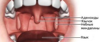
The structure of the lymphoid ring
Depending on their location, tonsils are divided into:
- Paired: palatal, tubal.
- Unpaired: lingual, pharyngeal.
Location of tonsils
In addition to this classification, in medicine it is customary to number the tonsils as follows:
- palatal – 1 and 2;
- pharyngeal (adenoids) – 3;
- lingual – 4;
- pipe – 5 and 6.
In addition, on the back wall of the throat there are small accumulations of lymphoepithelial tissue, they are called follicles. Together, these throat formations are called the Waldeer-Pirogov ring or lymphoid ring.
Types of throat diseases
The first symptoms of a sore throat are discomfort and pain. Having studied the appearance of the oropharynx, you can see certain changes:
- swelling and redness of the back wall of the pharynx and soft palate
. This indicates the presence of pharyngitis caused by bacteria and viruses; - sore throat, redness and swelling of the tonsils without any definite plaque
indicates the onset of a sore throat; later a yellow-white coating appears; - herpetic sore throat is characterized by the additional presence of small blisters
. When they open, they turn into sores; - purulent thick mucus draining from the nasopharynx
is a sign of inflamed adenoids, rhinitis or sinusitis; - caseous plugs
appear in the throat or
pus is released when you press the tonsils with a spatula
, this indicates chronic tonsillitis.
Caseous plugs are dense, pellet-like structures that make swallowing painful. They appear as white spots on the back of the throat and on the tonsils. They are not always immediately visible, since they are “hidden” in the almond folds;
- chronic cough in combination with enlarged follicles on the wall of the pharynx
can signal chronic hypertrophic pharyngitis; - a white coating covering the entire oral cavity
(gums, tongue, back of the pharynx, tonsils) indicates a fungal infection, which can occur when the immune system is weakened or after long-term use of antibiotics.
What are tonsils needed for?
A person is born with tonsils. In the first years of life they reach maximum development. From the moment sex hormones appear (15-16 years), the reverse process occurs, and they gradually atrophy and decrease.
All functions of the tonsils in the human body still remain not fully understood. Their main role is to protect and create local immunity against pathogenic microorganisms that enter the human body through airborne droplets.
In addition, the tonsils perform a hematopoietic function in young children and secrete enzymes that are involved in oral digestion.
Important! The tonsils can give a characteristic tone to speech and timbre of the voice. This point must be taken into account when removing them in patients whose work is related to the vocal apparatus (singers, announcers, etc.). The so-called “French pronunciation” can sometimes be a consequence of enlarged adenoids or hypertrophy of the palatine tonsils.
Appearance and location
Patients are often concerned about the location of the tonsils; many want to examine them in themselves or in their child. Unfortunately, you can only see the palatine tonsils or an excessively enlarged pharyngeal tonsil. Others are only accessible to a specialist using special tools.
To see all the structures with your own eyes, you can undergo a diagnostic examination using endoscopic equipment connected to a computer monitor. In this case, the doctor can easily see all the tonsils and show the patient on the screen where they are and what they look like.
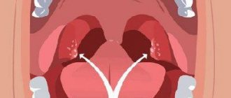
Palatine tonsils
Palatine tonsils
These lymphoid formations are located in the tonsillar niches between the two palatine arches. These are the only tonsils that the patient can see on their own, simply by opening their mouth wide.
The structure of the palatine tonsils is as follows: the free surface faces the pharynx and is covered with multilayered epithelium. Each palatine tonsil has deep slits of about 10-15, which are called lacunae (crypts). Patients may perceive these gaps as a kind of “hole”. Its other surface, with the help of a capsule, is tightly fused with the lateral surface of the pharynx.
Connective tissue bridges extend deep from the capsule. The lacunae branch and form a tree-like network in the thickness of the tissue. The epithelium, microbial waste, is rejected into the lumen of these lacunae, which serves as a substrate for the formation of almond plugs.
Nasopharyngeal or pharyngeal tonsil
It is better known as adenoids or adenoid vegetations (growths). This formation is located on the posterior fornix of the nasopharynx. You won’t be able to see for yourself where they are located and what they look like, unless they grow to such a size that they hang over the tongue.
Location of adenoids
The pharyngeal tonsil is the biggest problem for children and their parents. Enlarged adenoids interfere with normal breathing, contribute to hearing loss and the development of otitis media. They are observed and treated using conservative and surgical methods.
Tubal tonsils
Tubal and pharyngeal tonsils are located almost in the same place
Tubal tonsil steam room. It is very small in size and is located at the mouth of the auditory tube in the nasal cavity. Their schematic representation is presented in the photo.
An enlarged tubal tonsil can cause hearing problems and frequent otitis media, since during hypertrophy it blocks the communication between the nasal cavity and the middle ear.
Lingual tonsil
Location of the lingual tonsil
This formation is located at the root of the tongue. Externally it is lumpy and rough. Inflammation of the lingual tonsil causes sharp pain during conversations and when eating.
Types of inflammation and disease
The function of the tonsils is to protect the body from germs coming from the air. When immunity decreases and their functioning is disrupted, the following diseases may occur:
- Inflammation of the tonsils (angina). Sore throat usually means inflammation of the tonsils, since this disease is more common than others. If another tonsil becomes inflamed, then the diagnosis will sound like this: tonsillitis of the lingual tonsil or adenoiditis, etc.
- Hypertrophy (increase in size) of the tonsils. Tissue proliferation in itself is not a disease, but enlarged adenoids impair breathing and hearing, and hypertrophied palatine tonsils can interfere with normal eating and speaking. Whether it is a disease or not depends on the degree of hypertrophy and the presence of associated complications.
- Chronic tonsillitis. This is a complex autoimmune inflammatory restructuring of the tissues of the palatine tonsil, which can cause the development of diseases of other organs and systems (glomerulonephritis, rheumatism, endocarditis, etc.).
- Benign and malignant neoplasms.
Tonsil diseases
Many factors can impair the function of the tonsils, leading to the development of the disease. Among them are hypothermia, viral and bacterial infections against the background of weakened immunity, exacerbation of chronic diseases and others. With any inflammatory process in the tonsil, its structure, appearance, size and color are disrupted. The appearance of specific symptoms is influenced by the type of infection and the severity of the pathological process.
The formations may enlarge at the time of infection.
The tonsils in the throat are most likely to become infected. In this case, tonsillitis, scarlet fever, hypertrophy or chronic tonsillitis may occur. Inflammation of the palatine tonsil can be unilateral or bilateral, and can also be accompanied by the formation of purulent plaque and plugs on them, and the development of intoxication. The function of the tonsils is impaired, fever, weakness, headache appear, and the submandibular lymph nodes become enlarged. A person’s tonsils hurt not only when swallowing, but also at rest. To treat this condition, antibiotics, antipyretics and painkillers are given, and it is also recommended to gargle with antiseptic solutions, for example, such as:
- "Miramistin";
- "Furacilin".
This disease is dangerous due to its complications, which can appear after 3-5 days or weeks.
Enlargement of the pharyngeal tonsil most often occurs in preschool age. Impaired breathing and hearing, snoring, signs of hypoxia and rhinitis accompany adenoiditis. The severity of symptoms will depend on how enlarged they are. If the lingual tonsil is inflamed, the patient feels local pain when talking or eating. Diseases of the tubal tonsils in humans develop against the background of dysfunction of the tonsils in the body and damage to the adenoids.
Questions for the doctor
Are tonsils and tonsils the same thing or are they different concepts?
Tonsil and tonsil are the same concept, these words have different origins: the word tonsil means “gland”, and the word tonsil comes from the ancient Greek “almond”. In medicine, the first term is more often used, although “tonsils” is also correct.
What is the amygdala and where is it located?
The amygdala, or amygdala, is a collection of nerve cells in the temporal lobe of the brain. It contains the center of fear and pleasure. It has nothing to do with the usual tonsils located in the oropharynx, except for a similar name.
Why does a person need such a complex throat structure?
The main function of the tonsils is to protect against infection; in addition, they play a role in the development of immunity and hematopoiesis. This structure allows them to perform their role well and protect the body.
I discovered holes in my tonsils. Is it a disease or are they needed for something?
The so-called “holes” are lacunae of the tonsils; in some people they are more pronounced, in others they are less pronounced. Pathological contents (plugs) may accumulate in the lacunae; in these cases, it is removed by washing.
Why is it recommended to remove enlarged tonsils if it is not a disease?
Hypertrophy (enlargement) can affect other organs: cause otitis media, reduce hearing, or cause inflammation. In these cases, it is recommended to get rid of the tonsils.
In practice, the patient begins to be interested in questions about what tonsils in the throat are, what they look like, what they are needed for, when the tonsils become involved in the inflammatory process. Pathology of the tonsillar organs is accompanied by pain and sore throat, dysphagia, febrile fever, and signs of intoxication. In order to correctly assess subjective sensations, determine the specifics and location of pain, it is necessary to know the structure of the entire body, each organ separately.
Anatomy
Tonsils are accumulations of lymphoid tissue localized in the projection of the oropharynx and nasal cavity . There are 6 of them in total: two paired and two unpaired, which, together with other elements of the lymphatic system, form the Pirogov-Waldeyer pharyngeal ring.
What do tonsils in the throat look like? They have an external resemblance to a walnut (oval in shape, the surface is cut with depressions and grooves), only pink in color . The loose structure allows you to quickly recognize and prevent the active reproduction of microbial-viral-parasitic flora.
If you compare photos of tonsils in the throat under normal conditions and with inflammation, you can see how the structural elements of the pharyngeal ring change their appearance: they become hypertrophied, swollen and painful, purple-crimson in color with ulcerative-necrotic lesions of the mucous membrane.
The tonsils begin to form during the embryonic period of intrauterine development . At 4-5 weeks of gestation, components of the lymphoid organs are detected, which acquire their final structure closer to 7-8 weeks. In newborns, the tonsillar organs are poorly developed and do not function. At 2-3 months of a child’s life, when colonization of the respiratory tract by microbial communities begins, the tonsils acquire the function of a barrier and a component of the immune system of the respiratory sections.
For reference! The fastest to form are the pharyngeal tonsils, which reach their maximum size by 4-5 years, after which by 7-10 years their age-related reduction occurs with a decrease in parameters to the normal variant.
The classification forms of the tonsillar organ share a number of features:
- All vegetations are formed by lymphoid follicles , separated from each other by layers of connective tissue. Lymphoid follicles consist of combinations of T and B lymphocytes necessary to carry out lysis of damaged body structures at the cellular level.
- The tissue structures of the tonsil organs are supplied with large blood vessels and are innervated by the glossopharyngeal, lingual and vagus nerves.
- Participate in the production of immunoglobulin E , which is responsible for permanent immunity.
- The tonsils remain throughout life , but in the process of biological aging of the body they atrophy.
What is behind the tonsils in the throat? Since accumulations of lymphoid tissue are localized in the projection of the cavity behind the nose, which passes into the projection of the pharynx, behind the tonsils there is a canal connecting the oral cavity with the esophagus .
The tonsillar organ is a kind of “outpost” that ensures the capture and destruction of genetically foreign information (viruses, fungi, bacteria), thereby preventing the spread of infection to the lower parts of the respiratory tract.
Functional Features
In addition to the first-line protective mechanism of the respiratory system, the natural purpose of the tonsils is to generate immunity , providing a humoral and cellular immune response.
The immunomodulatory role of elements of the lymphoid ring has not been fully studied. On the one hand, they prevent irradiation by primary infection, on the other hand, the porous structure acts as a potential breeding ground for microbial associations, and the tonsils themselves play a key role in internal intoxication in chronic tonsillitis.
Nuance! Healthy tonsils in the throat, in addition to their barrier, immunogenic and hematopoietic functions, take part in the process of voice production. This fact must be taken into account by people whose professional activities are related to voice and speech data (actors, journalists, presenters, announcers, vocalists).
Pirogov-Waldeyer PHARYNGEAL LYMPHOID RING
TONSILS
Tonsils
— palatine and tubal (paired), lingual and pharyngeal (unpaired), forming the Pirogov-Waldeyer lymphoid pharyngeal ring, located in the area of the pharynx, root of the tongue and nasal pharynx. Tonsils are dense accumulations of lymphoid tissue containing small cellular masses - lymphoid nodules.
Lymphoid nodules
present in many organs of the immune system. The stroma of the nodule consists of reticular cells and reticular fibers, forming a three-dimensional network, in the loops of which there are many lymphocytes.
Many lymphoid nodules have light centers of reproduction, surrounded by a darker rim of small lymphocytes 5-6 microns in size, closely adjacent to each other. In the reproduction centers, lymphoblasts (about 12 µm in size) and medium-sized lymphocytes (about 8 µm in size) predominate, and mitotic figures are visible. Lymphoid nodules reach their maximum size by adolescence, their transverse size during this period reaches 1 mm. In children and adolescents, almost all lymphoid nodules have reproduction centers.
Palatine tonsil (tonsilla palatina )
- steam room, irregular in shape, located in the tonsillar fossa (bay), which is a depression between the palatoglossal and palatopharyngeal arches. The lateral side of the tonsil is adjacent to the connective tissue plate - the pharyngeal fascia, from which trabeculae (septa) extend medially into the lymphoid tissue of the organ.
On the medial free surface of the tonsil, up to 20 openings of the tonsil crypts, which are depressions of the mucous membrane, are visible. Some crypts have the shape of simply arranged tubes, others are branched deep into the amygdala. The mucous membrane is covered with stratified squamous non-keratinizing epithelium, which is infiltrated with lymphocytes.
In the diffuse lymphoid tissue of the tonsils there are dense accumulations of lymphoid tissue - lymphoid nodules (Fig. 101). The largest number of nodules is observed between the ages of 2 and 16 years.
Lymphoid nodules are located near the epithelial cover of the tonsil and near the crypts. In most lymphoid nodules, reproductive centers are visible. Around the nodules there is lymphoid tissue, which between the nodules looks like cellular strands up to 1.2 mm thick. The stroma of the tonsil is reticular tissue, the fibers of which form loops where lymphoid tissue cells are located. The connective tissue inside the palatine tonsil grows especially rapidly after 25-30 years, and the amount of lymphoid tissue decreases. After 40 years, lymphoid nodules in lymphoid tissue are rare, the size of the remaining nodules is relatively small (0.2-0.4 mm).
Blood supply
palatine tonsil. The palatine tonsil is supplied by branches of the ascending pharyngeal artery, the facial artery (ascending palatine artery), as well as the descending palatine (from the maxillary artery) and lingual arteries. Venous blood flows into the veins of the pterygoid plexus.
The palatine tonsil is innervated
due to fibers of the greater palatine nerve (from the pterygopalatine ganglion), the tonsil branch of the glossopharyngeal nerve and sympathetic fibers from the internal carotid plexus.
Age-related features of the palatine tonsil.
The palatine tonsils are formed at the 12-14th week of intrauterine development in the form of an accumulation
of mesenchyme
under the epithelium of the second pharyngeal pouch. In a 5-month-old fetus, the tonsil is represented by an accumulation of lymphoid tissue up to 2-3 mm in size. At this time, the forming amygdala begins to
Rice. 101.
The structure of the palatine tonsil: 1 - mucous membrane; 2 - stratified squamous epithelium; 3 - lymphoid tissue of the tonsil; 4 - lymphoid nodules; 5 - lumen of the crypt (according to I.V. Alma-
I also call L. S. Sutulova)
epithelial strands grow in, future crypts form. At the 30th week, the crypts still do not have a lumen, and lymphoid tissue is located around the epithelial cords. By the time of birth, the amount of lymphoid tissue increases, individual lymphoid nodules appear without reproductive centers, which form after birth. During the 1st year of a child’s life, the size of the tonsils doubles (up to 15 mm in length and 12 mm in width), and by the age of 8-13 they are at their largest and remain this way until about 30 years. After 25-30 years, pronounced age-related involution of lymphoid tissue occurs. Along with a decrease in the mass of lymphoid tissue in the organ, there is a proliferation of connective tissue, which is already clearly noticeable at 17-24 years of age.
Lingual tonsil (tonsilla lingualis)
- unpaired, lies in the lamina propria of the mucous membrane of the root of the tongue in the form of one or two accumulations of lymphoid tissue containing numerous lymphoid nodules. The mucous membrane above the tonsil has depressions - crypts, the walls of which are formed by multilayered squamous non-keratinizing epithelium infiltrated with lymphocytes.
The lingual tonsil is supplied with blood
branches of the right and left lingual arteries. Venous blood from the tonsils flows into the lingual vein.
The lingual tonsil is innervated
fibers of the glossopharyngeal and vagus nerves, as well as sympathetic fibers of the external carotid plexus.
Age-related features of the lingual tonsil.
The lingual tonsil appears in the 6-7th month of intrauterine development in the form of single diffuse accumulations of lymphoid tissue in the lateral sections of the root of the tongue. At the 8-9th month of fetal development, lymphoid tissue forms lymphoid nodules. At this time, small, irregularly shaped tubercles and folds are visible on the surface of the tongue root. By the time of birth, the number of lymphoid nodules in the tonsil increases. Soon after birth, reproductive centers appear in the lymphoid nodules. Subsequently, the number of lymphoid nodules increases until adolescence. In infants, there are an average of 66 nodules in the lingual tonsil. During early childhood, their number averages 85, and in adolescence - 90. The lingual tonsil reaches its largest size in children and adolescents. Starting from adolescence, the number of lymphoid nodules in the lingual tonsil gradually decreases. In old age and senility, the amount of lymphoid tissue in the lingual tonsil decreases, and connective tissue grows in it.
Pharyngeal tonsil (tt onsilla pharynget alis)
- unpaired, located in the area of the fornix and partly of the posterior wall of the pharynx between the pharyngeal openings of the right and left auditory tubes. In this place there are 4-6 transversely and obliquely oriented folds of the mucous membrane, separated by grooves, inside which is the lymphoid tissue of the pharyngeal tonsil. Between the folds in the grooves, the ducts of the glands open in the thickness of the folds. The free surface of the folds is covered with stratified squamous non-keratinizing epithelium; in the depths of the furrows there are areas of multi-row ciliated epithelium. Under the epithelium in the lamina propria of the mucous membrane there is diffuse lymphoid tissue and lymphoid nodules of the pharyngeal tonsil. The diameter of the nodules is 0.8 mm.
The pharyngeal tonsil is supplied with blood
branches of the ascending pharyngeal artery. Venous blood flows into the veins of the pharyngeal plexus.
The amygdala is innervated
nerve fibers extending from the branches of the facial, glossopharyngeal, and vagus nerves. Sympathetic fibers originate from the periarterial plexuses.
Age-related features of the pharyngeal tonsil.
The pharyngeal tonsil is formed in the 3-4th month of intrauterine life in the thickness of the developing mucous membrane of the nasal part of the pharynx. In a newborn, the tonsil is already well defined and measures 5-6 mm. Subsequently, the amygdala grows quite quickly. By the end of the year, its length reaches 12 mm and width - 6-10 mm. The tonsil reaches its largest size at 8-20 years of age, its length is 13-21 mm, and its width is 10-15 mm. Lymphoid nodules in the tonsil appear in the 1st year of life. After 30 years, the pharyngeal tonsil gradually decreases.
Tubal tonsil (tonsilla tubaria)
- steam room, located in the area of the tubal ridge, which limits the pharyngeal opening of the auditory tube from behind. The tonsil is an accumulation of lymphoid tissue in the lamina propria of the mucous membrane of the auditory tube near its pharyngeal opening. The tonsil contains single round lymphoid nodules. The mucous membrane is covered with stratified squamous non-keratinizing epithelium.
The tubal tonsil begins to develop at the 7-8th month of intrauterine life in the thickness of the mucous membrane around the pharyngeal opening of the auditory tube. First, separate accumulations of future lymphoid tissue appear, from which the tubal tonsil is subsequently formed. The tubal tonsil is already expressed in a newborn, its length reaches 7-7.5 mm, and it reaches its greatest development at 4-7 years.
In children, small tubercles are visible on the surface of the mucous membrane in the area of the tubal tonsil, under which there are lymphoid nodules. Lymphoid nodules and reproductive centers in them appear in the 1st year of a child’s life. Age-related involution of the tubal tonsil begins in adolescence and young adulthood.
The tubal tonsil is supplied with blood
branches of the ascending pharyngeal artery. Venous blood from the tonsil flows into the veins of the pharyngeal plexus.
Nerve fibers
they enter the amygdala as part of the branches of the facial, glossopharyngeal and vagus nerves, as well as from the periarterial sympathetic plexuses.
Location of the tonsillar organ
The location of the tonsils in the throat varies depending on the classification:
- The first and second are palatal lymphoid formations; they are assigned a place in the projection of the tonsillar niche between the palate and the tongue.
- The third - pharyngeal, occupies part of the projection of the nasopharynx.
- The fourth is lingual, concentrated in the mucous capsule of the lingual root.
- The fifth and sixth are tubal, focused in the cartilaginous part of the Eustachian tube and pharyngeal passage.
It is not always possible to demonstrate to the patient where the tonsils are located in the throat. For self-examination, with the oral cavity wide open, only the palatine tonsils or the inflamed, hypertrophied pharyngeal cavity are exposed. To visualize the remaining elements of the lymphoid pharyngeal ring, endoscopic equipment will be required.
For reference! How well the palatine tonsils are visible depends on their size and depth of location, the depth of the tonsillar bays.
How many tonsils does a person have in his throat?
In total, there are six elements of the Waldeyer lymphatic ring (two palatal, two tubal, one lingual, one nasopharyngeal). The structure of each has anatomical and functional features :
- Palatal . Binary vegetations of lymphoid tissue have variable sizes, ovoid external outlines, but are found in oblong, round, and lobular shapes. For visual viewing, a convex inner surface opens, while the outer surface faces the pharynx. A special feature of the structure is the presence of special canals – lacunae – throughout the entire area of the tonsils. They have a branched shape, the number varies from 15 to 20 in each tonsil. They begin to activate the immune defense, namely the involvement of pathogenic representatives of the microbiocenosis in the ducts, determining their negative impact on the body.
- Pipe . Paired formation, the main purpose of which is to provide the first line of protection of the larynx from the penetration of microbial communities, increasing the dynamics of immunological parameters. In the case of hypertrophy of the tubal lobes, a decrease in hearing acuity, otitis media are observed, and the functional activity of the excretory, cardiovascular and nervous systems is disrupted.
- Pharyngeal (unpaired) . The identical name is nasopharyngeal, Luschka's tonsil. The largest education. It is represented by transversely located folds of the mucous membrane, covered with ciliated epithelium, externally similar to the cock's comb. Adenoid vegetations with pathological enlargement cause difficulty in nasal breathing, hearing loss and other disorders. During a routine examination, the pharyngeal tonsil is not visible .
- Lingual . An unpaired structural element of the lymphadenoid pharyngeal ring in the form of two halves with a transverse stripe, looks like a coffee bean. Due to its small size it is barely visible visually. Controls functions that are significant for the body : cleans the inhaled air of pathogenic microorganisms, produces mucus that ensures painless passage of food, initiates the immune system to effects aimed at neutralizing antigenic foreign information. Hypertrophy causes symptomatic manifestations: pain when swallowing, spasmodic dry cough, nasal tone, snoring.
For reference! In adolescents, the lingual tonsil is enlarged. In the period from 15 to 18 years, its age-related involution begins. If the parameters of the organ have not reached the normal level, then the person is bothered by the feeling of aspiration of a foreign object in the mouth.
With the development of pathological processes of accumulations of lymphoid tissue (inflammation, hypertrophy), conservative treatment is indicated . The lack of positive dynamics requires surgical intervention. The main goal of the operation is to stop the inflammatory process and prevent complications in the entire body.
Complete removal of the tonsils or partial excision of the pathological focus should be carried out according to the doctor’s indications after dynamic diagnosis of the patient. A throat without tonsils heals within 15-20 days, after which the body’s defenses will be restored, and acute infections will proceed easier and faster .
TONSIL ANATOMY
Tonsils have a porous structure. On the outside they are covered with a mucous membrane, and on the back they are lined with a small layer of fiber. The basis of the tonsils is made up of fine mesh connective tissue, infiltrated with a large number of lymphocytes, which in some places form spherical clusters - follicles, which are located throughout the depth and on the surface. In the follicles, cells multiply and neutralize infectious and toxic substances that enter the pharyngeal mucosa.
Tonsils have different structures. The palatine tonsils are completely dotted with lacunae (crypts) into which the follicles open. The lacunae have a branched structure and penetrate through the tonsils. The number of lacunae in a person can vary from 10 to 20 in each tonsil. The tonsils have no other localizations of lacunae.
The size of the palatine tonsils varies in an adult: in the vertical direction from 15 to 30 mm, in the anteroposterior direction from 15 to 20 mm, in the transverse direction 12 – 20 mm.
In a healthy state, lacunae and follicles produce macrophages, lymphocytes and plasma cells in the required quantities. These cells are involved in fighting infections.
The blood supply to the tonsils is carried out by the branches of the external carotid artery - the facial, ascending pharyngeal, maxillary and lingual arteries. Venous outflow is carried out into the veins of the pterygoid and pharyngeal venous plexuses, into the lingual, facial and internal jugular veins. Lymph from the tonsils enters the internal jugular lymph nodes. The innervation of the tonsils is provided by the lingual, glossopharyngeal and vagus nerves, the pterygopalatine ganglion and the sympathetic trunk. The tonsils are very well supplied with nerves, which is why the process of inflammation of the tonsils during tonsillitis is so painful.
How to see and where they are
Glands are small specific formations of lymphoid tissue. There are 6 of them in total: two paired and two unpaired. Together they form the pharyngeal ring. The tonsils are located in the place where the nasopharynx passes into the pharynx. In terms of size, they can be compared to an average walnut. By the way, they received the name “tonsils” due to their external resemblance to it. Only their color is pink. Note that saying “tonsils and tonsils” is incorrect. It is the same. If inflammation occurs, they change their appearance. Why are tonsils needed? Mainly to protect the body.
When the lymph nodes in the neck become inflamed, they are often confused with the tonsils. To clearly understand what exactly bothers a person, you need to know where the tonsils are located.
To examine the tonsils in both the throat and mouth, a person is examined with endoscopic equipment that is connected to a computer monitor. Using a special apparatus, the doctor can easily examine each tonsil. He can even show the image to the patient, simultaneously explaining where they are located and what their appearance says.
Functions of the tonsils and their location
The tonsils are a collection of lymphoid cells located in the nose and oropharynx. Their main functions are protective
and
hematopoietic
. The tonsils prevent harmful microorganisms from entering the body. Lymphoid tissue produces antibodies that “fight” pathogenic microbes.
In addition, tonsils participate in the regulation of hematopoiesis, forming lymphocytes.
In humans
2 paired and 2 unpaired tonsils. They are located at the junction of the nasopharynx and pharynx, forming a pharyngeal ring.
Tonsils are pink. They are the size of a walnut.
What are there
It is customary to classify tonsils according to the location where they may be located. So they are:
- palatal (paired);
- pharyngeal or nasopharyngeal (unpaired);
- pipe (pair);
- lingual (unpaired).
Let's take a closer look:
- Palatal. In humans, these tonsils are located between a pair of palatine arches - in the tonsillar niches. As already mentioned, they are the only ones that are available for self-inspection. To see them, you just need to open your mouth wider.
- Pharyngeal (nasopharyngeal). Many people know about its existence under a different name. Very often you can hear that this tonsil is called adenoids. Its location is the posterior fornix of the nasopharynx. It is impossible to see where these adenoids are located and what they look like. They become visible when they grow excessively and hang behind the tongue. The pharyngeal tonsil is often a significant problem for children, as well as their parents. Enlarged adenoids do not allow you to breathe fully, because of them, hearing decreases, and otitis media begins to develop. Such babies are under the supervision of a doctor. Enlarged pharyngeal gland today is treated with conservative methods. If they are unsuccessful, they resort to surgery.
- Pipe. This paired tonsil is small in size. It should be located at the mouth of the Eustachian tube, in the nasal cavity. If the tubal tonsil increases in size, it can cause hearing problems and cause chronic otitis media. After all, the excessive growth of this accumulation of lymphoid tissue closes the communication between the middle ear and the nasal cavity.
- Lingual. It can be found near the root of the tongue. It has a rough and lumpy appearance. If the lingual gland is inflamed, a person will feel sharp pain while eating and talking.
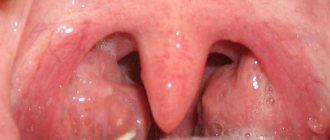
Appearance of healthy tonsils
Every person has experienced pain and discomfort in the throat at least once. When unpleasant symptoms appear, the patient independently tries to find the cause of their occurrence. However, many people mistake healthy tonsils for pathology and begin drug treatment without consultation. To avoid making this mistake, it is important to know what a healthy throat looks like.
The tonsils are small in size. They do not extend beyond the palatine arches (however, this description is not absolute. In some people, enlarged tonsils are the norm, a feature of the body);
- the tonsils are not fused to the palatine arches;
- the tonsils are pale pink in color, there is no redness or plaque on them;
- small elevations are visible on the surface of the tonsils;
- in other parts of the pharynx (soft palate, posterior mucous wall, uvula, tongue) there are no signs of inflammation;
- if you press on the tonsils with a spatula, liquid pus and caseous plugs are not released;
- the mucous membrane is non-edematous, without pronounced follicles and vascular pattern.
The criterion for a healthy throat is the presence of all of the listed signs together.
Tonsil device
All tonsils, both the structure and structure of which are almost the same, still have a number of features:
- The palatal ones are distinguished by the fact that they are pierced by special depressions (lacunae or crypts). In both tonsils there are approximately 10-15 such lacunae. Such recesses can be visually perceived as holes. With the second surface, through the capsule, the palatine tonsils are firmly fused with the side of the pharynx. The crypts form many branches that form an entire tree-like network within the amygdala. The lumens of the lacunae contain rejected pieces of epithelium and waste products of microorganisms. Thus, lacunae are peculiar traps for pathogenic bacteria and viruses, as well as a place for the immune system to “get to know” harmful microbes.
- The pharyngeal (nasopharyngeal) is represented by several transverse folds of the mucosa. The ciliated epithelium, located on the outer portion of the tonsil, forms its entire surface.
- The specific surface in the form of tubercles of the lingual gland is provided by squamous epithelium. It is divided in half by a septum and a groove passing through its center. Near it there is a depression where the ducts of the salivary glands exit.
- The tubal tonsils are the smallest. Their main task is to protect the hearing organ from infection. The structure of the tonsils is a continuous lymphoid diffuse tissue interspersed with nodules.
Each tonsil, both in the throat and in the mouth, has follicles throughout its entire surface, as well as inside. When the tonsils are healthy, the required number of plasma cells, macrophages and lymphocytes are actively produced in them and in the lacunae.
These cells fight infections affecting the upper respiratory tract. If a person gets a sore throat, then they, together with foreign microorganisms, are part of the pus contained in the lacunae and follicles.
What are tonsils needed for?
All people are born with a full set of 6 tonsils. The tonsils reach their peak development in the first years of a child’s life. But when sex hormones begin to appear (at about 15-16 years of age), their regression is observed - gradual atrophy and reduction in size of the tonsils occurs.
To this day, the tonsils and their functions in the human body have not been fully studied. However, their main role is defined. It consists of protecting and creating local immunity, which resists pathogenic microbes that enter the body through airborne droplets.
Nature has assigned several functions to the tonsils, which they, being healthy, successfully cope with:
- Barrier. Viruses and bacteria that enter the body or are already in it will certainly come into contact with the tonsils. The tonsils are necessary, first of all, in order to promptly eliminate many harmful microorganisms. They are destroyed by cells produced by lymphoid tissue (the tonsils are made up of it).
- Immunogenic. Tonsils are a mini-factory for the production of B lymphocytes, as well as T lymphocytes. It is this body that is responsible for such an important process. These cells are responsible for the functioning of the immune system.
- Hematopoietic. It is observed only in young children.
- Enzyme-producing. In babies, the tonsils secrete specific enzymes that take part in the process of oral digestion.
We emphasize that the tonsils perform all of the above functions in full only when they are in order. When their tissues are affected by inflammation, the entire body suffers. His ability to defend himself is significantly reduced. Because of this, the risk of developing various complications increases, which can adversely affect any organs and even their systems.
Interestingly, the tonsils sometimes give a certain tone to speech in general and voice timbre in particular. This nuance must be taken into account if their removal is indicated in patients working with voice (TV announcers, pop performers, teachers, and so on).
By the way, “French pronunciation” in some cases may be the result of enlarged adenoids or enlarged palatine tonsils.
Is removal necessary?
On the topic “Why do people need tonsils at all?” have been debated for many decades. Today, most doctors have concluded that the removal of tonsils should be resorted to only when their chronic, low-grade inflammation causes significant harm to the body and because of them, the lymph nodes in the neck periodically become inflamed. In addition, such an operation is justified if the patient is diagnosed with chronic tonsillitis that is not amenable to conservative treatment methods. With pathological growth of tonsil tissue in a person, it becomes difficult for a person to move food, and it becomes difficult for him to swallow. In this case, of course, there is no other way out.
The best otolaryngologists in the world do not recommend removing tonsils for children under 5 years of age. You need to wait until your immune system gets stronger. Therefore, up to 5 years, only conservative treatment is prescribed.
It is also undesirable to have tonsils removed at an early age because they appear to prevent food allergies from developing. According to statistics, 70% of children who have had their tonsils removed suffer from dysbiosis and food allergies.
If purulent plugs do not form on the tonsils, if they do not become inflamed at the first symptoms of a cold, and if they do not cause discomfort, and the lymph nodes in the neck are in order, there is no need for removal. If the tonsils are in perfect order, they bring only one benefit to the body.
Pathological signs
Unfortunately, tonsils cannot always fight off various infections. Then they take the blow, resulting in various throat diseases, the treatment of which becomes inevitable.
In the early stages of the disease, treatment with folk remedies can be used.
The following signs may indicate the development of a pathological condition:
- High fever and sore throat, along with inflammation of the tonsils and the appearance of plaque on them. They are signs of a sore throat.
- Redness in the pharynx, combined with redness and swelling of the soft palate, indicates the development of an acute form of pharyngitis.
- Mucus draining from the nasopharynx, which is purulent in nature, is a sign of rhinitis or sinusitis in adults. The same manifestations in children indicate inflammation of the adenoids. Treatment at home in this case is not always effective; you should urgently consult a doctor.
An equally serious problem is a cyst on the tonsils. This term means some cavity filled with liquid.
A cyst can appear as a result of damage to the tonsils by various infections. The cause may not only be hormonal imbalance.
The manifestation of chronic tonsillitis can also provoke the formation of a pathology such as a cyst.
This disease is usually accompanied by an unpleasant odor from the mouth and discomfort in the throat area. In some cases, the cyst may not cause any discomfort at all.
To prevent severe forms of disease, you need to regularly examine your throat. Having discovered any of the signs of the disease, you should not delay a visit to a specialist in order to cure the disease in time.
Let's summarize
Tonsils are an important part of the immune system. Their presence allows you to fully protect the body from the adverse effects of external factors. After all, it is the tonsils that take the first blow from pathogens. This is a kind of outpost of immunity.
In order for the tonsils to perform their functions efficiently, everything must be done to maintain their health. Unfortunately, many people don’t even think about why our body needs them and what their role is. That’s why they so easily agree to have it removed, despite the fact that the operation is absolutely unjustified. It is very important to try to preserve the tonsils. They can be removed only as a last resort.
The diagram shows the palatine tonsils
Tonsils
(lat. tonsillae) - accumulations of lymphoid tissue located in the area of the nasopharynx and oral cavity. Tonsils perform protective and hematopoietic functions, participate in the formation of immunity - they are a first-line protective mechanism against inhaled foreign pathogens. The full immunological role of the tonsils still remains unclear.
Together with other lymphoid formations of the nasopharynx, the tonsils form the pharyngeal lymphatic ring.
Tonsil development
Throughout life, the tonsils function with varying intensity. In newborn babies, these organs are poorly developed and practically do not perform their functions. Lymphoid tissue begins to “work” little by little only by 2–3 months of life. A sufficient level of functioning is established only by 1 year. Subsequently, the lymphoid tissue gradually increases in volume. The pharyngeal tonsil (adenoids) grows the fastest. The tonsils are fully developed only by the age of two. At this age, they are covered inside with narrow ducts (lacunae). It is the narrowness of the gaps that promotes the development of pathogenic microbes and inflammation in them. All lymphoid tissues of the pharynx reach their maximum size by 5–7 years, and it is at this age that children most often get sick and need protection from microbes. Subsequently, there is a gradual decrease in the amount of lymphoid tissue in the tonsils. Over time, it is replaced by connective tissue. Normally, having fulfilled its role in the formation of immunity, at the age of 12–14 years the lymphoid tissue of the tonsils begins to atrophy, and by the age of 18–19 only small particles of the lymphoid ring remain, and often they disappear completely.
TONSIL DISEASES
All reactions that occur in the tonsils can be divided into two groups: Primary - the tonsils themselves are damaged directly. Secondary - when the inflammatory process covers the nose, oral cavity and pharynx. The most common diseases of the tonsils are:
- tonsillitis (acute inflammation);
- chronic tonsillitis;
- hypertrophy of the nasopharyngeal tonsil (adenoids);
- hypertrophy of the palatine tonsils.
Acute inflammation of the tonsils is called acute tonsillitis or tonsillitis. Sometimes the inflammatory process becomes chronic (chronic tonsillitis). This ongoing inflammatory process reduces the protective properties of the tonsils and they themselves become a source of infection, so chronic tonsillitis requires constant monitoring and treatment by an otolaryngologist.
Inflammation of the pharyngeal tonsil is called adenoiditis, it occurs mainly in children, and the tubal tonsil is usually involved in the inflammatory process. Due to the anatomical proximity of the auditory tube, such children often develop otitis media.
Children are very often susceptible to hypertrophy of the tonsils (enlargement), which is accompanied by various changes, usually not of an inflammatory nature. Sometimes the formation of various tumors and defects of the tonsils is observed, requiring the closest attention and timely treatment.
Isolated diseases of the lingual tonsil are much less common. They can occur in middle-aged and elderly people and are accompanied by an abscess (an abscess limited by the membrane) of the lingual tonsil. This inflammation occurs with high fever, severe pain when protruding the tongue, difficulty swallowing and speaking.
Tumors of the tonsils can be benign or malignant. Benign tumors include epithelial tumors - papilloma and non-epithelial tumors - fibroma, lipoma, angioma, neuroma, myoma. Most of them are characterized by slow growth and compaction over a long period of time. The initial sign of a tumor is difficulty swallowing, a sensation of a foreign body in the throat, and enlarged tonsils. Later, pain occurs when swallowing, radiating to the ear, lower jaw, and neck. Benign tumors are removed, malignant tumors are treated in accordance with all rules for the treatment of malignant neoplasms.
A tonsil cyst is a benign formation, a small cavity filled with fluid (mucus), located either on the surface or inside the tonsil. The causes of its occurrence may be an infectious lesion of the tonsil, hormonal imbalance and others. The appearance of a cyst may indicate the presence of chronic tonsillitis. In some cases, the cyst does not cause any discomfort, but sometimes it can cause discomfort. In addition, the presence of this formation causes bad breath.
Tuberculosis of the tonsils, as a rule, is secondary and is observed in patients with pulmonary tuberculosis. It often occurs hidden under the guise of chronic tonsillitis. The diagnosis is made taking into account the medical history on the basis of morphological and bacteriological studies.
Syphilitic lesions of the tonsils can occur at any stage of the disease. In primary syphilis, one tonsil is affected. Secondary syphilis can manifest as syphilitic sore throat (tonsils are enlarged, with sharply defined bluish-red lesions or oval plaques with a red rim). With tertiary syphilis, the formation of gummas is possible. The diagnosis is made on the basis of bacteriological and serological studies.
Classification
Location of the pharyngeal tonsil.
Tonsils are divided into:
- paired palatines
- in the recess between the soft palate and the tongue (first and second tonsils). - tubal
- in the area of the pharyngeal opening of the auditory tube (fifth and sixth tonsils)
- pharyngeal
(nasopharyngeal) - in the area of the fornix and the back of the pharyngeal wall (third tonsil, Luschka's tonsil). Pathological enlargement of this particular tonsil is called adenoids
- under the surface of the back of the tongue (fourth tonsil)
The following numbering of the tonsils is accepted:
- the first and second are palatal;
- third - pharyngeal (nasopharyngeal);
- fourth - lingual;
- the fifth and sixth are the tubal tonsils.
Palatine tonsils
Innervated by branches of the glossopharyngeal nerve.
Pathology
Defects: additional lobules, bifurcations
Damage (injury): burn, wound, introduction of foreign bodies.
Hypertrophy. As a rule, it occurs in childhood and affects the palatine and pharyngeal tonsils. In the absence of functional impairment, treatment is not required. If necessary, treatment begins with conservative treatment; if it is ineffective, surgical intervention is required - tonsillotomy (removal of part of the palatine tonsils) or tonsillectomy (removal of the palatine tonsils along with adjacent connective tissue). This anomaly is usually found in representatives of the Negroid race.
Inflammation
Acute tonsillitis - inflammation of the tonsils; a substance that accumulates on the surface of the tonsils.