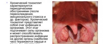
Inflammation and formation of a purulent cavity in the tissues around the tonsils is called a peritonsillar abscess. This pathology is quite common among adults than among children. The peritonsillar abscess was assigned an ICD code of 10 J36. Peak incidence rates occur between 15 and 35 years of age. Both men and women are affected with equal frequency. In 75-80% of cases, a paratonsillar purulent cavity is formed against the background of chronic inflammation in the adjacent tonsil.
Peritonsillar abscess: classification
Experts classify paratonsillitis according to the location of inflammation.
This is the main division of the peritonsillar abscess, which determines the symptoms and further treatment of the disease. Other existing classifications are less significant and are not widely used in clinical practice. Classification by localization of the pathological process:
- Directly below the tonsil is an inferior paratonsillar abscess. Its symptoms are pain when moving the tongue. A visual examination reveals moderate redness of the throat and enlarged tonsils.
- Near the oral cavity, above and in front of the tonsil, an anterosuperior (anterior) paratonsillar abscess is localized. The soft palate on the affected side is swollen, has a bright red tint and is tense.
- Posterior paratonsillitis is localized closer to the larynx and behind the tonsil. It is difficult to diagnose this form of damage, since it manifests itself only as an enlargement of the tonsil due to inflammation. Also with posterior paratonsillitis, a typical symptom is hoarseness and pain when swallowing and speaking.
- In the tissues of the pharynx, along the lateral line from the tonsil, there is external (lateral) paratonsillitis. This form is rare, but has a more severe course.
The bilateral development of the pathological process is also highlighted. Bilateral paratonsillitis is rare and indicates a severe weakening of the body's immune system. Also, this pathology can be detected after a chemical burn to the throat or injury to the tonsil mucosa.
Unilateral paratonsillitis is equally likely to affect each tonsil. Left-sided paratonsillar abscess is detected as often as right-sided one.
Peritonsillar abscess causes
First of all, it is necessary to point out that a paratonsillar abscess of the throat, as a rule, occurs as a result of the spread of the inflammatory process from the palatine tonsils to the paratonsillar tissue.
Complications of paratonsillar abscess
For the most part, this complication occurs with chronic tonsillitis rather than with recent acute tonsillitis. As for the causative agent of the inflammatory process, the motivating factor is often the same flora that caused the onset of sore throat or chronic tonsillitis.
The route of infection spread is mostly tonsillogenic, that is, when the infectious flora penetrates the tissue from the upper poles of the tonsils, where the lacunae are deeper and more convoluted. There may be either a right-sided peritonsillar abscess or a left-sided peritonsillar abscess.
Peritonsillar abscess diagnosis
Separately, it is necessary to indicate that the cause of paratonsillitis can be caused by trauma to the pharynx by a foreign object, including improper eruption of a wisdom tooth. At the same time, it must be noted that the penetration of infection alone is not enough for the occurrence of a pathological process. That is why external factors are of great importance: hypothermia, stress, metabolic disorders, etc.
Peritonsillar abscess: history of the disease and types
Currently, a distinction is made between inferior, anterosuperior and posterior paratonsillar abscesses.
In Russia, in 85–90% of cases, anterior superior paratonsillar abscess occurs. This is due to the fact that the upper pole of the tonsil is very poorly drained. Moreover, the localization of this type of paratonsillar abscess takes place between the upper part of the palatoglossal arch, the upper pole of the tonsil and the anterior-superior part of the so-called. supramalminal fossa. It is worth noting that the swollen and hyperemic soft palate on the side of the lesion protrudes anteriorly. After only 5 - 7 days, a purulent protrusion becomes noticeable in the upper part of the palatoglossal arch - you can view a photo of a peritonsillar abscess on the Internet. The abscess may then open spontaneously.
Posterior peritonsillar abscess
Posterior peritonsillar abscess, in contrast to the anterior superior abscess, is observed much less frequently (only about 10% of the total number of peritonsillar abscesses). However, it must be borne in mind that the symptoms of this type of peritonsillar abscess have their own specifics. At the same time, this abscess is dangerous with a high risk of developing laryngeal edema with further acute stenosis.
Inferior paratonsillar abscess
Inferior peritonsillar abscess is very rare in our country. Its occurrence is due primarily to odontogenic causes. This type of peritonsillar abscess is localized in the tissue behind the lower third of the palatine arch between the lingual and palatine tonsils.
In our other reviews, read about other aspects of peritonsillar abscess.
Disease paratonsillar abscess diagnosis code according to ICD-10 (ICD is the International Classification of Diseases)
What is the name of the doctor who treats peritonsillar abscess?
If symptoms of a peritonsillar abscess occur, you need to make an appointment with an otolaryngologist (ENT doctor) at a hospital, clinic or medical center via the Internet, by phone number or visit a medical institution.
A peritonsillar abscess is a pus-filled cavity located in the tissues surrounding the tonsil. A purulent cavity (abscess) develops at the last stage of inflammation of the peritoneal tissue that occurs after a sore throat or exacerbation of chronic tonsillitis.
Leave a request and within a few minutes we will find you a good specialist and help you make an appointment with him.
| Peritonsillar abscess | |
| ICD-10 | J36 |
| ICD-9 | 475 |
| DiseasesDB | 11141 |
| eMedicine | emerg/417 |
| MeSH | D000039 |
In most cases, only one side is affected, but bilateral peritonsillar abscess also occurs.
Causes and symptoms
Peritonsillitis as an independent nosology is quite rare. Basically, it is a complication of severe chronic tonsillitis. Also, a purulent cavity can form against the background of a chronic carious process and after damage to the mucous membranes of the tonsil.
The occurrence of a paratonsillar abscess is associated with the penetration of pathogenic microorganisms into the tissue near the tonsil.
The main ones are:
- Staphylococcus
- Streptococci
- Haemophilus influenzae
- Escherichia coli
- Mushrooms
- Klebsiella
Factors that can provoke the onset of the disease and increase the risk of developing paratonsillitis are:
- Local or general hypothermia of the body
- Reduced protective functions
- Congenital anomalies of the tonsils
- Bad habits (smoking, alcohol abuse)
Also, some chronic diseases increase the risk of the formation of a peritonsillar purulent cavity. These include: oncology, diabetes mellitus, anemic conditions, immunodeficiency.
In the process of development of paratonsillitis, successive stages can be distinguished.
- The first is edematous, in which the tissues near the tonsil swell somewhat, but there are no obvious clinical symptoms.
- Infiltration. A person feels a sharp pain when swallowing. Visually, you can notice hyperemia of the mucous membranes of the tonsil and tissues near it.
- The final stage of paratonsillitis on days 4-7 is the formation of a cavity with pus. Upon examination, the pharynx was severely deformed and hyperemic. Fluctuation of tissues near the tonsil is determined.
Peritonsillar abscess after opening. Treatment after the procedure
Therapy after opening the abscess occurs with the help of antibacterial drugs.
In the treatment regimen, the doctor will prescribe a penicillin drug: Amoxicillin (what reviews exist about Amoxicillin for sinusitis can be read in this article) and Cephalexin. They are taken 4 times a day, 200 or 500 mg. The duration of therapy will be 10 days. If the patient has an allergic reaction to penicillin, then macrolides are prescribed - Erythromycin, Clarithromycin (but this article will help you understand whether the antibiotic Clarithromycin Teva treats bronchitis) Clarithromycin
Antibiotics are also prescribed for external use. This includes ointments such as Levosin, Mafedin and Levomekol. They have a local effect, since they act exclusively on the affected area and are not absorbed into the blood.
Levomekol
Also, after opening, it is necessary to treat the treated wound. It is necessary that its edges do not stick together until granulation of the cavity from the depths occurs. A tampon with Vishnevsky ointment or Vaseline oil is left in the tissues that have undergone surgery. Change your tampon every 2-3 days. Regarding how granulation develops, it is necessary to remove the tampon from the depths. After opening, it is worth cauterizing the excess granulation, while making sure that the epithelium growing along the edges of the wound is not injured. If the wound heals slowly, you will need to stitch it.
This article will help you understand what white dots on the tonsils look like in the throat and what medications can cure this disease.
Why there is a sore throat, white plaque on the tonsils and how to determine which disease you have yourself is described in great detail in this article.
How white plaque on the tonsils is treated and whether it can be cured at home is described in this article.
- Yuri, 32 years old: “I had an autopsy performed when I did not cure an acute sore throat in time. Then I was on a business trip, so it was not possible to seek help from a doctor; I treated myself, as a result of which pustules began to form. Because of them, I could not eat or drink normally. The pain was simply unbearable. When I went to the doctor, he immediately sent me for an autopsy. Afterwards I went to get a dressing done and also took the antibiotics prescribed by the doctor.”
- Anna, 43 years old: “I have chronic tonsillitis, so I have exacerbations 2-3 times a year. At the same time, severe pain appears that does not allow me to eat, drink, or even speak normally. One day, during another exacerbation, I had an abscess. I consulted with a specialist, and he said that I need to wait 4 days, since during this period the abscess can open spontaneously. But this did not happen, so they opened my abscess in the hospital. After that, my temperature dropped, the pain disappeared and my overall health improved.”
- Ksenia, 23 years old: “I developed an abscess due to acute tonsillitis. But I was not able to visit a doctor to autopsy it, since this process happened spontaneously for me. Of course, this cannot be called a pleasant thing. But I felt a long-awaited relief. Then I went to the doctor anyway, because they treated my wound with an antiseptic and also prescribed antibiotics. I worried for a long time so that no complications would arise (after all, everything happened at home, not in the hospital), but everything ended well and after 2 weeks I completely recovered.”
Symptoms of paratonsillar abscess
Peritonsillar abscess in adults and children manifests itself with the same symptoms. Initial symptoms can be felt already on the 3rd or 5th day after suffering an upper respiratory tract disease of infectious etiology, for example, after a sore throat. But in older people, the formation of a purulent cavity in the tissues near the tonsil can occur within 24 hours after infection.
We also recommend reading Tonsil abscess: causes, symptoms, treatment
The general symptoms accompanying peritonsillar abscess are quite mild. The patient's temperature initially rises to subfebrile levels, then to febrile levels. The person feels decreased ability to work, general fatigue and weakness, as well as severe headaches. As paratonsillitis progresses, bad breath and shortness of breath appear.
Peritonsillar abscess is characterized by characteristic local symptoms. But it is also detected in diseases such as phlegmonous tonsillitis or retropharyngeal abscess. This complicates subsequent diagnosis.
- Severe pain in the throat, usually on one side, radiating to the ear area or lower jaw.
- Trismus of the masticatory muscles from the side of the formation of a purulent cavity. This is a movement disorder in the jaw joint in which the patient cannot open his mouth wide.
- A feeling of the presence of a foreign body in the throat, which prevents the patient from speaking and eating.
- Increased saliva production.
- Enlarged lymph nodes in the submandibular region from the side of the formed purulent cavity.
- Increased pain when turning the head.
During a medical examination of a patient with a peritonsillar abscess, the doctor may notice hyperemia of the tonsil due to the disease. Also, in some cases, the purulent cavity can open on its own, after which there is a decrease in symptoms and an improvement in well-being.
Symptoms
With a paratonsillar abscess, the patient complains of a sore throat on one side, which intensifies on the second and third days of the disease, swallowing becomes too difficult.
There is increased salivation, the saliva is viscous and thick, so it is difficult to spit out, the mouth is half open, and there is a pained expression on the face. The patient's head is tilted down and to the painful side to create conditions for weakening the neck muscles on the painful side and to reduce soreness in the throat. This is a defensive reaction to pain.
From the fourth or fifth day of illness with paratonsillar abscess, the general condition worsens:
- sore throat gets worse
- swallowing is impossible
- food often ends up in the nasal throat and nasal cavity due to the spread of the inflammatory process to the soft palate.
Accumulations of saliva, bad breath, and overt nasality are observed.
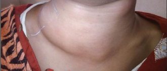
The visible symptoms of a peritonsillar abscess depend on the location of the inflammation and the stage of the disease. Upon examination and palpation of the neck, enlarged and painful lymph nodes are detected.
Pharyngoscopy (visual examination of the mucous membrane of the throat) shows redness, filling with blood and swelling of the mucous membrane of the anterior arch and protrusion of the soft palate with anterosuperior localization of the abscess.
In case of peritonsillar abscess, the uvula is swollen and hyperemic, often displaced in the opposite direction from the diseased side.
The palatine tonsil, the source of infection in peritonsillar abscess, is enlarged due to edema, but is often not visible because it is covered from the front by the edematous anterior arch. The opposite tonsil is smaller and has noticeable redness. If an abscess forms, a yellowish spot is visible at the level of the base of the uvula.
As a rule, with a peritonsillar abscess it is difficult to open the mouth completely.
In case of posterosuperior paratonsillitis
hyperemia and edema are visible, as well as swelling of the palate above the tonsil and in the area of the upper third of the posterior arch leading to impaired mobility of the soft palate. The palatine tonsil is clearly visible, so there is almost no enlargement of the anterior arch. In the case of posterior paratonsillitis, a very thickened spindle-shaped posterior arch is visible. The uvula is hyperemic and swollen. The palatine tonsil is imprinted in advance and is clearly visible. Trismus of the muscles is usually absent or slightly pronounced.
Anterior paratonsillitis
is localized between the palatine tonsil and the lower two thirds of the anterior arch and is characterized by infiltration and swelling of the anterior arch. The soft palate is slightly infiltrated and swollen.
Lower paratonsillitis
located between the palatine and lingual tonsils behind the lower third of the anterior arch, under the palatine tonsils. In this area, pronounced swelling is visible, which extends to the lower pole, and the tonsil of the palate extends upward. The soft palate is little changed.
The patient complains of severe pain during swallowing and tongue movements. The movement of the lower jaw is difficult.
External paratonsillitis
characterized by the formation of an abscess in the peritonsillar tissue outside the tonsil, in the depths of the niche. Pharyngoscopy (visual examination of the mucous membrane of the throat) shows increased blood supply to the mucous membrane of the anterior palatine arch, but there is almost no swelling and edema.
There is asymmetry of the pharynx due to the fact that the tonsil of the palate is shifted to the midline. Pain when swallowing is insignificant, muscle trismus is not pronounced. In patients with an abscess of this form, pronounced changes are observed in the angle of the lower jaw. Noticeable swelling of the soft tissues, spreading downwards along the anterior edge of the sternocleidomastoid muscle, severe pain on palpation.
Blood test in patients with peritonsillar abscess:
- pronounced leukocytosis (10-14*109/l) with a shift of the leukocyte formula to the left,
- erythrocyte sedimentation rate (ESR) increased to 40 mm per hour,
- eosinophilia (increased number of granulocytic leukocytes - eosinophils in the blood).
In a urine test with a peritonsillar abscess, diuresis is reduced, the majority show albuminuria (the release of albumin, a blood protein in the urine), the number of leukocytes increases, and red blood cells appear.
So, the main ones
symptoms of peritonsillar abscess
:
- severe sore throat, especially from an abscess that has formed
- inability to swallow or open the mouth due to severe pain (it’s difficult to even brush your teeth)
- drooling and bad breath
- tilting the head to the painful side
- visible neck swelling and tenderness
Diagnostics
The clinical picture of the disease with the formation of a purulent cavity is well differentiated. Therefore, to make a preliminary diagnosis, it is enough for a specialist to collect anamnestic data, a visual examination of the pharynx and the result of pharyngoscopy.
The difficulty is in the differential diagnosis of paratonsillitis with diseases such as retropharyngeal abscess and phlegmonous tonsillitis. The symptoms and clinical picture of the diseases are similar, but the treatment tactics are different.
Diagnostic measures include the main points:
- The doctor carefully collects anamnesis and main complaints, fills out a medical history. For example, a peritonsillar abscess on the left or right most often forms 4 days after acute or chronic inflammation of the palatine tonsil.
- Medical checkup. A visual examination of the throat is carried out, and body temperature is measured. The doctor palpates regional lymph nodes.
- Pharyngoscopy. This is the most informative method that allows you to determine the location of the peritonsillar abscess. A distinctive sign of a purulent cavity in the tissues on pharyngoscopy is fluctuation (oscillation) of the hyperemic pharyngeal wall near the tonsil.
- Hardware visualization. In order to differentiate paratonsillitis and other diseases of the tonsils and surrounding tissues (tumors, phlegmonous tonsillitis, scarlet fever and others), specialists prescribe ultrasound of the neck, CT and radiography, which allows one to determine the exact localization of the purulent cavity. Also, these examinations, when diagnosing peritonsillar abscess, reveal possible complications of the disease.
- Laboratory examination. A general blood test is taken from the patient, in which signs of inflammation are manifested by a high level of neutrophils and ESR. In some cases, a puncture is performed followed by bacteriological culture to determine pathogenic microflora.
Peritonsillitis should be differentiated from diphtheria, infectious mononucleosis, oncology, syphilis, scarlet fever and leukemia. In case of diphtheria, the patient has no characteristic trismus against the background of swelling of the tonsil, and Loeffler's bacilli are detected in the sputum analysis. Scarlet fever - characteristic rashes are noticeable on the body.
With syphilis, in addition to enlarged tonsils, a positive Wassermann reaction is determined, and infectious mononucleosis is manifested by leukocytosis and the accumulation of atypical monocytes in a blood test. Tumor damage to the tissues of the pharynx does not cause a sharp rise in body temperature and severe pain like paratonsillitis.
Peritonsillar abscess under general anesthesia. Methods for effective treatment of peritonsillar abscess
A peritonsillar abscess is a cavity filled with pus, which is a type of acute paratonsillitis - inflammation of the tissue around the pharyngeal tonsils. This disease develops if chronic or acute tonsillitis is not treated in time.
Causes
Abscesses of paratonsillar tissue can develop as a result of acute tonsillitis, but much more often they are a consequence of chronic inflammation of the tonsils. The transition of the disease to such a purulent form has several causes:
- Weakening of the general immunity of the body due to long-term illness of any organ. It is accompanied by a decrease in local immune defense of the mucous membrane with long-term inflammation in the pharynx.
- A decrease in these protective factors leads to the activation of opportunistic bacteria that normally inhabit the throat and do not lead to disease. They are contained in a biofilm covering the mucosa. In the process of a chronic disease, the biofilm grows, and the bacteria in its composition multiply and cause a purulent form of the disease.
- Antibiotics used to treat tonsillitis do not work against opportunistic pathogens.
- An anatomical feature of some people, aggravated by frequent sore throats, is enlarged tonsils with very deep crypts. It is easier for germs to linger in them.
- Scars and adhesions on the tonsils as a result of frequent illnesses make it difficult to heal, which aggravates the situation.
- Odontogenic genesis is possible - the spread of inflammation from a carious tooth to the throat.
- Weber's glands promote the spread of inflammation from the tonsils to the surrounding tissue.
Treatment of peritonsillar abscess
Therapeutic tactics for paratonsillitis depend on the localization of the inflammatory process and its stage. But it is not possible to carry out therapy at home. All measures are prescribed under the supervision of the attending physician and in a hospital setting.
In the early stages, when the cavity has not yet formed, and paratonsillitis is manifested by swelling and redness of the mucous membrane of the tonsil and the tissue around it, conservative treatment is indicated. It includes medications and physical therapy. The main drugs prescribed by specialists:
- Antibacterials (such as amoxicillin, macrolides, or second- and third-line cephalosporins)
- Local analgesics
- Immunostimulants and vitamin complexes
- Sanitation of the throat and tonsils with solutions containing antibiotics and decoctions of medicinal herbs
- Antipyretic treatment for high fever
If conservative treatment methods are ineffective, as well as in the later stages of paratonsillitis, surgical treatment is indicated. When a cavity with purulent contents has formed, it can be opened. After opening the peritonsillar abscess, the clinical symptoms regress and the patient’s well-being improves.
Surgical treatment of paratonsillitis can be palliative or radical. Gentle methods of opening a purulent cavity include puncture of the abscess followed by suction of the pus. As well as directly opening the cavity using a scalpel. This procedure is performed under local anesthesia, but is dangerous due to the complication of gluing the drainage hole with purulent exudate. This may lead to relapse of the disease.
Radical surgical treatment includes bilateral tonsillectomy. Doctors often have to resort to this operation for lateral (side) paratonsillar abscess.
In the postoperative period, it is necessary to adhere to the basic principles and measures:
- Bed rest, diet
- Treatment of the postoperative area with antiseptic drugs
- Antibacterial and anti-inflammatory treatment
- Painkillers
- Antihistamine treatment
- Immunostimulants and vitamins
With early diagnosis and adequate treatment, peritonsillar abscess is highly treatable. Therefore, when the first warning symptoms (redness and swelling of the throat) appear, it is recommended to seek help from a qualified specialist.
Treatment
Peritonsillar abscess is treated in three ways:
- Conservative
- Surgical
- Complex
Conservative treatment is prescribed when symptoms are detected in a timely manner, when paratonsillitis is at the initial stage. Conservative treatment is of two types:
- General, involving the use of antibiotics, vitamins, painkillers and immunostimulating agents. In this case, antibiotics of the penicillin group Amoxicillin, Amoxil are prescribed; in case of an allergic reaction, as well as the ineffectiveness of penicillin, they are replaced with Macrolides (Azithromycin) or Cephalosporins (Cefodox, Ceftriaxone). For children with diseases of the tonsils and throat, Amoxiclav, Ormax, Cefodox in suspensions are prescribed.
- Local, using gargles and mouth gargles with Furacilin, Chlorhexidine, local antiseptics in spray form, for example, Bioparox.
Surgical treatment is possible in two types:
- Puncture. A puncture is performed in the area of the peritonsillar abscess to suction out the pus;
- Opening a paratonsillar abscess, when the abscess is cut with a scalpel. There are cases when the incision sticks together after surgery and the intervention needs to be carried out again, but this time draining the area where paratonsillitis has formed. Under no circumstances should you open a peritonsillar abscess at home; this can be extremely dangerous due to the onset of blood sepsis, and as a result, death.
After a number of treatment procedures have been carried out without any results, the peritonsillar abscess is treated by surgical removal of the tonsils. This is a radical method, the last hope for recovery. But, as a result of the operation, the peritonsillar space is cleared and the peritonsillar abscess is removed along with the source of infection in the tonsil.