Functions
The nasal cavity is the first filter through which inhaled air passes.
It reacts to changes in the environment and prevents the inhalation of extremely dry or humid air. Partial destruction of bacteria occurs in the nasal cavity. The nasal mucosa traps dust particles and removes them into the external environment. Due to the peculiar structure of the cavity, when inhaling, the air is moistened, warmed and, already purified, moistened, warm, enters through the pharynx and trachea into the lungs.
In the mucous membrane of the nasal cavity there are primary sensory cells - the olfactory zone. These cells are the first to catch all the odors of the environment. The olfactory zone is located deep in the nasal cavity and is very closely connected with the emotional function of the brain. A pleasant, familiar smell can lift your spirits and vice versa.
Structure
The walls of the nasal cavity are separated by the nasal septum, dividing it into two cavities, each of which opens outside the nostrils.
Each cavity consists of a vestibule and a respiratory surface. The bony cavities of the nose contain the sinuses (sinuses). Thanks to the bones of the skull and cartilage, the walls of the cavity are solid. This feature allows the walls not to collapse when inhaling. The vestibule of the cavity is lined with flat epithelium, under which there are sebaceous glands, and the inner walls are lined with ciliated epithelium. The surface of the epithelium is lined with mucous membrane.
The nasal cavity contains the olfactory and respiratory areas. In the thickness of the mucous membrane of the nasal cavity there are a large number of blood vessels. The submucosal surface contains glands, nerve and choroid plexuses, and lymphoid tissue. Lymphatic follicles located in the vestibule of the nose perform an immune function.
Diseases of the nasal cavity and treatment
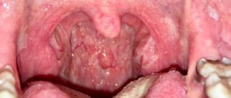
Acute rhinitis is an acute inflammation of the nasal mucosa, which can occur as a consequence of other infectious diseases or as an independent disease. In acute rhinitis, hyperemic and swollen mucous membrane of the nasal cavity. There is a feeling of heat and nasal congestion, accompanied by headaches, impaired nasal breathing, increased secretion, and lack of smell.
At the first signs of acute rhinitis, aspirin is prescribed. Warming, hot tea, and effects on reflexogenic zones are indicated. Drug treatment consists of prescribing vasoconstrictors and antihistamines. Vasoconstrictors are indicated for severe swelling of the mucous membrane. For inflammatory processes in the mucous membrane, antibacterial agents are prescribed.
Disease of the nasal mucosa. Clinically, chronic rhinitis is manifested by nasal congestion, difficulty breathing through the nose, mucus secretion, and headache. Chronic rhinitis can cause sinusitis, pharyngitis, tonsillitis, etc.
There are several types of chronic rhinitis: vasomotor, allergic, hypertrophic, medicinal. Vasomotor rhinitis occurs due to decreased vascular tone in the nasal cavity. The individual reaction of the body to irritants becomes the cause of allergic rhinitis. When the connective tissue of the nasal cavity grows, hypertrophic rhinitis develops. Long-term use of vasoconstrictor drugs causes drug-induced rhinitis.
The cause of ozena is atrophy of the nasal mucosa. Clinical manifestations of ozena: thick foul-smelling discharge from the nasal cavity, impaired nasal breathing, dry throat, lack of smell, formation of dry crusts.
Treatment is carried out with medication. Immunity enhancing drugs, antibiotics, and vitamins are prescribed. Local treatment is aimed at softening and removing crusts from the nasal cavity. In severe cases, surgery is performed.
The causes of a deviated septum are:
- Uncoordinated development of facial skeletal structures
- Polyps
- Hypertrophied nasal concha
- Injuries
- Tumors
A deviated nasal septum makes nasal breathing difficult and causes congestion, mucous or purulent discharge, and headaches. Treatment is usually done surgically.
Fusions of the nasal septum and the lateral wall of the nasal cavity are called synechiae. Obstruction of the nasal passages (congenital or acquired) is called atresia.
The narrowing of the nasal passages due to fusion causes disruption of nasal breathing. In some cases, adhesions cause sinusitis. Treatment of adhesions is carried out surgically.
Hematomas are formed as a result of the accumulation of blood between the periosteum and the bone of the nasal septum. A hematoma can cause narrowing of the nasal passage, impaired nasal breathing, pain, and swelling. Sometimes the hematoma festeres and turns into an abscess, which is dangerous for intracranial complications (brain abscess, meningitis, etc.). An abscess of the nasal septum is manifested by severe swelling and pain.
Treatment of a fresh hematoma is limited to puncture and suction of blood. For an abscess, surgery is performed.
Anatomy of the inner nose
The nasal cavity is a space that is limited from above by the bones of the facial skeleton, from the side it reaches the eye sockets, below it comes into contact with the palatine part of the oral cavity, and from behind it communicates with the nasopharynx through special openings (choanae).
The structure of the nasal cavity is complex, this explains the variety of functions it performs and the severe course of some diseases in this area and the paranasal sinuses associated with it.
The vertical septum divides the entire nasal cavity into two approximately equal halves. They are not completely symmetrical due to the fact that the vast majority of people have deviations or defects in the bony part of this septum. Nature invented the division into 2 halves for more uniform heating, purification and humidification of incoming parallel air flows. A slight curvature of the septum, which does not interfere with normal breathing, is considered normal. With significant defects resulting from injuries or severe inflammation, turbulence in air flows, drying out of the mucous membrane, and blocking of the openings connecting the nasal cavity and sinuses occur, which leads to respiratory failure and the occurrence of chronic inflammation.
It will also be interesting: Features of catarrhal sinusitis and its treatment
Each half of the internal nasal space has 5 walls, more about them:
- The medially located wall is the septum itself, which consists of a posterior bone and anterior cartilaginous part.
- Small processes from the frontal and ethmoid bones participate in the formation of the upper wall; here are the openings for individual branches of the olfactory nerves.
- The structure of the lower wall consists of two plates, one from the upper jaw, the other from the hard palate.
- The posterior wall is present only in the upper section; it consists of part of the sphenoid bone, below which are the choanae.
- The most difficult is the lateral or side wall. Its composition is represented by the nasal bones, maxillary processes, lacrimal plate, part of the ethmoidal labyrinth, plate of the hard palate and part of the sphenoid sinus. Along this wall of the nasal cavity, the turbinates are suspended inward in three rows, between which are the nasal passages.
This important part of the nose deserves detailed consideration.
Structure of the nose
Disease Prevention
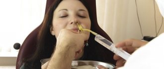
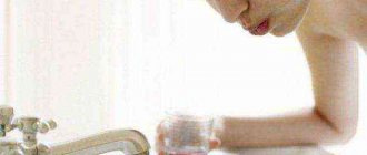
In order for the nasal cavity to perform its functions, it is necessary to regularly carry out hygiene. Infectious diseases can be avoided by washing. In addition, rinsing prevents dry mucous membranes.
Washing for preventive purposes is recommended in unfavorable environmental conditions and epidemics of acute respiratory viral diseases.
FAQ
Hair in the nasal cavity acts as a filter, trapping dust, toxins, viruses and germs. The thicker the hairline, the less often a person suffers from respiratory diseases. Among people with thick nasal hair, there are three times fewer people with allergies.
Small hairs or cilia can trap and trap potentially harmful germs and substances in the mucus that is secreted in the nasal cavity. The cilia are constantly in motion, moving mucus towards the oral cavity.
Due to exposure to a respiratory infection, damage to the olfactory receptors is possible. In other cases, when an acute inflammatory process occurs in the nasal cavity, the disappearance of the sense of smell is due to swelling of the mucous membrane.
Due to swelling of the mucous membrane, the inhaled air cannot pass through the olfactory zone. In order to smell, sufficient aeration, the absence of inflammatory processes and a sufficient degree of moistening of the mucous membrane are necessary.
The modern environmental and epidemiological situation constantly endangers the nasal mucosa. As a result, it ceases to cope with its basic functions. As a result, frequent colds and allergies occur. Therefore, it is necessary to love your nose, regardless of its shape, and constantly take care of the mucous membrane.
Nasal narrowing may be congenital and may result in atresia of the nostrils or absence of the choanal openings. There are also cases of partial closure of the choanae with bone or scar tissue, and this kind of anomaly can be unilateral or bilateral. Acquired narrowing of the nose is the result of injury, ulcerative processes in the nose or nasopharynx, and sometimes as a consequence of careless cauterization and surgical interventions.
In such cases, so-called synechiae usually form in the nasal passages, which in the form of scar bridges connect the opposing surfaces of the nose. For the most part, these types of adhesions are formed between the nasal turbinates and the nasal septum. Sometimes synechiae represent bone formations. Such adhesions are observed mainly in the posterior parts of the nose, near the choanae.
The narrowing of the nasal lumen, depending on its degree, causes more or less pronounced difficulty in nasal breathing. Diagnosis usually does not present any difficulties. The presence of adhesions in the nasal cavity, atresia of the nostrils or obstruction of the choanae can be easily detected using rhinoscopic examination.
Treatment of nasal constrictions, both congenital and acquired, can only be surgical. Surgery for synechiae usually comes down to dissection of the scar bridges. Along with this, it is advisable to resect the corresponding area of the nasal concha or nasal septum in order to exclude the possibility of secondary formation of scar adhesions.
Choanal atresia of a connective tissue nature can be destroyed using a galvanocauter or a knife. Bone atresias require the use of a chisel to create the required hole size.
Nostril atresia can be corrected surgically. The wing of the nose is separated using an incision along the naso-buccal line and pulled upward with a hook. A triangular flap is cut out from the adjacent area of the skin of the cheek, with the apex facing upward and the base downward, i.e., towards the upper lip. This flap is shifted to the midline and placed on the bottom of the nasal cavity, and the wing of the nose is transferred outward and connected with sutures to the skin of the cheek along the incision line that separated the flap. A drainage tube is inserted into the lumen of the formed nostril. This plastic surgery gives a very good result.
Deviation of the nasal septum.
Various types of curvature and thickening of it. Curvatures and dislocations of the nasal septum in many cases cause a narrowing of the nasal cavity and difficulty in nasal breathing. The nasal septum is relatively rarely an absolutely flat plane located strictly along the midline. In most cases, there is some displacement, thickening or curvature of it. No less often, there are also spines and ridges of a bone and cartilaginous nature of various sizes, located mostly in the lower part of the nasal septum. All of these defects are not considered a pathological phenomenon since they do not cause any clinical symptoms. With a pronounced curvature of the nasal septum or dislocation of its cartilaginous part, a narrowing of the nasal cavity occurs, causing respiratory distress to varying degrees.
A deviated nasal septum can be caused by traumatic injury to the nose or be the result of abnormal development of the facial skeleton. Heredity also plays a significant role in the development of this deficiency. There are known families where a deformation of the nasal septum has been observed in a number of generations. Dislocation of the cartilaginous part of the septum is usually of traumatic origin.
Symptoms of a deviated nasal septum boil down to more or less obstruction of the nose and difficulty in nasal breathing, which in turn can cause a number of general disorders. In some cases, the pressure of a spike or protrusion of the nasal septum on the lateral wall of the nose can cause the development of reflex phenomena. The diagnosis is very simple and is made on the basis of rhinoscopic examination data.
Deviated nasal septum
Since a deviated nasal septum causes disturbances in the normal anatomy of the nasal cavity, all conservative measures (vasoconstrictor drops, tablets, breathing exercises) have a temporary and not always pronounced effect. For clinical manifestations of a deviated nasal septum, surgical treatment is performed - endoscopic septoplasty . During the operation, no incisions are made on the face. As a result of its implementation, the shape of the external nose does not change. The operation lasts on average from 30 minutes to 1 hour and can be performed under either local or general anesthesia. The operation ends with the installation of special silicone plates into the nasal cavity - the so-called. splints and gauze swabs, which are removed the next day after surgery. Thus, the patient only needs to stay in the hospital for 1 day, after which we send him home. For 5-7 days after the operation, it will be necessary to attend special dressings to speed up healing and prevent the formation of adhesions.
Currently, the only method of treatment for all types of deformities of the nasal septum should be considered submucosal resection. Isolated resection of ridges and spines should be used only in rare cases. Firstly, there are usually combined curvatures, and secondly, with modern technology, a typical resection of the nasal septum is technically much easier than an isolated resection of ridges and spines.
Some authors suggest that in elderly people, instead of submucosal resection of the nasal septum, end-to-end excision of all its layers is performed. Still, in our opinion, even in old age one should prefer submucosal resection, which does not significantly complicate the operation.
Indications for resection of the nasal septum . Surgery on the nasal septum is indicated in cases where there are certain disorders listed above, which with sufficient clarity can be put in a causal relationship with existing deformities of the nasal septum. The curvatures themselves, accidentally discovered, no matter how pronounced they are, usually do not serve as an indication for surgery. However, if there is a pronounced deformation of the nasal septum with moderate respiratory distress at a young age, then it must be taken into account that in the future, due to age-related weakening of cardiovascular activity, respiratory muscle tone, etc. these septal deviations can cause the onset of functional disorders. It is more difficult to operate in old age, and an operation designed to functionally reconstruct a complex respiratory apparatus and adapt the entire body to correct nasal breathing may not provide sufficient effect at this age. Therefore, in such cases, it is better to eliminate the deformation of the septum in youth. In our opinion, it is also necessary to operate if a young person has complete or almost complete obstruction of one half of the nose due to a curvature of the nasal septum, while the patient, thanks to free breathing through the other half of the nose, does not complain.
Regarding the permissible age for resection of the septum, we completely agree with L.T. Levin, who performed this operation with success for both children and adults, but how. This author rightly points out that in children and people over 48-50 years of age, the indications for this operation should be significantly narrowed.
Very often, with more or less significant curvatures of the nasal septum, there is simultaneously hyperplasia of the inferior or middle concha (or concha bullosa), or both of these conchas on the side opposite to the curvature. Often it is on this side that the difficulty in breathing is most severe. This can also be established objectively by the size of the stain from the steam that settles during exhalation on a cold spatula placed at the nasal openings. If in such cases we limit ourselves to only resection of the nasal septum, then we will not get an improvement in nasal patency not only on the side where there is hypertrophy of the conchae, but also on the side of the curvature, since the hypertrophied conchae, pressing on the septum that has become mobile after the operation, will not allow it to take a sagittal position Therefore, in such cases, simultaneously with resection of the septum, a conchotomy (or partial resection of the concha bullosa) should be performed. This is easier and better to do immediately after resection of the septum, unless unusual bleeding or the risk of subsequent synechiae, due to gross violation of the integrity of the mucous membrane of the septum during surgery, forces the conchotomy to be postponed to another session (in a month).
Often, when the anterior sections of the nasal septum are bent, hypertrophy of the posterior end of the lower concha on the narrowed side is observed (this is established using posterior rhinoscopy before resection of the septum or with anterior rhinoscopy at the end of this operation). If this hypertrophy is pronounced, then it is better to eliminate it immediately.
If, with a curvature of the nasal septum, the narrow side is more or less satisfactorily passable for air, and the other side is obstructed by hypertrophied turbinates, then it is better to first perform only a conchotomy. If the effect is insufficient, resection of the nasal septum is performed after 2-3 months.
If there is hypertrophy of the soft tissues of the nasal septum, then they should be excised with scissors (if they are hanging) or (with cushion-shaped hypertrophies) destroyed with a galvanocauter, if possible submucosally. Elimination of hypertrophy of the soft tissues of the posterior sections of the vomer often presents great technical difficulties. They usually become accessible only after resection (or mobilization) of the nasal septum. The destruction of these tissues by galvanocauter must be done with extreme caution, without simultaneously cauterizing the shells in order to avoid subsequent synechiae. It is better to use conchotomes for this purpose.
Often, when there is a curvature of the nasal septum, there is an asymmetry in the structure of the ethmoid bone. On the side where the septum forms a concavity, the ethmoid labyrinth is increased in size compared to the opposite side.
In such cases, it is necessary, simultaneously with the operation on the nasal septum, to remove part of the corresponding ethmoidal labyrinth, without removing the middle concha, if possible, but only placing it in a more lateral position.
In addition to the above indications for resection of the nasal septum, this intervention must also be used as a preliminary measure for performing other operations or to ensure better results of these operations.
Such operations include opening the frontal sinus, ethmoid cells and main sinus, operations on the lacrimal sac, etc.
In rare cases, resection of the nasal septum is performed in order to be able to insert an ear catheter to clear the eustachian tube.
Causes of the disease
Experts name several reasons contributing to the development of the disease. This is mainly due to functional disorders of the central and autonomic parts of the nervous system, changes in the functioning of the endocrine system.
- These changes lead to malfunctions of the neuro-reflex mechanisms and change the reaction of the nasal mucosa to external stimuli.
Vascular tone in the nasal turbinates is disrupted under the influence of internal processes (hormonal disorders in the body, emotional experiences, diseases of the endocrine system) and external influences, for example, tobacco smoke, strong odors, spicy or hot foods. The blood vessels dilate, causing swelling of the nasal mucosa.
It is important to know that uncontrolled use of vasoconstrictor drugs in the form of sprays or nasal drops for a long time (more than 1 month) ALWAYS ends in the development of vasomotor rhinitis.
You should not choose medications under the influence of advertising. With a prolonged runny nose, only a specialist can identify the real causes of difficulty breathing and clearly prescribe appropriate treatment.
Causes
Swelling of the nasal mucosa:
- infectious diseases of the upper respiratory tract;
- granulomatous diseases;
- rhinitis (allergic or chronic vasomotor);
- sinusitis (sinusitis, ethmoiditis)
Anatomical disorder:
deviated nasal septum;- hypertrophy of the nasal concha;
- obstruction of the nasal passage (congenital defect);
- entry of a foreign body into the nasal cavity;
- Choanal atresia.
Excessive tissue growth , which leads to tumors in the nose, adenoids and polyps.
The body's reaction to vasodilators in the blood (a side effect of the drug or a hormonal disorder in the body).
Violation of respiratory function through the nose can bring not only discomfort, but also other consequences:
- difficulty in nasal breathing requires breathing through the mouth, while the air that enters is not heated and does not pass through nasal filters, therefore, it can lead to infection of the pharynx and lower respiratory tract;
- can lead to a lack of oxygen, which primarily affects the brain. Headaches, confusion and memory impairment begin;
- Constant swelling in the nose can lead to hearing loss because the auditory tube becomes inflamed.
Symptoms of vasomotor rhinitis
Swelling of the mucous membrane leads to obstruction of the nasal passages, which is manifested by nasal congestion.
- Upon examination, an increase in the size of the nasal concha is revealed, the mucous membrane is swollen, slightly bluish, and sometimes pale. Due to the enlarged turbinates, it can be difficult to inspect the posterior parts of the nasal septum.
Vasomotor rhinitis has one characteristic feature - congestion in only one nostril. A person lying on his side has a blocked nostril on the exact side he is lying on. When you turn over to the other side, breathing in the first nostril is quickly restored, but the second, on the contrary, swells and soon stops letting air through.
Exacerbations of the disease are accompanied by mucous discharge from the nasal cavity, sneezing and tearing. Initially, nasal discharge is colorless and quite abundant. Accumulated mucus causes a feeling of a lump stuck in the throat. This increases discomfort and affects the general condition of the body.
The chronic course of the disease is accompanied by weakness, increased fatigue, frequent headaches, insomnia, excessive sweating, and rapid heartbeat.
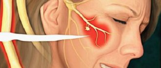
Dacryocystitis or obstruction of the nasolacrimal duct
Dacryocystitis is an inflammation of the lacrimal sac as a result of obstruction of the nasolacrimal duct. The eye is washed with tears, which prevent the eye tissue from drying out and pathogenic bacteria from multiplying in it. This process, necessary for the eyes, is disrupted when the lacrimal canal is poorly patted. Dacryocystitis is manifested by constant lacrimation and swelling of the lacrimal sac. In this case, you definitely need to consult an ophthalmologist.
The causes of dacryocystitis in newborns and adults are usually different. Partial or complete blockage of the nasolacrimal duct, leading to dacryocystitis in adults, is most often caused by atherosclerotic phenomena or infections. Dacryocystitis in newborns occurs as a result of the persistence of the gelatinous film that protected the nasolacrimal canal during the prenatal period. Normally, this film should rupture with the baby’s first breath. Sometimes its rupture occurs during the first 2 weeks of the child’s life. About 5% of newborn babies require treatment for dacryocystitis in the first months of life due to the fact that the independent rupture of the gelatinous film has not occurred. Dacryocystitis in newborns caused by infection is much less common.
Chronic lacrimation and swelling of the lacrimal sac, located in the fossa near the inner corner of the eye, is the main symptom of dacryocystitis. When pressing on this area, purulent or mucopurulent discharge appears. The conjunctiva of the eyelids (the inner part of the eyelids adjacent to the eye) also looks swollen. In the acute course of dacryocystitis, there is a sharp reddening of the lacrimal sac area and a pronounced narrowing of the palpebral fissure. Within a few days, a fistula forms in the inner corner of the eye, which can open on its own, releasing purulent contents. With a long-term chronic course of dacryocystitis, severe stretching of the lacrimal sac is possible, during which the skin covering it becomes thinner and becomes bluish. Dacryocystitis is very dangerous for eye health, as it can lead to infection of the cornea, damage to it and vision problems.
Only an experienced ophthalmologist can distinguish dacryocystitis from a number of other eye diseases, including conjunctivitis, and, therefore, prescribe adequate treatment. Dacryocystitis in a child cannot go away on its own. For treatment, massage, drug therapy, and in some cases surgical treatment are used.
The standard treatment for dacryocystitis in newborns is the systemic use of massage, it is carried out for at least 2 weeks up to 10 times a day. The purpose of the massage is to break the gelatinous film. With regular and correct massage, dacryocystitis in most cases can be cured without the use of surgical treatment. All manipulations are carried out at home by parents. Particular attention should be paid to hand cleanliness. Only an ophthalmologist teaches parents how to properly massage. The massage session begins with squeezing out the contents of the lacrimal sac. Then an antiseptic solution of Miramistin is instilled into the eye cavity and the purulent discharge is completely removed. And only then can you proceed directly to the therapeutic massage. After the massage, disinfectant drops should be placed into the eye.
If treatment of dacryocystitis in a newborn with massage turns out to be ineffective, the child needs to undergo probing of the nasolacrimal canal. Surgical treatment of dacryocystitis in newborns is recommended at the age of 2-3 months. In our clinic, probing is performed under medicated sleep conditions and is painless for the child.
It is also possible to use low-traumatic laser dacryocystorhinostomy in newborns.
In children over 3 years of age, dacryocystorhinostomy is recommended. The operation consists of modeling an artificial nasolacrimal duct connecting the lacrimal sac with the nasal cavity to restore the outflow of tears into the nasal cavity.
Treatment of vasomotor rhinitis
Treatment of vasomotor rhinitis begins with identifying the causes of the disease, is carried out comprehensively and is aimed at eliminating the underlying disease, reducing swelling and discomfort of the patient, and generally improving the condition. Only a doctor can prescribe a set of measures taking into account the characteristics of the body and the general course of the disease.
- It is necessary to exclude the possibility of allergic rhinitis, since its main symptoms are very similar to vasomotor rhinitis, while the treatment methods are completely different.
Folk remedies in the treatment of vasomotor rhinitis
- If you suffer from severe swelling of the mucous membrane of the nasal passages, then a diuretic mixture, which consists of rose hips, about 15 g, 10 g of horsetail and a string of fresh, 5 g of dry birch leaves, 3 g of lingonberry leaves, will help alleviate the condition.
- The prepared dry mixture should be brewed in a proportion of 1 tbsp. spoon per glass of boiling water. The collection is infused in a thermos directly for 8-9 hours. Take half a glass of infusion on an empty stomach 3 times a day for a month.
- Tea according to this recipe will also bring relief if taken regularly. To prepare it, take 20 g of chamomile, yarrow herb, crushed nettle and raspberry leaves and 10 g of thyme.
- Brew the resulting mixture with boiling water at the rate of 4 tbsp. per liter and boil for 10 minutes. It is advisable to drink this tea for a month, 150 ml of tea half an hour before meals.
— You can rinse the nasal cavity with herbal infusion using a small syringe. To prepare the infusion, prepare a dry collection of medicinal herbs. 15 g of horsetail, 30 g of oak bark, 20 g of rowan fruits and the same amount of mackerel leaves, 10 g each of peppermint and sage.
- Pour two tablespoons of the prepared mixture into a glass of just boiled water and simmer for ten minutes over low heat. Rinse with cooled broth. It is advisable to boil the syringe before use and lubricate the spout with Vaseline.
— A very popular folk remedy is juice from Kalanchoe leaves. Traditional healers recommend instilling 6 drops of juice no more than 4 times a day.
- Lubricating the mucous membrane with 10% walnut septum ointment, or calendula ointment with Vaseline will bring relief. The components for the last option are taken in equal quantities.
Treatment
Most people, going to extremes, constantly use vasoconstrictor drugs at the slightest swelling to make their breathing at least a little easier. This is not an option, since this can cause harm to yourself.
Vasoconstrictor drops for edema:
Short-term action drops based on naphazoline.- Drops that act for a longer time (they contain phenylephrine).
- If the drops are based on xylomethosine, then easy breathing is ensured for 6-8 hours, but no more.
- Drops that act for as long as 10 hours must necessarily include oxymetazoline.
Precautions when using vasoconstrictor drops:
- such drops are not medicinal, they only help ease breathing;
- cause addiction with prolonged use, which is later very difficult to get rid of;
- Without medical supervision, drops can be used for up to 5 days;
- if the nasal breathing process has not stabilized after 5 days, consult a doctor;
Methods for treating difficulty breathing in other diseases :
- acute rhinitis requires auxiliary procedures, such as nasal massage, taking a hot foot bath and using mustard plasters;
- to cleanse the nasal cavity of bacteria and moisturize the mucous membrane, carry out the procedure of rinsing the nose with saline solution (prepared at home) or ready-made pharmaceutical preparations based on sea salt (these can be Aqua ENT or Aquamaris);
- to soften the nasal mucosa, you should instill herbal remedies that will have an oily base (Pinosol or peach and mint oils);
- if purulent discharge is observed during difficulty breathing, an antiseptic (miramistin or dioxidin) is used;
- as an adjuvant, inhalation procedures are carried out based on essential oils (eucalyptus is an excellent remedy for colds, pine or cedar oils);
- if allergic rhinitis occurs, use antiallergic sprays (Allergodil, Nasonex), as well as oral medications (Loratadine or Suprastin);
- Difficulty breathing through the nose also requires physiotherapeutic procedures (electrophoresis with calcium chloride).
Treatment methods for anatomical disorders of the nose or other disorders:
- hypertrophied mucous membrane is cauterized with a laser or special chemicals;
- septoplasty (nasal septums are broken and then straightened);
- vasotomy (to prevent the cavernous tissue from swelling, it must be partially destroyed);
- removal of polyps and adenoids.
Folk recipes
For colds, as well as sinusitis, you can use sinus rinsing procedures at home (infusions based on propolis, salt and medicinal herbs).
Also, using drops based on menthol oil, sea buckthorn and rosehip oil will help clear the nasal passage and restore irritated nasal mucosa.
Tampons with garlic juice are used for purulent diseases of the nose. It is widely known that treatment at home involves inhalation of potato decoction, which has a thinning effect, and when propolis is added, it also has an antimicrobial effect.