Maxillary sinusotomy
The second name of the method is sinusotomy. A traditional operation consisting of opening the maxillary sinus and then removing all its pathological contents. Varieties:
- Classic. They enter the paranasal cavity by excision (cut) of tissue in the upper jaw area. The fungus is removed mechanically or with a laser. Disadvantage: risk of complications, long recovery period.
- Endoscopic. An endoscope is inserted into the nasal sinus through the natural passages; no incision is required. Then the mycetoma tissue is burned out with a laser. Plus - quick rehabilitation; after removal, relapses are rare.
The choice of operation method depends on the level of the clinic and the doctor. The procedure time varies from 30 to 60 minutes; in the classic version, general anesthesia or local anesthesia is required.
How the problem manifests itself
A number of symptoms indicate the development of fungal inflammation in the maxillary zone. This:
- recurrent/constant headache;
- discomfort in the upper jaw and teeth;
- difficulty breathing;
- edema;
- discharge with an unpleasant odor;
- blood in nasal discharge.
The disease can manifest itself 1-2 years after the onset of the inflammatory process. Sometimes this happens after 20-30 years, when the ball occupies the entire space, leaving practically no room for air.
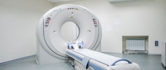
A fungal infection can only be detected using computed tomography. Other methods (X-ray, MRI) do not provide a complete picture. Due to the fact that the vital activity of the fungal formation is initially secretive, the disease takes on a chronic form.
What medications will help with fungal sinusitis?
Non-invasive forms of sinusitis do not require antifungal medications. But to eliminate mycetoma and treat allergic sinusitis, surgical intervention is often initially performed. The air cavity can be opened through the mouth using the Denker method. The technology of maxillary sinusotomy and minimally invasive endoscopic cleaning of the sinuses from pus is used.
At the next stage, a course of conservative therapy is prescribed:
- Antifungal agents, which are aimed at preventing complications after surgery and the development of the disease in an invasive form. Itraconazole is prescribed. If necessary, you can use Amphotericin B or Ketoconazole.
- Glucocorticosteroids, characterized by the presence of immunomodulating and anti-inflammatory effects. With their help, you can prevent relapse after surgical procedures. Topical agents, nasal drops and sprays – “Flixonase”, “Nazarel”, “Polydex” - will help relieve symptoms. In other cases, it is advisable to use systemic drugs in the form of tablets, for example, Prednisolone or Berlicort. Time limit – no more than 7 days.
- Immunotherapy to enhance the body's protective properties. Experts suggest using Amiksin IC or Arbidol. Echinacea, Eleutherococcus, and Aralia are prescribed as immunomodulators with a stimulating effect.
- Antibiotics are rarely prescribed for fungal diseases. With their help, the question of how to treat sinusitis in the presence of bacterial infection or the presence of serious suppuration is resolved.
Causes and prerequisites of the disease
Pathogenic fungi and other microbes often settle on the human mucosa. A disease such as fungal sinusitis is easily provoked by various conditions.
- Sudden entry of foreign objects into the paranasal passages (this phenomenon is more typical for young children).
- Regular colds or pathological conditions of a viral nature that affect the upper respiratory organs.
- Mechanical injuries (bruises, fractures) of facial bones, congenital curvature of the nasal septum.
- Uncontrolled use of antibacterial agents.
- Hormonal disbalance.
- Allergy.
Pathological changes caused by the settlement of microbes of fungal etiology in the nasal passages are provoked by the above factors or their combinations. Symptoms and treatment for fungal sinusitis in adults are determined by your doctor. Complex therapy must be supplemented with folk remedies. Doctors also note the invasive route of fungal “attack” of the nasal passages. This is possible with a significant decrease in a person’s protective functions. Often, this happens as a reaction of the body to serious illnesses - diabetes, HIV, after chemotherapy. Many people are interested in how to cure fungi, who has had fungal sinusitis, how long the therapy will last. Everything depends on the initial state of health of the patient, on his immunity.
Fungal sinusitis symptoms, treatment:
- constantly stuffy nose;
- frequent painful spasms in the head area;
- a white mass is released from the nasal passages, resembling cottage cheese in appearance;
- snot is discharged with bloody streaks and contains pus;
- the mucous membrane of the nasal passages acquires a blue tint;
- swelling of the face appears.
If the disease is not treated in a timely manner, fungal microorganisms will begin to multiply and complications will begin.
Therapeutic measures aimed at combating fungal sinusitis are long-term. The disease is often resolved with surgery, as traditional treatment is often ineffective. Antifungal drugs are often powerless, but in advanced forms of the disease, there is a threat to human life.
Types of treatment for fungal sinusitis.
- Puncture. This operation is performed on an outpatient basis. The nasal sinus is pierced with a thin needle, purulent accumulations are pulled out with a syringe, then the nose is washed. After the procedure, the patient is prescribed antifungal medications. The result will be noticeable after a week.
- Drug therapy. There are different types of treatment for fungal pathology. As a rule, the patient is prescribed steroid, immunomodulatory, and antifungal medications. They will help prevent relapse of the disease. Immunomodulators will help strengthen the body's protective properties. Such medications include Anaferon and Arbidol. Tinctures of ginseng and rosehip will also be effective.
Fungal sinusitis cannot be treated with antibiotics. The effects of such medications increase the growth of fungi. To recover from the disease faster, you should improve your diet, spend more time in the fresh air, and do exercises.
Classification
In fact, mycetoma is a collective term that indicates several pathological processes:
- eumycetoma, the causative agent of which is mold and exotic species of fungal organisms;
- actinomycetoma - a disease caused by fungi of the genus actinomycetes.
Since some pathogenic organisms are saprophytes, they can remain on human skin for a long time and tolerate heat well, which explains such a prevalence of the disease.
According to localization, mycetoma has no clear restrictions, however, it most often occurs:
- foot mycetoma;
- pulmonary mycetoma;
- mycetoma of the uterus.
Nasal mycetoma, in turn, is divided into the following subtypes:
- mycetoma of the maxillary sinus;
- mycetoma of the maxillary sinus.
It is necessary to understand that other localization of the pathological process is not excluded.
Epidemiology
| The style of this section is non-encyclopedic or violates the norms of the Russian language. The section should be corrected according to Wikipedia's stylistic rules. |
The true incidence and geographic distribution of mycetoma throughout the world is not precisely known due to the nature of the disease, which is usually painless, slowly progressive, and may result in death in patients. Mycetoma is distributed throughout the world, but extremely unevenly. It is endemic in tropical and subtropical regions with the highest rates on the African continent. It is located in the so-called mycetoma belt, stretching between latitudes from 15 in the south and 30 in the north. The belt includes Sudan, Somalia, Senegal, India, Yemen, Mexico, Venezuela, Colombia, Argentina and others.
Mycetoma, or as it is also called madurmycosis or Madura foot, was first described in Sanskrit in Vedic texts in India. The first modern source in English was written much later as a result of explorations in the Madras area.
The geographic distribution of individual mycetoma fungal organisms shows considerable variation, which can be convincingly explained on an ecological basis. The areas in which mycetoma predominates are relatively dry areas with a short rainy season and relative humidity.
The organisms are usually present in the soil in the form of grains. Infection occurs when the fungus enters the host tissue through a wound in the skin caused by sharp objects, such as pricks from thorns, stones or shrapnel.
Clinical manifestations of the disease
The insidiousness of mycetoma lies in the difficulty of diagnosing it in the early stages. And in the case of a chronic illness, only an attentive doctor can identify a fungal ball, paying attention to the low effectiveness of classical conservative treatment regimens and the nature of the discharge.
The symptoms of the disease are:
- Frequent headaches, which are localized in the projection area of the sinuses, forehead, bridge of the nose, and temples.
- Feeling of pressure in the sinus, upper teeth.
- Difficulty breathing, frequent congestion.
- Constant nasal discharge mixed with blood and an unpleasant odor.
- Edema.
- The change in the position of the eyeball in the orbit becomes more pronounced over time.
Impaired sense of smell and vision are often observed, and patients complain of diplopia. The first pronounced symptoms can be detected 1-2 years after fungal invasion, and in some, mycetoma is detected 20-30 years after the appearance of the fungus in the sinus.
The most reliable method of detecting the disease is CT scanning and sputum collection for culture.
Diagnostic measures
Already during the initial examination, after collecting an anamnesis, an experienced otolaryngologist can make a diagnosis. It is quite difficult to independently grasp the connection between the manifestation of pain in the occipital area and distinguishing the signs of sphenoiditis. The patient’s complaints will help to suspect inflammation of the sphenoid sinus, but with a chronic course of the pathology, the patient is unlikely to be able to accurately identify the day of onset of the disease. Symptoms of the inflammatory process at the initial stage may not be expressed.
Inflammation of the main sinus can be confirmed by rhinoscopy. If pathology is present, swelling of the cavity in the area of the mouth will be noticeable. X-rays are used extremely rarely to confirm the diagnosis when sphenoiditis is suspected, this is due to the fact that the cavity is located quite deep and its shadow is covered by many dark spots.
The newest and most accurate method for diagnosing sphenoiditis is CT. The study allows you to accurately assess the condition of the cavity and definitively confirm or refute the presence of inflammation. Magnetic resonance imaging of the sinuses is used less frequently for these purposes.
Treatment with folk remedies
To treat fungal sinusitis, home rinsing is often used. This helps remove contents from the maxillary sinuses.
The following are used as rinsing compositions:
- solutions based on propolis and salt (15 drops of propolis and a teaspoon of salt per glass of water), iodine (a teaspoon of salt, 2 drops of iodine) or a weak solution of potassium permanganate;
- carrot and beet juices added to the saline solution, they reduce swelling of the mucous membrane and contribute to its overall health;
- traditional solution of salt and soda;
- decoctions of chamomile, sage, horsetail.
You can relieve inflammation in the maxillary sinuses and suppress fungal infections with the help of self-made drops and turundas. The composition of the drops, as well as the composition of the washing solutions, may include propolis, vegetable juices, aloe juice, as well as onions and garlic, which have an active antibacterial effect, and honey.
Oil drops have a softening effect. To treat fungal sinusitis, sea buckthorn oil, menthol or rosehip oil are instilled into the nose. To prolong the effect of the drops, turundas soaked in the appropriate composition are placed in the nose.
Softening the contents of the maxillary sinuses can be achieved by using hot and cold inhalations. For hot inhalations, jacket potatoes, propolis, honey or fir oil are used. For cold ones – grated onion or horseradish.
Notes
- Motswaledi HM, Mathekga K, Sein PP, Nemutavhanani DL (August 2009). "Paecilomyces lilacinus eumycetoma." Int. J. Dermatol. 48
(8):858–61. DOI:10.1111/j.1365-4632.2008.04047.x. PMID 19659864. - Brownell I, Pomeranz M, Ma L (2005). "Eumycetoma". Dermatol. Online J. 11
(4): 10. PMID 16403382. - ↑ 1 2 3
Hamilton Bailey's
Demonstrations of Physical Signs in Clinical Surgery
ISBN 0-7506-0625-8 - Hemashettar BM, Siddaramappa B, Munjunathaswamy BS, et al.
(December 2006).
"Phaeoacremonium krajdenii, a cause of white grain eumycetoma." J. Clin. Microbiol. 44
(12): 4619–22. DOI:10.1128/JCM.01019-06. PMID 17005754. - Filamentous Fungi. Archived from the original on August 1, 2012.
- Severo LC, Oliveira FM, Vettorato G, Londero AT (March 1999). “Mycetoma caused by Exophiala jeanselmei. Report of a case successfully treated with itraconazole and review of the literature". Rev Iberoam Micol 16
(1):57–9. PMID 18473595. - Vilela R, Duarte OM, Rosa CA, et al.
(November 2004).
"A case of eumycetoma due to Madurella grisea in northern Brazil". Mycopathologia 158
(4):415–8. DOI:10.1007/s11046-004-2844-y. PMID 15630550. - Ahmed AO, Desplaces N, Leonard P, et al.
(December 2003).
“Molecular detection and identification of agents of eumycetoma: detailed report of two cases.” J. Clin. Microbiol. 41
(12):5813–6. DOI:10.1128/JCM.41.12.5813-5816.2003. PMID 14662990.
Prevention of mycetoma
Preventing a disease from occurring is better than treating it. In addition, untreated fungus can spread to other organs. It is possible to reduce the risk of developing mycetoma if:
- a person uses the services of one trusted dental clinic;
- the patient treats all colds in a timely manner and does not lead to the need for antibiotic therapy;
- there is an opportunity to reduce work-related stress;
- do not give up your annual vacation and spend it in environmentally friendly places;
- do not use personal belongings of a sick person - his cosmetics, nasal drops or handkerchiefs;
- the patient takes antibiotics only as prescribed by the doctor;
- a person heals fungal infections of the oral cavity, respiratory system and even eyes in a timely manner;
- After undergoing removal of a mycetoma, the patient carefully complies with all doctor’s orders and does not refuse to wash the paranasal sinuses and nasal cavity.
These tips cannot be called a panacea against fungal infection of the paranasal sinuses. However, the likelihood of getting mycetoma is significantly reduced. In addition, mycetoma is fraught with some complications. They usually concern the transition of the infectious process to a chronic form, as well as damage to the eyes, throat, mouth and other organs and tissues of the human body by Aspergillus.
Development and danger of mycetoma
We can say that mycetoma is sinusitis caused by a fungus. The pathogen can enter the nasal cavity with a current of air, in which, under certain conditions, there are many spores.
For example, an air conditioner that has been uncleaned for a long time is a breeding ground for micelles; you can also become infected by being in a poorly ventilated, damp room with a persistent smell of mold, and so on.
Schematic representation of a "fungus ball"
Getting through the narrow canal between the sinus and the nasal cavity, the spore does not always grow. A healthy person with good immunity copes with the disease, but if the defenses are reduced, the fungal seeds pose a serious danger.
The following category of people should be alert to the development of the disease:
- long-term users of antibacterial drugs (they can cause dysbiosis of the nasal sinuses - a violation of the ratio of pathological flora to the own flora of the nose, as a result - a local decrease in immunity);
- endocrinological patients (patients with diabetes, thyrotoxicosis, and so on);
- cancer patients (including cancer of the maxillary sinus);
- young children (newborns, preschool children) - with an unformed immune response;
- older people – reduced immunity with age;
- living in unfavorable conditions (environmental pollution, insufficient supply of nutrients from food, and so on);
- antisocial category of persons (alcoholism, drug addiction);
- smokers (tobacco smoking, hookahs and even vapes) and so on.
In healthy people with strong immunity, the disease can form when zinc enters the maxillary sinus; this occurs as a result of filling the teeth of the maxillary row. Zinc has a beneficial effect on fungal reproduction and spore germination.
First of all, intensive growth of micelles occurs. They form a kind of ball, which grows quite quickly in volume and fills the space of the paranasal maxillary sinus, often a person is of little concern.
Photo of maxillary sinus cancer obtained during maxillary sinusoscopy.
Developed clinical symptoms begin when the fungal ball causes inflammation of the mucous membrane, narrowing the anastomosis or clogs it itself. This leads to disruption of the outflow from the sinus and the formation of acute sinusitis.
The fungus is a strong allergen, this is the main danger of mycetoma of the maxillary sinus. Prolonged contact increases the content of immunoglobulin E and other markers of allergization of the body. This increases the risk of developing allergic rhinitis, dermatitis, urticaria, Quincke's edema, and most importantly, bronchial asthma.
Mycetoma of the Maxillary Sinus on CT
Switch to English registration. Phone or email. Another's computer. Passed through sinusitis. View topic 9
Maxillary sinuses: causes of inflammation. Treatment
Fungal sinusitis manifests itself in two forms - mycetoma and allergy to the fungus. In fact, it is mycetoma that is considered to be true sinusitis of fungal etiology, since all the symptoms of this disease are caused by abnormal growth and reproduction of fungi.
In the second case, the reason that symptoms characteristic of sinusitis appear is an atypical reaction of the body to the presence of fungi in the air and nasal mucosa. However, let's look at the signs, symptoms and treatment of these two forms of fungal sinusitis.
Mycetoma is one of the forms of mycosis in which fungi, growing in the maxillary sinuses, form a spherical body consisting of polyps.
The main pathogens are considered to be fungi of the genus Exserohilum, Alternaria, less common is Aspergillus, which has more than a hundred varieties of moldy fungi. WATCH THE VIDEO ON THE TOPIC: Deciphering a CT scan of the sinuses - how to decipher a CT disk
Mycetoma of the maxillary sinus
Fungal body, or mycetoma, is a special form of inflammation of the paranasal sinuses of a fungal nature. This disease is quite rare and is asymptomatic for several years.
The cause most often is a part of the root filling of a tooth that is removed into the maxillary sinus, around which a fungal colony grows over time, resembling the consistency of plasticine.
This problem undoubtedly requires a solution, since if the growth of the fungal colony is not stopped, over time the entire sinus will be filled with this fungal body, which leads to complications such as sinusitis and impaired nasal breathing function.
Mycetoma is a fungal disease that affects the paranasal sinuses. In the vast majority of cases, fungal infection of the maxillary sinuses occurs; the pathological process is usually one-sided; mycetoma is extremely rarely found in both maxillary sinuses.
Mycetoma is a fungal disease that affects the paranasal sinuses. In the vast majority of cases, fungal infection of the maxillary sinuses occurs; the pathological process is usually one-sided; mycetoma is extremely rarely found in both maxillary sinuses.
A fungal ball is an accumulation of mycelium in the sinus of fungi of the genus Aspergillus, and therefore in the medical literature one can find another name for the disease - aspergillosis.
In recent decades, fungal diseases have been diagnosed much more often than before, but this is partly due to the fact that over time new opportunities for diagnosis have appeared and its quality has improved.
Mycetoma of the maxillary sinus photo
Mycetoma of the maxillary sinus is an infectious and inflammatory disease of fungal origin. Recently, there has been a tendency towards an increase in the incidence of this pathology in patients of various age groups.
As a rule, there is a unilateral lesion. But bilateral inflammation of the paranasal sinuses is also possible.
You can also find another name for this disease - aspergilloma of the paranasal sinuses, since the causative agent of the infection is fungi of the genus Aspergillus.
WATCH THE VIDEO ON THE TOPIC: Restoration of the maxillary frontal sinus
Mycetoma of the maxillary sinus
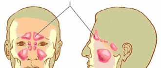
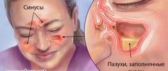
The maxillary sinuses in humans refer to the paranasal cavities located near the nasal cavity. In addition to them, the facial skull contains frontal, ethmoidal and sphenoid sinuses, which perform several functions.
First of all, they warm the air entering the upper respiratory tract. Also, the ciliated epithelium removes small particles, pathogens, allergens, dust, directing it to the nasopharynx area.
Maxillary sinuses location: where are they located Sinuses hurt: possible causes Diagnosis Subtotal darkening of the maxillary sinuses: what does it mean? Mycetoma of the maxillary sinus: what is it? Filling material in the maxillary sinus: consequences Tooth root in the maxillary sinus: what to do? Perforation of the bottom of the maxillary sinus during tooth extraction treatment How to clean the maxillary sinuses at home and is it possible?
What is mycetoma
In the nasal cavity of any person there are different types of fungi on a regular basis, and the person does not experience discomfort or complain of unpleasant sensations. As soon as the body’s defense system experiences stress and falls, the fungus becomes active and begins to sharply increase its number.
Concentrating in the area of the maxillary sinus, it causes fungal sinusitis. The disease is caused by fungi of the genus Candida, Aspergillus and the like. When the human mucosa is examined, this fungus is almost always found on it, in small quantities.
What factors contribute to the development of the disease? Fungal sinusitis is characterized by the fact that in the early stages it is difficult to detect - the symptoms are quite scarce.
Mycetoma of the paranasal sinus is a form of mycosis, a fungal disease that affects the maxillary sinuses. Basically, mycetoma is localized in the maxillary and maxillary sinuses; in rare cases, the presence of a fungal body is diagnosed in the frontal or sphenoid sinuses.
Fungal sinusitis: endoscopic surgery is always needed
page Random page. CT and MRI findings are nonspecific for the common manifestations of sinusitis. Due to the small size of fungal elements, they are detected only with special staining methods [36].
This is probably due to the fact that it is interpreted as a banal polyposis process, and the complexity of laboratory confirmation and ignorance of specialists are the reason for the insufficiently accurate diagnosis.
Until recently, the fungal body fungal ball was the most well-known and widespread form of non-invasive mycosis of the SNP.
VIDEO ON THE TOPIC: mushrooms of the maxillary sinus
What is a fungal body in the maxillary sinus?
International clinic for the treatment of low back pain. Cincinnati Children's Hospital Medical Center. Group of private clinics PremiQaMed. Fungal infections of the paranasal sinuses are quite rare. As a rule, they affect people with weakened immune systems.
Source: https://redstarcharity.ru/e3/5250-mitsetoma-gaymorovoy-pazuhi-na-kt.php
Mycetoma of the lung
Have you been trying to cure FUNGUS for many years?
Head of the Institute: “You will be amazed at how easy it is to cure fungus by taking it every day...
Read more "
The disease pulmonary mycetoma in the initial stage of development can be confused with pulmonary pneumonia. Because:
OUR READERS RECOMMEND!
Our readers successfully use Tinedol to treat nail fungus. Seeing how popular this product is, we decided to bring it to your attention. Read more here...
- the disease does not show any special symptoms;
- X-rays during this period do not show changes in the lung tissue;
- When listening, no noise in the lungs is observed.
All these anti-symptoms without a sputum test lead to the standard treatment of pneumonia - therapy with antibiotic drugs. At this stage, this is extremely undesirable, since antibiotics provoke the growth of fungal colonies, which worsens the patient’s condition.
Symptoms
Symptoms of the disease are divided into stages:
- chest pain;
- increased body temperature;
- dry cough;
- cough with phlegm;
- coughing up blood;
- swelling of the lungs;
- infiltration;
- pulmonary fistulas.
Conventionally, damage to the lungs by a fungal strain is divided into three stages:
- Characteristic chest pains are noted, the temperature rises and a dry cough appears. It is these symptoms that are confused with pneumonia.
- Further, the disease moves to the stage of gradual exacerbation, the cough becomes sputum, sometimes with blood. If treatment is neglected, the lungs swell and increase in volume from the accumulation of cells.
- The last stage of a fungal disease is fistulous tracts formed as a result of the accumulation of exudate, a waste product of the fungus.
Causes
Mycetoma of the lung is caused by fungi of the genus Aspergillus. It is impossible to say with one hundred percent certainty what exactly is the cause of fungal infection in the lungs.
The probable causes of the disease may be the following:
- decreased immune system;
- intestinal dysbiosis;
- long-term therapy with corticosteroids, antibiotics;
- chronic diseases;
- acute respiratory tract disease.
Damage to the lungs can provoke the presence of a fungal infection in other parts of the body:
Together with a decrease in immunity, the disease progresses, causing a chronic course of the disease.
Untimely medical care can cause the infectious agent to enter the bloodstream and result in death.
Treatment
Pulmonary aspergillosis rarely affects all tissues; lesions that develop in the lower part of the organ tissues, as with pneumonia, are more common. Therefore, diagnosis at the first stage of the disease is complicated by similar symptoms.
Direct treatment is prescribed during diagnostics, which includes taking sputum samples and an immunodiagnostic test.
Therapy for pulmonary mycosis consists of:
- antifungal drugs;
- immunomodulatory drugs;
- drugs to inhibit the growth of fungi in the gastrointestinal tract;
- vitamin complexes;
- physiotherapy.
Physiotherapy for lung damage involves a course of inhalations with an antifungal drug that acts locally on the lesion. In some cases you may need:
- administration of glucose drips;
- blood transfusion.
Surgical intervention is carried out in case of an advanced condition - the appearance of fistula tracts. In this case, the affected part of the lung tissue is removed.
As a rule, it is advisable for treatment to be in a hospital setting to monitor the adequacy of the treatment used.
Mycetoma of the maxillary sinuses
Infection of the maxillary sinuses by fungal strains is also called mycetoma of the maxillary sinus. The disease mycetoma of the maxillary sinus does not grow, which means that it does not cause an increase in the localization zone, but is limited to it. This gives a positive prognosis for treatment.
A fungal infection is an accumulation of a fungal strain and its decay products in a limited space in the maxillary sinuses. The causative agent of the lesion is mycosis of the genus Aspergillus, which, when multiplying, forms mycelium in the area of the nasal sinuses. The fungal body, or otherwise called mycetoma, is in practice called mycotic (fungal) sinusitis.
The cause of the disease is the ingestion of dental material during the treatment of damaged teeth. Other causes of the disease are not clear.
Course of the disease
Infection of the maxillary sinuses with a fungus has some symptoms that make it possible to identify a suspicion of a fungal infection.
Symptoms:
- nasal congestion;
- purulent discharge from the nostrils;
- temperature increase;
- local painful sensation.
Fungal infection of the sinuses is rarely bilateral; often inflammation occurs only on one side. Therefore, purulent discharge protrudes from one nostril.
Diagnosis of the lesion is as follows:
- visual examination;
- clarification of patient complaints;
- CT scan of the maxillary sinus.
As a rule, examination is not decisive in diagnosis; accurate diagnosis is carried out only with the help of a computer tomogram showing a fungal infection in the form of compactions.
Symptoms of the disease
Mycetoma is asymptomatic for a long time. Fulminant rapid progression is quite rare and occurs only in individuals with significant immunosuppression. The clinic manifests itself with the development of inflammation and obstruction (blockage) of the excretory anastomosis of the maxillary sinus.
The patient notes the following symptoms:
- severe headache, sometimes accompanied by neurological symptoms (damage to the facial and trigeminal nerves, facial pain in the sinus projection area, imitating toothache);
- visual impairment (double vision, darkening of the eyes, pain when moving the eyeballs);
- fever (often fibrile - 38-38.5˚C);
- intoxication syndrome (lethargy, fatigue, drowsiness, weakness);
- sensation of an unpleasant odor, while the sense of smell itself may be lost;
- nasal discharge does not appear immediately, has a cheesy-jelly-like, sometimes even brown-brown appearance, often with a bloody component;
- allergization of the body (appearance of signs of neurodermatitis, urticaria, allergic bronchial obstruction).
In children with mycetoma of the maxillary sinus, impaired nasal breathing is noted - they breathe through the mouth. Parents note swelling of the face. The eyes turn red, tears flow, often the development of allergic conjunctivitis, dermatitis, and a high risk of developing bronchial asthma. The child quickly loses weight and becomes weaker.
Symptoms and characteristic signs
Mycetoma in the maxillary sinus tends to slowly but steadily progress. A person may not suspect the presence of a pathological process for a long time. The first symptoms appear when the fungal ball reaches a large size and grows throughout all the cavities. Signs:
- the appearance of discomfort, pain in the upper jaw;
- nasal discharge with a curd-like consistency and a foul odor;
- bleeding;
- congestion;
- loss of smell (anosmia);
- increased body temperature;
- headache.
Whether the eye can hurt due to sinusitis is discussed in this article.
Diagnostics
When the first symptoms appear, you should contact an otolaryngologist. Diagnosis of sinusitis includes:
- patient interview;
- rhinoscopy;
- X-ray.
If necessary, the patient is sent for a CT or MRI.
After the examination, the doctor makes a diagnosis: decreased pneumatization of the maxillary sinuses. This is sinusitis.
X-ray and CT scan of the sinuses
X-ray of the sinuses for sinusitis is one of the safest research methods.
If necessary, a contrast examination is performed. It involves the introduction of an iodine-containing solution into the sinuses.
On an x-ray, sinusitis looks like darkening of the sinuses. This indicates the presence of fluid in the maxillary sinuses. Another sign of acute inflammation is thickening of the mucosa.
A CT scan of the maxillary sinuses helps to study the layer-by-layer structure of this part of the skull. X-rays are passed through hollow structures. After computer processing, their 3-D model is created.
CT scan can detect polyps in the maxillary sinus. They are benign growths arising from the mucosa.
CT helps to assess the development of polyposis from the very beginning, when the size of the tumors is 1 mm. A porous mass is found on one or both sides in the sinus area. The polyp itself is also detected. Its structure is heterogeneous.
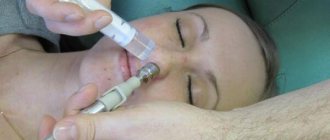
Ultrasound
Ultrasound of the maxillary sinuses is a safe and informative research method.
It does not carry radiation exposure and is used even during pregnancy. This method is used in emergency cases when the result is needed urgently. No special preparation is needed. The algorithm is as follows:
- the patient sits or lies down on the couch;
- a special conductive gel is applied to his skin;
- the image is displayed on the monitor;
- the doctor examines the condition of the sinuses;
- The patient is given a conclusion.
The duration of the study is 10 minutes.
What is subtotal darkening of the sinuses?
Sometimes, after an X-ray of the paranasal sinuses (PSN), the doctor writes in the conclusion: subtotal darkening of the maxillary sinuses. This indicates the presence of chronic sinusitis in the acute stage. Sometimes the detection of dark spots signals the growth of a cyst.
Will an MRI of the brain show sinusitis?
X-ray examination cannot be completely replaced by new technologies. Magnetic resonance imaging is an additional examination method.
An MRI of the brain will show sinusitis. This study detects pathology at an early stage. It also detects any non-inflammatory complications and allows you to assess the condition of nearby tissues.
ESR
A general blood test helps to comprehensively assess the condition of the body. If the pathology is acute or purulent, the erythrocyte sedimentation rate accelerates. This is a normal reaction to inflammation. ESR for sinusitis is often 50 mm/1 hour.
Symptoms of the presence of parasites in the nasal cavity
When roundworms are localized in the nose, they can crawl into the paranasal sinuses and provoke the development of acute or chronic disease of the paranasal sinuses. In addition to parasites in the human body, the presence of a fungal infection allows roundworms to form polyps, adenomas and cysts in the sinuses.
In X-ray practice, every tenth examination of the head, done for various reasons, revealed the presence of a cyst of the paranasal sinuses. Most of these cysts were an accidental discovery, because the patient did not feel any manifestations of sinusitis. Such cysts are formed when roundworms are present in the sinuses.
Also, parasites in the human body, including roundworm, can penetrate the pharyngeal opening of the auditory tube, causing the patient such troubles as ringing in the ears, hearing loss and dizziness, seasickness syndrome, and the possible development of Minière's syndrome.
Methods for detecting mycetoma
The main and most informative method for detecting mycetoma is CT scanning (computed tomography). The fact is that the density of Aspergillus mycelium coincides with the density of the metal, and the source of infection is clearly visible on the resulting tomograms. It is easy to recognize mycelium, which is surrounded by masses whose X-ray density is reduced.
Mycetoma consists of multiple polyps that are formed from spherical mycelium of Aspergillus. Although sometimes the causative agents of the disease are fungi of the genus Candida. Doctors may also see “cellular detritus” - this is a viscous discharge that has a different color.
Moreover, mycetoma affects the maxillary sinuses much more often than the sphenoid or frontal sinuses. Aspergillosis is considered a non-invasive type of fungal infection of the sinuses. It turns out that the mycelium of the fungus develops inside the affected cavity, but does not affect the mucous membrane of the sinus.
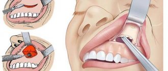
The essence of the operation
With an intraoral gairotomy, the otolaryngologist makes a horizontal incision in the soft tissues of the upper gum and moves them. Then, using special instruments, part of the bone plate on the facial wall of the maxillary sinus is removed. Thanks to the hole made, the sinus cavity becomes accessible for manipulation, and the otolaryngologist removes the pathological contents using a sharp spoon.
After scraping the mucous membrane, an anastomosis is formed with the nasal cavity, smoothing out the bone defect. A flap is made from the remaining free mucosa and placed on the bottom of the maxillary sinus to restore epithelization of its walls.
At the end of the operation, a tampon is placed in the sinus, which is removed into the nasal passage, and the wound on the side of the mouth is sutured. Using a tampon, sterility of the sinus is achieved and the accumulation of blood and pus is prevented.
After 48 hours, after preliminary anesthesia, the tampon is removed, and the patient is prescribed vasoconstrictor drops and sinus lavage. After a week, the sutures in the mouth are removed.
The essence of endoscopic maxillary sinusotomy
This type of operation is performed under local anesthesia using special endoscopic equipment through the nose.
The indication for endoscopic maxillary sinus, as well as for conventional surgery, is a severe purulent process in the maxillary sinus, for which conservative treatment has proven ineffective.
The duration of endoscopic maxillary sinusotomy is 20 minutes. The opening of the maxillary cavity occurs through the nose, and not an incision is made, but a minimal puncture with a diameter of about 5 mm. An endoscopic tube is inserted into this incision, and foreign objects and pathogenic material are removed using suction. All manipulations are performed under the control of an endoscope.
The following methods of endoscopic access to the maxillary cavity exist:
- through the middle nasal meatus;
- through the lower nasal passage;
- through the anterior stack of the sinus;
- through the alveolus of the tooth (if there is a fistula);
- through the tubercle of the maxilla.
The use of endoscopic maxillary sinusotomy helps reduce trauma to surrounding tissues and speed up rehabilitation after surgery.
With the help of endoscopic maxillary sinusotomy, it is possible to avoid the feeling of numbness in the teeth on the operated side, which the patient experiences during a conventional operation.
Highly qualified otolaryngologists of the Multidisciplinary Clinic "MedicCity" will help you in solving various ENT problems. We possess all modern methods of diagnosing and treating diseases of the ear, nose and throat.
Main causes of inflammation
Inflammation in this cavity is often provoked by viruses and bacteria of the cocci group, Haemophilus influenzae, and Moraxella catarrhalis. It is important to remember that with sphenoiditis, a mixed nature of the pathogen is often observed, when at the time of examination it is discovered that the prerequisites for the development of pathology were created not only by bacteria and viruses, but also by fungi.
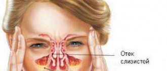
Anatomically, the cavity is constructed in such a way that any, even minor, swelling of the mucous membrane causes a disruption in the normal air exchange process. In this case, the outflow of fluid from the sinus is impossible; due to the concentration of pathogenic contents, pathogenic microorganisms rapidly multiply, and the inflammatory process progresses.
Among the factors that increase the risk of developing pathology are:
- small sizes and irregular shapes of the sinuses;
- narrowness of the anastomosis;
- anomalies in the form of absence of an anastomosis;
- swimming in a pool or open pond with heavily polluted water;
- decreased immunity;
- hormone therapy;
- chemotherapy and radiation therapy;
- the presence of additional partitions in the cavity;
- deviated septum of the sphenoid sinuses as a result of trauma;
- decreased patency of the anastomosis;
- if affected by tuberculosis or syphilis;
- the presence of polyps or cysts in the cavity;
- foreign bodies penetrating the cavity.
Bacteria that enter the wedge-shaped cavity begin to actively multiply on the surfaces of the mucous membranes, which offer active resistance. At this moment, a significant amount of toxins and fragments of destroyed epithelial cells are formed, a secretion occurs, which soon acquires a purulent-mucosal character.
If sphenoiditis does not cure within 3-4 weeks, and the disease periodically manifests itself, a chronic course of the pathology is diagnosed. Chronic sphenoiditis quite often occurs as a result of skull trauma.
The main sinus is filled with mucous contents, which are released through channels into the nasal cavity. In this case, the patient's symptoms of rhinitis predominate. The nasal mucosa swells, significantly increasing in size, blocking or completely closing the nasal passages. It is because of this that the symptoms of the pathology are rapidly intensifying.
Characteristic signs of mycetoma (clinical picture)
Let us recall that the causative agent of mycetoma is fungi belonging to the genus Aspergillius. For some time, the fungus practically does not bother the patient in any way. Doctors discover it by chance when ordering skull x-rays for other reasons. Moreover, the duration of the disease is almost impossible to determine.
- on the affected side the nose is stuffy;
- a person is tormented by pain in the sinus area;
- foul-smelling pus discharges from the nose;
- increase in body temperature of a sick person.
Such signs can easily be confused with purulent maxillary sinusitis of bacterial etiology. Therefore, doctors only assume the presence of a fungal ball after listening to the complaints of the sick person. The examination also does not allow making an accurate diagnosis, since the mycetoma itself is located deep inside the sinus.