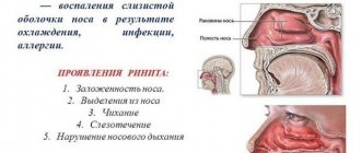
Importance of complication
The maxillary cavities are located above the upper jaw, they are designed to warm the air entering through the nose and clean it from dust and debris. When treating teeth and insufficient thickness of bone tissue, filling material often penetrates into these cavities through damaged tissue.
The possibility of curing a foreign object over time is completely excluded, and patients are also unable to carry out therapy on their own. The therapy involves self-penetration into the sinuses and removal of the filling material stuck there.
This is technically impossible for the patient to do on his own; often the pieces are sharpened at the border of the gum and tooth, and only a professional doctor can handle them.
Therefore, it is important to detect it in time, make a reliable diagnosis and provide quality treatment. Such an operation is not particularly difficult for a doctor. Depending on the specific medical institution, the patient may be discharged either on the same day or the next. The operation is performed using local anesthesia.
Treatment methods
As mentioned earlier, a foreign body found in the maxillary sinus is removed through the intervention of a surgeon. Removal in modern medicine is carried out in two ways:
Endoscopic. An endoscope is used, which is inserted through the nasal passages; in this case, the removal of the foreign body occurs together with the growth of the fungus; it is important to remove it all to prevent its proliferation; The sinuses are also pierced to cleanse them of pus.- Laparoscopic. The surgeon makes an incision in the upper jaw.
The procedures are performed using local anesthetics and last about 15-25 minutes. Patients may require an additional 2-4 days of hospital stay depending on the severity of the surgery and the severity of symptoms.
It is believed that the first method of removing a foreign body from the maxillary sinus is the most convenient and less traumatic for the patient. But this method cannot always be used for a number of reasons. One of which is an abnormal structure of the nasal septum (congenital or acquired). In this case, it is impossible to get into the maxillary sinus using an endoscope.
After any type of surgery, antibiotic therapy is required.
Based on the results of laboratory tests, the type of fungus that led to inflammation is determined. Only after this is a specific drug prescribed to stop the proliferation of microorganisms.
Both operations are performed in specialized clinics. It is better to choose a modern healthcare facility that has all the necessary equipment to perform the operation with less trauma and greater precision in performing the manipulations.
A cyst is also considered a foreign body, although it does not enter the cavity from the outside, but is formed there. The method of its removal is surgery: classical or endoscopic.
The first method means an incision under the lip. Afterwards, the anterior wall of the sinus is opened. The final stage is when the surgeon removes the formation through a small incision.
This traumatic manipulation is performed under local anesthesia. Disadvantage: disruption of the physiological composition of the mucous membrane. Which can subsequently lead to a number of diseases.
Endoscopic removal is less dangerous. Complications after such manipulation are observed in extremely rare cases.
Reasons for appearance
Many patients do not understand how filling material intended for use in dental treatment gets from the gums into the paranasal cavities?
Medicine knows cases of penetration of foreign substances under the following conditions:
- prosthetics,
- installation of seals.
Sometimes the walls between the gums and sinuses are quite thin, so pieces of filling material can easily get into the tissue through their damage. Injuries often occur during the dental treatment process.
The bone at the treatment site can be very thin, and in this case the roots will be located close to the paranasal cavities.
If there is not enough bone tissue at the treatment site, then in some cases the dentist artificially builds up the material. This is necessary for reliable dental treatment. In this situation, damage to the walls and penetration of a piece of dental substance into the sinuses is also often observed.
Filling material, once in the gum layer, often causes suppuration; a person feels severe pain while chewing food, discomfort after touching the damaged area.
The case when a foreign substance does not cause any reaction in the body is quite rare, but sooner or later they end in suppuration.
Foreign body in the maxillary sinus code according to ICD-10
A foreign body in the maxillary sinus (ICD-10 code – T17.1) can be identified by a number of signs. This phenomenon is often disguised as inflammatory diseases of the nasopharynx, which cannot be treated with antibacterial drugs.
Patients visit specialists for a long time, not suspecting that the cause lies in the maxillary sinus. In addition to the filling material, tooth fragments, parts of dentist's instruments, and heads of cotton swabs can get there. The disease does not manifest itself for a long time until the body begins to reject foreign tissue. During this process, opportunistic flora raises its head, immunity decreases (the body is busy fighting a foreign object).
Reasons for the appearance of filling material in the maxillary sinus
The reason for the appearance of a piece of filling is always the same - a visit to the dentist. Such an incident may arise under the influence of the following factors:
- Incorrect filling technique. The dentist may make a mistake in his work, as a result of which dust from the material penetrates into the sinus cavity.
- Thinning of the maxillary sinus as a result of chronic diseases of the nasopharynx and oral cavity. If you self-medicate for a long time, unpleasant consequences arise, one of which may be a foreign body.
- Physiological features of the structure of the jaw, as a result of which the wall of the sinuses is initially thin. When installing dental implants, the doctor has to artificially thicken it.
Symptoms and signs
The severity of symptoms depends on the size of the foreign body, its structure and the patient's health.
You can suspect that filling material has entered the maxillary cavity based on a number of symptoms:
- Feeling of heaviness in the nasal cavity. There may be no pain: the patient feels that something is preventing him from breathing.
- Painful chewing. The pain is localized in the upper jaw, but can spread throughout all the teeth (with an advanced form of inflammation).
- Decreased or complete lack of appetite. Such a symptom does not necessarily indicate a problem in the field of otolaryngology. Usually it is a consequence of general intoxication.
- Painful swallowing, reminiscent of tonsillitis. This occurs due to the spread of bacteria into the mucous membrane of the tonsils.
- Prolonged runny nose that cannot be treated. Neither folk remedies nor antibiotics help the patient.
- An increase in body temperature occurs during a purulent process. Antipyretic drugs provide a temporary effect.
- General weakness, drowsiness. Symptoms that occur when bacteria release waste products.
- Purulent nasal discharge with a putrid odor should alert a person and prompt immediate medical attention.
If you notice several symptoms, you can safely suspect a foreign body, which is a reason to consult a doctor.
Diagnostic methods
There are several diagnostic methods that can be used to determine the presence of filling material. Diagnostic stages:
- An otolaryngologist studies the patient’s complaints and medical history.
- Visual inspection of the nasal passages using a mirror. This helps determine inflammation of the mucous membrane.
- Laboratory research. A general and biochemical blood test will give a general picture of the patient’s condition, help identify the presence of an inflammatory process in the body and determine the degree of its severity.
- Rhinoscopy using an endoscope. The condition of the mucous membrane is examined, where large particles are visualized. At this stage, the doctor can remove them at a convenient location.
- Computed tomography allows you to study tissue in detail and detect a foreign object.
- Radiography is used to assess the condition of tissues and their changes.
Based on the research results, the otolaryngologist makes an accurate diagnosis and begins treatment of the patient.
Clinical picture
Anatomically, the maxillary sinuses are very close to the roots of the upper teeth.
Damage is likely in the maxillary sinus, where the roots of the following teeth are located:
- first molars or second,
- second premolars.
Risk factors:
- acute periodontal inflammation of the maxillary cavity above the upper teeth,
- osteomyelitis,
- formation and suppuration of cysts in the upper jaw,
- traumatic damage to teeth,
- building up bone material inside the gums during dental treatment,
- procedure for installing dentures,
- improper tooth extraction, resulting in suppuration of the upper jaw,
- caries, which causes inflammation of the roots of the tooth.
Entering as a foreign body, it causes tissue irritation, and when a hole is formed, oral microflora can also penetrate through it into the cavity, which can cause the formation of pus.
Consequences of a foreign body entering the maxillary cavity
Mycetoma and fungal sinusitis are the most common consequences of filling agents getting into the maxillary sinus. We should not forget about other adverse consequences.
Possible development:
- cysts of the upper jaw;
- periodontal disease;
- osteoperiostitis;
- fistula formation;
- inflammatory processes of the palatine tonsils;
- lesions of the middle and inner ear;
- spread of infection into the nasolacrimal duct;
- inflammation of the membranes of the brain.
You will be interested in the article - Features of the procedure for removing a maxillary sinus cyst.
Removing a foreign body from the maxillary sinus will help avoid complications. Therefore, it is important to remove the filling material even when nothing bothers the person.
Symptoms of the disease
A person can assume the presence of a foreign body in several situations.
You can try light tapping on your face. They need to be drawn along the facial bone under the eyes next to the nose. If this is accompanied by pain, then there is one of the signs.
If you have a nagging, severe headache, there may be various reasons for this. But in combination with other signs, this indicates that a foreign body has entered the maxillary sinuses.
Another alarming sign is a persistent runny nose. During a seasonal cold, this symptom is easily treatable, and in combination with other manifestations, this speaks in favor of the occurrence of the disease.
Patients complain of a specific aching pain in the upper jaw, which tends to intensify when chewing.
With complications, there is an increase in temperature and profuse, painful nasal discharge that is purulent in nature and is characterized by a specific odor.
The detection of several symptoms from the list forces the patient to go to the hospital for a comprehensive examination. This approach will allow you to exclude secondary complications, plan the method of treatment and preventive treatment.
When a foreign object remains inside human tissue for a long period of time and does not provoke any symptoms, a threat to the patient also exists. Stressful situations, hypothermia, vitamin deficiency, trauma are catalysts for the inflammatory process.
Types of foreign bodies entering the maxillary sinuses
Penetration of a foreign object into the maxillary sinus is impossible through the hole connecting it to the nasal cavity. This requires certain conditions: injury or dental procedures. Since the upper row of teeth is close to the maxillary sinus, and the lower wall of the upper jaw is the thinnest, pressure can cause its perforation (we recommend reading: tooth root perforation: what is it, how to treat it?). The cavity that appears in the maxillary bone can be penetrated by:
- piece of tooth root
- seal,
- material for bone augmentation (when preparing the site for implant installation),
- a fragment of an instrument used for examination or treatment.
Diagnostics and its features
The most effective way to make a correct diagnosis is computed tomography, but this does not exclude the use of other, more traditional methods:
- conducting a patient interview,
- use of rhinotherapy to study the maxillary sinus,
- x-ray examination,
- MRI,
- puncture of the maxillary sinus.
An experienced doctor can reliably assume the presence of such a disease, but it can be confirmed using computed tomography.
How much does it cost to remove foreign bodies from the nose?
The procedure for removing a foreign object is offered by many medical centers and clinics in Moscow. Prices for these services vary in each clinic, since the pricing policy of each medical institution is individual.
Our doctors
Job title:
ENT doctor
Experience:
20 years
Education:
Moscow Medical Academy named after. THEM. Sechenov Experimental secondary school No. 600 of the Research Center for Aesthetic Education of the Russian Academy of Education.
Zaitsev Vladimir Mikhailovich
Additional merits:
- Head of the clinic
- Doctor of the highest category
- Candidate of Medical Sciences
- Author of the invention patent
- Author of more than 20 scientific papers, manuals, and methodological recommendations
Specialization:
- Diagnosis and treatment of ENT diseases
- Treatment of children from the age of three
- Hearing diagnostics
- Repositioning of the nasal bones
- Non-puncture treatment of sinusitis
Make an appointment
Position:
ENT doctor
Experience:
6 years
Education:
Pediatric Faculty of the Dagestan State Medical Academy.
Magomedov Murad Magomedovich
Specialization:
- diagnosis and treatment of ENT diseases;
- performing outpatient operations and surgical procedures;
- nasal polypotomy;
- repositioning of the nasal bones;
- puncture of the maxillary sinuses;
- radio wave destruction;
- stopping nosebleeds;
- non-puncture treatment of sinusitis;
- septoplasty;
- coagulation of the Kisselbach zone;
- endoscopic infundibulotomy;
- microsinusrotomy;
- tracheostomy;
- eardrum bypass.
Make an appointment
Position:
ENT doctor
Experience:
3 years
Education:
Graduated from the State Budgetary Educational Institution of Higher Professional Education “Russian National Research Medical University named after N.I. Pirogov. Ministry of Health and Social Development of the Russian Federation in 2020 “Faculty of Medicine”.
Korzhova Anna Sergeevna
Specialization:
- hearing diagnostics (audiological examination);
- treatment of children from the age of three;
- diagnosis and treatment of ENT diseases;
- performing outpatient operations and surgical procedures;
- diagnostic and therapeutic punctures of the maxillary sinuses;
- nasal polypotomy;
- opening of paratonsillar abscess, nasal septum, abscesses of the external auditory canal;
- opening of suppurating cysts of the tonsils;
- repositioning of the nasal bones;
- puncture of the maxillary sinuses;
- puncture of the maxillary sinus with catheterization;
- removal of small benign tumors of the ENT organs;
- radio wave destruction of the inferior turbinates with vasomotor rhinitis;
- submucosal vasotomy of the inferior turbinates;
- stopping nosebleeds;
- cauterization of the vessels of the nasal cavity;
- non-puncture treatment of sinusitis.
Make an appointment
Position:
Phoniatrician
Experience:
26 years
Education:
Moscow Medical Academy named after. THEM. Sechenova (graduated with honors)
Payganova Natella Ernestovna
Additional merits:
- Doctor of the highest category
- Candidate of Medical Sciences
- Author of three patents for inventions
- Author of more than 120 scientific papers, manuals, and methodological recommendations
Specialization:
- Phoniatrics
Make an appointment
at the ENT Clinic of Doctor Zaitsev, we offer to quickly, efficiently and without complications carry out all the necessary manipulations at the best prices in Moscow. Our ENT doctors will tell you in detail about how much the services cost at your initial appointment. At the same time, the price will not change during the treatment process: we do not impose unnecessary procedures and manipulations.
To make an appointment, please call the reception numbers; You will be booked in at a convenient time
We will definitely help you!
Advantages of this type of service
- Effective treatments
- Professional equipment
- Certified specialists
Treatment procedure
Regulations for actions during treatment of a pathological condition:
- The foreign body must be removed; this is the main task of treatment.
- With such a lesion, inflammatory processes regularly develop. They are generated not only by filling material that has found its way into an unusual place, but also by oral microflora or pus from inflammatory processes in the gums that have penetrated into the resulting hole. They need to be cleaned.
- The patient needs to undergo treatment of both the maxillary cavities and the oral cavity, eliminating the causes of the disease.
Removal of a foreign body always occurs only through surgery. In this case, the cavity is penetrated through a hole and the object is directly removed. There are two such methods:
- laparoscopic,
- endoscopic.
The first method is more traditional. In this case, an incision is made between the upper lip and gum and the material is removed through the resulting hole. In the second case, the paranasal sinus is punctured in the place where the bone is thinnest and a foreign particle is captured through the resulting hole with a special tool and then brought out.
The operation is performed under local anesthesia and lasts no more than thirty minutes. The second method is most widespread. Laparoscopy is usually used in situations where, for some reason, endoscopy is not recommended.
If granulomas arise as a result of the disease, in addition to removing the object, the mucous membrane of the maxillary sinus is scraped.
After this, drug treatment of the inflammatory process is carried out. For this, immunostimulants or antibiotics, as well as other medications, can be used. Additionally, antibacterial solutions or an antiseptic are used to wash the damaged paranasal sinus.
Vitamin complexes are also used in treatment.
About the proximity of teeth and sinuses
The consequences of filling material in the maxillary sinus are unpredictable: they can lead to surgical intervention or the development of dangerous and even chronic complications.
The proximity of the maxillary sinus and the upper row of teeth is dangerous for human health due to the individual structure of the tooth roots. They can have different lengths and sizes. Therefore, in the case of wisdom teeth treatment, the dentist must monitor his actions especially carefully.
The area of the maxillary sinus.
Let us recall that the maxillary sinus is located above the human upper jaw. It occupies about ten centimeters in the nose area, and the bottom of the sinus is in contact with the wisdom teeth.
The sinus is covered with a mucous membrane, which is susceptible to various inflammatory infections and more serious diseases, such as sinusitis.
This area performs several important functions . The maxillary sinus is responsible for air circulation and the resonance of your own voice.
Therefore, any infection or even a filling getting into the maxillary sinus is a serious and dangerous situation. Let's consider several reasons for foreign bodies getting into this cavity:
- First of all, a foreign object can end up in the dentist’s office.
- Negligent attitude towards health and ignoring processes in the oral cavity.
- In special, but extremely rare cases, there is a lack of hygiene.
Inflammatory disease of the maxillary sinus usually appears on only one side, but in some situations there is immediate bilateral inflammation.
This disease affecting the upper teeth is called odontogenic sinusitis.
Signs of foreign body entry
To confirm the presence of a foreign object in the maxillary sinus area, it is enough to know a few signs. They are usually symbolized by inflammatory processes in this area:
- When tilting the head down, a person experiences pain in the sinus area. The patient feels as if the facial bone is being squeezed out under pressure.
- After sleep, a person may experience a feeling of a heavy head. This feeling can also cause lack of sleep.
- Loss of appetite associated with problems chewing foods. Patients often experience pain when swallowing food.
- In special cases, a person develops purulent or watery discharge. They have an unpleasant odor.
- Increased body temperature, up to 39 degrees Celsius.
- Severe swelling of the cheeks.
- Increased fatigue, general weakness.
- Nausea and vomiting.
The listed symptoms are symptoms of a disease such as sinusitis. In any case, the patient needs to listen to the body’s signals and consult a qualified doctor for diagnosis and treatment.
Unfortunately, the entry of a foreign object into the maxillary sinus is always unexpected . The size of the impact of a filling or even a whole tooth depends on the individual characteristics of the person’s structure.
To determine the location of an object, it is necessary to take a photograph using X-rays. In addition, be examined using computed tomography and other elements that help determine the location.
If you do not start freeing the sinus from the extra object at the very first symptoms, a person may experience incredible headaches and loss of sleep. In more severe cases, memory loss is noted.
How a foreign object can get into the maxillary area
According to statistics, more than forty percent of foreign objects entering the maxillary sinus area are associated with unprofessional dental treatment . Since the upper row of teeth is in close proximity to the maxillary sinus, when filling teeth, perforation of the upper jaw may occur.
In this case, the filling material easily falls into the described area. Due to the naturally thin wall of the upper mouth, a foreign object can easily pass into the walls of the maxillary sinus.
Perforation of the bottom of the maxillary sinus after tooth extraction.
The appearance of a hole between the teeth and the maxillary sinus is dangerous to human health. Therefore, in this case, urgent surgery is necessary.
Other reasons for the appearance of holes include large tooth roots . Remember that not only a filling can get into the maxillary area, but also small objects that could be used to conduct an external examination of the tooth.
But getting foreign objects is not always associated with a visit to the dentist. There are cases of exacerbation due to endodontic treatment.
In all cases, it is necessary to perform an operation to remove the foreign object. If an experienced specialist works, this problem can be noticed already in the dentist’s chair.
Consequences
Finding a foreign object inside the maxillary cavity is very dangerous due to its possible consequences. Doctors include acute symptoms:
- Sinusitis in bacterial or fungal form.
- Encapsulation of material in the form of a cyst or benign disease; often in such situations, an exacerbation is diagnosed; it turns into a purulent form.
- Sometimes granuloma formation occurs.
Minor symptoms:
- Meningitis disease.
- Cold-related inflammatory disorders of various types.
- Hypertrophy of the mucous membrane is possible; the pathology is accompanied by impaired nasal breathing.
- The source of inflammation can transmit coccal infection to other parts of the body. One result may be myocarditis of the heart muscle.
- Other complications.
Taking care of your dental health is the best prevention. In addition, you need to avoid facial injuries, not subject your body to severe stress, organize a healthy diet and lead an active lifestyle.
Regular dental checkups will allow you to fully monitor the health of your teeth.
Symptoms indicating abnormalities
As mentioned above, a foreign body can be located in the maxillary sinus for many years without specific symptoms, until it leads to the development of a fungal infection or inflammatory process. Then the following symptoms appear:
nagging pain in the nose and cheeks;- runny nose;
- a certain heaviness in the head, which is accompanied by pain when tilting the head forward;
- toothache may develop;
- discomfort in the jaw area;
- headache;
- increase in body temperature up to 38 degrees;
- formation of pus in the nasal cavity.
All these symptoms are very similar to sinusitis. Indeed, when a foreign body, for example, part of a filling, gets into the maxillary sinus, a fungus grows around it, followed by an inflammatory process. Of course, all this does not happen quickly, but the consequences are quite serious.
There are complications of finding a body in the sinus:
purulent sinusitis;- fungal sinusitis;
- meningitis (inflammation of the brain);
- due to the inflammatory process, infectious and inflammatory diseases of other ENT organs may occur.
Doctors, without performing an x-ray, can treat such patients for sinusitis for a long time to no avail until they discover the true cause of the disease. After all, a foreign body cannot be treated; only its removal can make a difference in the patient’s well-being.