Tooth root and maxillary sinus.
Each person has individual characteristics of the structure of the jaw and skull bones.
Sometimes structural features cause a lot of inconvenience to a person and contribute to the development of various diseases. Doctors have been treating hypothetical diseases unsuccessfully for a long time, without even suspecting that the cause is not pathological, but structure. Dentists also often encounter such problems, as do ENT doctors. You don’t even suspect how such a terrible disease as sinusitis and teeth are connected. The upper jaw is designed in such a way that the roots of the teeth in some places are separated from the lower paranasal sinus by literally a partition a millimeter thick or even less. Some teeth, especially in the depths of the dentition, may have very long roots that these roots can penetrate into the maxillary sinus. The root of the tooth and the maxillary sinus are in a dangerous neighborhood, especially if dental problems often arise. If the root begins to become inflamed, or periodontal disease occurs, then the infection can enter the maxillary sinus and provoke sinusitis. If such a tooth is removed, a hole in the maxillary sinus will remain and infection can penetrate through it. The way out in such a situation is to eliminate the hole itself and carry out antiseptic measures. In some cases, dentists close the perforated hole and rinse the sinus before doing so. but it is still necessary to notice the problem in time, and this is not always possible. More often, exacerbations of nasal diseases are observed after tooth extraction, and sometimes real sinusitis begins abruptly and for no apparent reason. Chronic periodontitis can lead to thinning or complete resorption of the bone septum separating the root from the sinus. The reverse process is also possible. Sinusitis can provoke inflammation of the bone tissue, and pus can get into the tooth socket. A characteristic sign of this phenomenon can be the outflow of air from the tooth socket when exhaling through the nose. In this case, a characteristic whistle may be heard. The sound intensifies if you hold your nose and try to exhale air in the same way. The first sign of perforation of the wall of the maxillary sinus during tooth extraction is the flow of blood from the hole in which air bubbles are present. Let's consider two scenarios for the development of events. If perforation is just discovered and no inflammatory processes are observed in the sinus, you can try to close the hole by suturing the hole and closing it with a turundula. But until the connective tissue grows in this place, the implant cannot be placed. It is possible to close the hole with a bone plate or composite materials. But these methods are usually used for large holes. You can build up the bone using a titanium mesh, or you can install a bone block. There are various methods, not all of them are available in clinics, some are quite expensive due to the high precision of the work. If no complications are observed and there is no inflammation, then after three months the base for the implant can be installed. If there is inflammation in the sinus or in the tissues of the jaw, then it must first be treated, and only then the hole must be closed.
goldstarinfo /2013/02/04/zuby-koren-gajmorova-pazuxa/» goldstarinfo
Oral diseases.
08/03/2018 admin Comments No comments
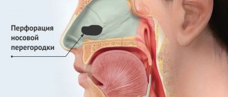
Penetration of bones and tooth roots into the maxillary sinus indicates perforation of the latter. A similar complication may be accompanied by treatment at the dentist. Perforation of the maxillary sinus occurs quite often. From this article you will learn the causes of this pathology, signs of perforation, methods of diagnosis and treatment, as well as prevention. Photos of tooth roots in the maxillary sinus can also be seen in the sections of the article.
How and why can a tooth end up in the maxillary sinus?
The maxillary sinus is the largest in size of all the sinuses of the nose. Its volume in an adult can reach 10 cubic centimeters. The sinus is a steam room and is located to the right and left of the wings of the nose. Visually, its projection falls on the cheek, which becomes slightly swollen and red with sinusitis.
The oral cavity and maxillary sinus are separated from each other by a layer of bone, which is lined on both sides with mucous tissue. However, both the maxillary sinus and the oral cavity are not hermetically sealed. That is why the appearance of an infection on one side of a thin bone septum (for example, pulpitis) quickly entails inflammation in the adjacent chamber.
There are several factors that significantly increase the likelihood of infection spreading:
Anatomically, the roots of premolars and molars are located very close to the sinus. The thickness of the bone layer between them in some patients can be only 1 mm instead of the ideal 10. In some lucky patients, the roots of the teeth are generally located in the sinus itself, being separated from its internal space only by a thin layer of mucous membrane. What to do if your tooth roots are actually in the maxillary sinus? First of all, don't panic! And secondly, carefully monitor their health and regularly take a panoramic photo, which allows you to identify the slightest sign of trouble. The bony septum may become thinner over time. This occurs due to persistent inflammatory diseases - periodontitis, pulpitis and others.
Therefore, if sinusitis is suspected, timely treatment and removal of teeth, including problematic wisdom teeth, is so important. Loose bone tissue as a result of systemic diseases, such as osteoporosis.
Symptoms of perforation of the maxillary sinus
The symptoms of the disease are specific, especially if they occur after tooth root removal. In this case, the patient can note it virtually immediately after the intervention. It is as follows:
- The appearance of air bubbles in the blood that is released from the tooth socket. The number of bubbles increases with a sharp exhalation of air through the nose;
- The appearance of bloody discharge from the nose is noted. They can be scanty in nature and are detected from the side of the maxillary sinus perforated by the dentist;
- Sore throat, presence of nasal sound, the patient additionally notes a state of nasal congestion;
- Patients may also complain that they feel air passing through the hole. There may be a feeling of heaviness in the projection of the upper jaw. The oppressive feeling does not disappear, but only intensifies over time.
If such symptoms appear, you should immediately seek help from a doctor, who will examine the patient and do an X-ray examination, based on which it will be possible to judge the degree of perforation.
The dentist may suspect perforation of the maxillary sinus during implantation based on certain signs. The main one is the failure of the pin, which it is simply no longer able to strengthen at the base of the bone tissue. The doctor may also suspect the presence of pathology based on the following characteristic signs:
The appearance of small air bubbles in the hole;
- Infection of the cavity. As a rule, this happens because the diagnosis of perforation was not carried out completely or was not done completely;
- The patient complains of sharp, unpleasant and aching pain in the sinus area;
- Hypertrophy and swelling of the mucous membrane;
- The patient has difficulty breathing, with the development of an infectious process the appearance of purulent discharge.
General symptoms include fever, weakness, chills, headaches, which may accompany the inflammatory process.
Symptoms
When perforation and entry of the tooth root into the maxillary sinus occurs during dental resection procedures, characteristic signs of rupture appear, including:
- Bleeding from the tooth socket with small air bubbles, the number of which increases when you try to exhale sharply through your nose.
- Bloody nasal discharge from the side of the ruptured sinus.
- A sharp change in the timbre of the injured patient’s voice, characterized by nasal sound.
If there is a tooth root in the maxillary sinus, the symptoms cannot go unnoticed. Sometimes, after resection, the patient begins to complain about difficult passage of air through the resulting hole, as well as pressure and heaviness in the projection of the maxillary sinus.
When perforation occurs during implantation or endodontic therapy, signs of such a complication include:
- Specific failure of the instrument or material for implantation after some effort to move it into the jaw.
- Changing the position of the instrument used in the resulting wound.
- The appearance of small air bubbles and blood discharge from the hole.
Perforation may not be detected and repaired immediately after a sinus rupture, which leads to infection of the sinus cavity and is accompanied by signs of acute sinusitis or sinusitis. The following symptoms are typical for such pathologies:
- Acute intense pain in the sinus area of the upper jaw.
- Swelling of the nasal mucosa on the side of the injury, accompanied by difficulty in nasal breathing.
- Discharge of purulent secretion from the nose.
Systemic complications and symptoms of intoxication appear, which are characterized by chills, headaches, weakness and increased body temperature. But how to detect the root of a tooth in the maxillary sinus?
Diagnosis of the type of perforation and therapy
If the therapist diagnosed sinusitis, and the person had his teeth treated shortly before, the cause-and-effect relationship is quite clear. Instrumental research will help establish an accurate picture and locate the source.
The endodontist probes the hole left after tooth extraction, determining the presence or absence of a bone bottom in it
The procedure is performed with extreme caution
X-rays of the nasal sinuses will provide significant assistance. If darkening in the cavity is visible on the film, it means that there is an accumulation of blood inside. Also in the picture it is possible to see fragments of the tooth roots, the location of the pins, and the protruding material of the intracanal filling. X-rays using contrast will help dispel doubts.
The disadvantage of X-rays is that they are two-dimensional. Stereo images are easy to obtain when undergoing computed tomography. The doctor will review the scan result using a special program, examine the tooth from all sides and choose a treatment plan.
Diagnosis of perforation of the floor of the maxillary sinus during tooth extraction is based on a typical clinical picture. In doubtful cases, as well as when such a complication is suspected during implantation or endodontic manipulations, it is necessary to use instrumental diagnostic methods:
- Probing the socket of an extracted tooth or perforated canal with a thin probe. This allows us to determine that there is no bone bottom in the wound. In this case, the instrument passes freely through soft tissues and does not encounter obstacles along its path.
- X-ray of the sinus area. In this case, the pictures can reveal both darkening of the cavity due to the accumulation of blood in it, as well as fragments of dental roots, implants or filling material. Sometimes it is advisable to conduct radiography with contrast, when a contrast agent is introduced into the cavity through a perforation fistula.
- Computed tomography, which allows you to determine perforations and the presence of foreign bodies in the sinus with maximum accuracy.
- If old perforations are suspected, general clinical blood tests are performed, the results of which may indicate the presence of an active source of infection in the body.
Treatment of a tooth cyst in the maxillary sinus
Warming up and physiotherapy during treatment will not give results and can even cause harm - under their influence the cyst will degenerate into extensive sinusitis. Also, sprays, drops, gels, and solutions will not have a therapeutic effect. The patient is only indicated for surgical intervention.
The main methods for removing a cyst in the maxillary sinus:
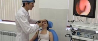
Endoscopy
The most popular and gentle method for removing tumors. Advantages - no incisions on the face, faster recovery of the sinuses. It is performed under local anesthesia.
- Treatment of the nasal passages with an anesthetic. Subsequent injection of the nose with an anesthetic drug (Ultracaine).
- A special tube is inserted into the cavity with the cyst through the nasal passage. The cyst is punctured.
- In some cases, an additional incision is made above the upper teeth on the side of the mouth (the cyst is removed through this hole).
Removal of a cyst in the maxillary sinus
Caldwell-Luke method
Used more rarely. It is performed under local anesthesia.
- First, the doctor makes an oblique incision.
- Next, a trepanation (hole) is performed in the maxillary sinus. Its size depends on the size and location of the cyst.
Disadvantages – long recovery period due to the risk of injury to the anterior wall of the sinus.
Operation Denker
A hole (trepanation) is made through the anterior wall of the sinus. General anesthesia.
- The incision is made from the wisdom tooth to the frenulum.
- Next, the soft tissues are moved apart, the bones are exposed and trepanation is performed.
This is the most traumatic operation, which is prescribed only when absolutely necessary.
Otolaryngologists are still arguing about the need to remove the cyst. Some doctors say that if the tumor does not interfere or cause discomfort, it does not need to be removed. Others insist that even a painless cyst can cause dangerous complications. In most cases, the tumor must be removed. In public clinics the procedure is free of charge.
What can happen from self-medication?
The answer to the question depends on what you are trying to treat. Normally, this should not be allowed; if you decide to try to get rid of the pathology using a conservative method that you prescribed for yourself, the consequences can be unpredictable.
If you start treating yourself and leave the disease unattended, there can be unpredictable and dangerous consequences.
The spread of infection to the surrounding bone tissue will provoke osteomyelitis of the upper jaw. Inflammation can spread to other sinuses, creating an additional health threat.
The formation of abscesses, phlegmons and purulent foci are consequences if perforation is left without treatment and medical supervision.
This disease is classified as dangerous, capable of causing serious pathologies in the body, some of which (for example, meningitis, if inflammation affects the brain) can cause death.
Prevention measures
To prevent a critical condition, you need to go for a routine examination at least once a year. There are no specific and precise means that will forever protect the body. Doctors recommend adhering to general rules of prevention and taking your own health carefully and seriously.
- All nasal diseases must be promptly treated with high-quality and complete therapy.
- Regular visits to the dentist. Detection and treatment of dental diseases - caries, periodontal disease. Strengthening gums.
- If you have allergic reactions, limit contact with allergens and take antihistamines on time.
- Strengthening the immune system, vitaminization, adherence to the regimen and principles of healthy eating. These are general measures that will be relevant for any pathology.
Methods for diagnosing perforation of the maxillary sinus floor
During diagnosis, a comprehensive study and analysis of the typical clinical picture observed with perforation of the maxillary sinus is used. The condition of the tooth socket, the presence of air bubbles, nosebleeds, and pain are taken into account. X-ray diagnostics are also performed, which show the area of perforation with high accuracy.
The dentist can perform a probing procedure for a perforated canal or an extracted tooth using a thin medical probe. This helps to reliably understand that there is no bone bottom in the wound. The instrument itself must pass freely through soft tissue without encountering obstacles along the way.
X-rays of the sinuses can detect characteristic darkening in the images, which are formed as a result of the accumulation of blood clots. Among other things, X-rays can show filling material, implants, and fragments of dental roots. X-rays with a contrast agent are often taken to help clearly visualize the condition of the bone tissue and surrounding tissues.
In such situations, a contrast agent is introduced into the cavity through a perforation fistula. An additional method of instrumental diagnostics is computed tomography, which determines the degree of perforation and the presence of foreign bodies, such as tooth fragments or residual filling material. If perforation is suspected, general clinical tests are required, which can show the presence of a focal infection in the patient’s body.
Diagnosis: which doctor to go to
If you have pain in the maxillary sinus and teeth, then the following types of diagnostics will be the most informative:
- Visual inspection and probing of the hole if the wound is open.
- Radiovisiography or panoramic photograph of the tooth.
- X-ray.
- CT scan of teeth and sinuses.
In case of a sharp deterioration in health, a general clinical blood test is indicated to identify systemic infections.
The diagnosis of damage to the sinus floor is based on the clinical picture. Among the instrumental examination methods:
- Probing - the perforated canal is examined using a thin probe to confirm the absence of a bone bottom in the wound.
- X-ray reveals darkened areas represented by blood accumulated in the sinus. Fragments of roots, implant or filling material may also be found. To clarify problematic issues, radiography with contrast can be performed.
- Computed tomography - determination of the location of the perforation and foreign bodies. Gives the most accurate information.
- General clinical blood test - in advanced cases.
Computed radiography procedure
Treatment of the pathological process
Treatment depends on the severity of the observed pathological process. Typically, surgical intervention is not required in situations where the perforation was carried out during the extraction of a diseased tooth and was detected at the same moment. In this case, X-ray data should show the absence of an infectious process in the maxillary sinus.
If there are no fragments, filling material or infection in the wound, then in this situation the dentist tries to form a blood clot in the hole after tooth extraction and make sure that the resulting cavity is not susceptible to infection. For such purposes, a small gauze swab can be used, which is soaked in iodine solution. Iodine itself has good bactericidal properties, allowing you to quickly eliminate all pathogenic microorganisms.
Most often, such a tampon can self-fix in the resulting wound cavity after tooth extraction. Sometimes stitches are required on the gums, which are done directly by the dentist. Treatment with an iodine solution is carried out for six to seven days until granulation tissue forms, which allows the perforation to close.
The dentist can temporarily close the defect using a special hypoallergenic plate made of plastic. The plate itself is securely fixed to adjacent teeth using clasps. It carefully separates the cavities of the maxillary sinus and mouth, which in turn promotes rapid healing and regeneration of healthy tissue in the perforation area.
It is important to carry out preventive measures aimed at preventing the development of any inflammatory complications. After all, after tooth extraction during perforation, pathogenic microorganisms can enter the wound surface and can cause a serious inflammatory process. Prevention lies in the mandatory use of anti-inflammatory drugs and antibiotics prescribed by the doctor; agents that have a vasoconstrictor effect are also used to stop bleeding.
Prevention lies in the mandatory use of anti-inflammatory drugs and antibiotics prescribed by the doctor; agents that have a vasoconstrictor effect are also used to stop bleeding.
If foreign bodies penetrate into the maxillary sinus during perforation (fragments of tooth roots, filling materials, implant perforation), treatment is carried out in a hospital setting. In severe cases, surgical interventions can be performed on the maxillary sinus with its further opening, during which tissue is cleansed and foreign bodies are removed. After sanitation, plastic closure of the resulting perforation is performed.
Treatment
The choice of treatment for maxillary sinus perforation depends on the changes that occur as a result of the rupture. Non-surgical treatment seems possible in case of violation of the integrity of the tissues during tooth extraction, when the pathology was detected immediately, and according to the results of the radiographic examination it is clear that there is no infection of the sinus cavity and there are no foreign bodies in it. With such a clinical picture, the doctor, as a rule, tries to preserve the blood clot formed in the hole as much as possible. In addition, preventative measures are taken to prevent wound infection.
For this purpose, a small gauze swab soaked in an iodine solution is inserted into the hole. As a rule, the tampon is self-fixed in the wound cavity. However, sometimes it may be necessary to place a stitch in the gum. Treatment with an iodide solution should be carried out for 6-7 days until the defect disappears and full-fledged granulations are formed. It is not recommended to remove the tampon from the socket during treatment, as this can damage the clot and lead to infection of the wound.
When a tooth root is found in the maxillary sinus, everyone should know what to do. In some cases, the doctor decides to close the defect with a special plastic plate, which is secured to adjacent teeth with clasps. Thus, it is possible to achieve separation of the sinus and oral cavity.
Features of therapy
Treatment methods for perforation of the maxillary cavity depend on the speed of its detection. If damage can be noticed during tooth extraction or during prosthetics, therapy can be performed without surgery. However, in case of old pathology, surgical intervention is necessary. The procedure is also carried out if an x-ray confirms the penetration of foreign objects into the adnexal cavities.
Treatment without surgery
After a perforation is detected, the patient is given an x-ray. If no signs of infection, particles of dental tissue or filling material are found in the cavity, the doctor treats it without surgery.
He treats the hole with an antiseptic as carefully as possible so as not to touch the blood clot that forms in it. The open channel is closed in one of the following ways:
- Place a tampon soaked in iodine solution into the hole. If it does not stay in the hole, the gum is sutured. The tampon is not removed for 7 days. After a week it is removed. By this time, scarring occurs in the canal.
- A septum is placed over the damaged area. It is attached to adjacent dental units using special devices. The plate is removed after the hole has completely healed.
To prevent inflammation and infection, the patient is prescribed antibiotics, anti-inflammatory drugs, and local vasoconstrictors for the nose. During therapy, the patient remains at home, but goes to the doctor every day for examination.
Surgical intervention
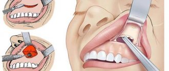
The operation is aimed at removing foreign objects and dead tissue, cleaning the cavity from pathogenic microorganisms, and plasticizing the oroantral fistula. Before the procedure, the patient is given anesthesia. Access to the maxillary cavities is opened through the oral cavity. Operation stages:
- Opening of bone voids.
- Elimination of foreign bodies, infected and dead tissue.
- Rinsing with an antiseptic solution. For the procedure, a syringe with a thin needle is used.
- Plastic surgery of the damaged bottom of bone voids. The hole is closed using tissue cut from the oral mucosa.
After the operation, the patient is prescribed antibiotics. Swelling is relieved with antihistamines. Physiotherapy is indicated to speed up healing.
Complications
Rupture of the maxillary sinus is a serious consequence of dental procedures, the treatment of which most often occurs in an inpatient setting. What is the risk if the tooth root has grown into the maxillary sinus?
Self-treatment of the problem using traditional medicine methods can lead to even more serious complications, including:
- Severe inflammatory process in the sinus cavity with further infection of adjacent tissues. Then osteomyelitis develops in the upper jaw.
- Transition of the inflammatory process to other cranial sinuses, including the sphenoid, frontal and ethmoid.
- Loss of healthy teeth located in the perforation area.
- Formation of foci of pus, phlegmon and abscesses.
Due to the immediate proximity of the brain, and the fact that the tooth root has gone into the maxillary sinus and the latter has ruptured, the occurrence of an infectious lesion of the meninges cannot be ruled out. The next stage of serious complications may be meningitis or meningoencephalitis, which are life-threatening for the patient.
Removal of a tooth with a root in the maxillary sinus.
Because You are not logged in. To come in.
Because you are not a trust user. How to become a trustee.
Because The topic is archived.
Because You are not logged in. To come in.
Because you are not a trusted user (phone number is not verified). Enter and confirm your phone number. Read more about trusts.
Because The topic is archived.
Because You are not logged in. To come in.
Because you are not a trusted user (phone number is not verified). Enter and confirm your phone number. Read more about trusts.
Because The topic is archived.
Because You are not logged in. To come in.
Because you are not a trusted user (phone number is not verified). Enter and confirm your phone number. Read more about trusts.
Because The topic is archived.
Because You are not logged in. To come in.
Because you are not a trusted user (phone number is not verified). Enter and confirm your phone number. Read more about trusts.
Because The topic is archived.
Because You are not logged in. To come in.
Because you are not a trusted user (phone number is not verified). Enter and confirm your phone number. Read more about trusts.
Because The topic is archived.
Because You are not logged in. To come in.
Because you are not a trusted user (phone number is not verified). Enter and confirm your phone number. Read more about trusts.
Because The topic is archived.
Because You are not logged in. To come in.
Because you are not a trusted user (phone number is not verified). Enter and confirm your phone number. Read more about trusts.
Because The topic is archived.
Because You are not logged in. To come in.
Because you are not a trusted user (phone number is not verified). Enter and confirm your phone number. Read more about trusts.
Because The topic is archived.
Because You are not logged in. To come in.
Because you are not a trusted user (phone number is not verified). Enter and confirm your phone number. Read more about trusts.
Because The topic is archived.
the person who was operated on 12 years ago is still sleeping, I can’t ask) just yesterday I went to see the surgeon again, the cat then referred me to 39, because something had come out in the same place and was hurting. cut, squeezed, sewed up.
and then.. well, it was when little was invented, so it took a long time, with complications, etc.. tune in for the best..
Did you have compulsory medical insurance at the maternity hospital? I again forgot the last name of this guy) he’s so memorable, he’s a surgeon..
Are you being monitored in sadko or pomc?
Because You are not logged in. To come in.
Because you are not a trusted user (phone number is not verified). Enter and confirm your phone number. Read more about trusts.
Because The topic is archived.
Because You are not logged in. To come in.
Because you are not a trusted user (phone number is not verified). Enter and confirm your phone number. Read more about trusts.
Because The topic is archived.
no, regional, 56 on Volodarsky, male surgeon, over 40 years old, his last name is funny, you can’t go wrong.
go as if in acute pain, according to compulsory medical insurance. and then you get there...
Because You are not logged in. To come in.
Because you are not a trusted user (phone number is not verified). Enter and confirm your phone number. Read more about trusts.
Because The topic is archived.
Because You are not logged in. To come in.
Because you are not a trusted user (phone number is not verified). Enter and confirm your phone number. Read more about trusts.
Because The topic is archived.
I wish you good luck so that everything works out and you manage to avoid surgery.
I myself removed the same tooth in the summer, but the dental surgeon warned me about the possible consequences and I first tried to save the tooth, took a tomogram, tried to cure it, but unfortunately it was not possible, the canals turned out to be very complex and were not sealed properly at the time squint-handed dentist.
The result is still deletion, but I was lucky, without consequences.
Because You are not logged in. To come in.
Because you are not a trusted user (phone number is not verified). Enter and confirm your phone number. Read more about trusts.
Causes and symptoms of perforation of the maxillary sinus
During dental procedures, perforation of the maxillary cavity may occur. The reasons may be:
Tooth extraction. If the dentist acts carelessly and unprofessionally during the extraction of the upper molars, the bone separating the oral cavity and the sinus may perforate. If the roots enter the cavity, a through hole remains during removal. Root filling. With excessive expansion of the root canals or excessive compaction of the filling material, the filling can go beyond the canal and perforate the lower wall of the sinus. Introduction of the implant. When installing implants, the dentist must carefully examine the patient's jaw. If implanted incorrectly, the pins pierce the separating bone. Root resection. Careless movement of the instrument during resection of a hilar cyst can pierce the sinus (see also: consequences of resection of a dental cyst)
Such consequences are the mistake of the dentist who did not study the location of the cyst at the apex of the root and did not pay attention to its proximity to the cavity.
INTERESTING: what are the symptoms of a left maxillary sinus cyst and how to treat it?
What symptoms indicate that dentists have damaged a bone:
- blood with air bubbles comes from the alveolar socket;
- blood comes from the nasal cavity;
- failure of a dental instrument into a cavity.
READ ALSO: why there is bleeding from the mouth: reasons
Immediately at the moment of perforation, the patient does not feel pain, because dental procedures are performed under anesthesia. Subsequently, the pain will become noticeable, and a feeling of fullness will appear in the molar.
Frequent cases
Perforations of the maxillary sinus occur, as a rule, as a result of the dentist’s work during dental treatment. Tissue ruptures are typical for the following situations:
- Tooth extraction.
- Dental implantation.
- Endodontic therapy.
- Tooth root resection.
If perforation occurred during a tooth extraction operation, this may be a consequence of unskilled and excessively rough work of the dentist, as well as the individual characteristics of the structure of the maxillary sinus of the patient himself. If the dental roots are located directly in the sinus cavity, perforations during the removal of molars are almost inevitable.
One of the complications during endodontic therapy can also be perforation of the tooth root, which in most cases is associated with perforation of the sinus floor. This situation can occur as a result of excessive expansion of the root canals, as well as when using brute force when installing pins and compacting cement for fillings. In this case, most often not only the root of the tooth may be in the maxillary sinus, but also its fragments and particles of filling material. And this is dangerous for the patient.
When a rupture occurs during installation of an implant or during filling a root canal, as well as as a result of inserting pins into the root of a tooth, this in the vast majority of cases is a therapeutic error by a specialist. Sometimes you can find the roots of a wisdom tooth in the maxillary sinus.
Perforation of the bottom of the maxillary sinus, diagnosis and treatment
A perforation of the bottom of the maxillary sinus sometimes occurs to a patient in the dentist's chair, the reasons being the peculiarities of the anatomical structure of the skull or a medical error. According to ICD 10, this situation is not classified as a separate disease, but is considered a complication of the treatment.
When does perforation of the maxillary sinus floor occur?
In ordinary life, perforation is impossible; it always becomes a complication of any dental manipulations with the upper teeth. Perforations occur during root removal, during installation of implants, and during the treatment of pulpitis. At the same time, a gross violation of treatment tactics by the doctor, as well as the special structure of the skull and the anatomy of the teeth can become a source of trouble for a person.
For example, the risk for the patient increases many times if preliminary radiography reveals too small a distance between the apex of the tooth root and the bottom of the maxillary sinus.
There is also danger if the doctor tries too hard to expand the root canals or fills them with excessive compaction. The filling material, transforming, is able to go beyond the apex of the tubule; subsequently, the sinus is perforated in almost one hundred percent of registered cases.
A breakthrough can also occur when installing a pin or inserting an implant. In the latter cases, it is always the dentist's mistake. Such an accident significantly complicates further procedures when performing prosthetics.
The jaw bones of patients who have lost their teeth long ago and applied late for implantation are extremely vulnerable. If a tooth is removed, the processes of tissue degeneration accelerate.
Perforation also occurs during root resection if the dentist did not bother to carefully study the patient’s examination data in advance and does not know the size of the bone plate that separates the maxillary sinus and the inflamed cyst on the tooth. Inaccurate movement and perforation occurs. It can also be caused by surgery to remove a significant amount of jaw bone.
Treatment of perforation of the maxillary sinus
Non-surgical intervention is possible only in the case of instantly detected perforation, when it is diagnosed during the procedure of tooth extraction or treatment of pulpitis. The doctor immediately sends the patient for an x-ray and, based on the results, determines whether an infection of the maxillary sinus has developed.
For example, a cavity wall was perforated during tooth extraction. The dentist noticed the defect immediately. In this case, the doctor makes efforts to preserve the blood clot in the hole where the tooth was previously located.
The therapy involves the application of tampons with iodine, and the tampon is tightly fixed in the formed depression and is not removed from there during treatment - so as not to disturb the clot that seals the wound.
It usually takes up to a week for the breakout to granulate and disappear.
If it was not possible to avoid the penetration of foreign bodies into the maxillary sinus or the infection began to spread, the sinus will need to be opened and cleaned. The resulting wound is closed plastically.
Old perforations
It happens that the patient does not go to the clinic after treatment due to accidental perforation. At first the pain is sharp and pronounced, but gradually its character changes and the sensations smooth out. A fistulous tract is formed between the affected sinus and the outer part of the gum.
Everything that happens is accompanied by the development of symptoms of typical sinusitis: pain from the perforated intramaxillary cavity, constant nasal congestion, outflow of pus from the nasal passages (and sometimes the fistula on the gum begins to fester).
Some patients complain of swelling of the cheek on the side where the sinus is damaged.
Unfortunately, non-invasive techniques will not help here. An operation is required to remove foreign matter and foci of infection from the sinus. The fistula is cut out, and the opened defect is repaired. After the procedure, a course of antibiotics is prescribed with accompanying physiotherapeutic treatment and the use of drugs that reduce the risk of allergic reactions.
New methods for plastic surgery of perforations of the maxillary sinus floor using a beeswax membrane and platelet-rich plasma in combination with an allogeneic bone matrix
According to the literature, perforations of the maxillary sinus (MS) occupy a leading place in the etiology of odontogenic sinusitis - from 40 to 80% [1,2,4-6].
Pathogenetic treatment of perforated sinusitis involves closing the oroantral communication with a duplication of the mucous membrane, ensuring optimal conditions for directed bone regeneration [3]. Currently, there are many known methods for diagnosis, plastic closure and rehabilitation of patients with perforation and foreign bodies in the maxillary sinus (MS) [1,5-11,14-16]. However, literature data convinces us that this problem remains relevant to this day. Existing methods of surgical treatment of patients with perforation and foreign bodies in the upper jaw do not always provide a favorable outcome; complications often occur (32.4-80.0%) [14]. The purpose of the work is to clinically substantiate the use of a membrane made of beeswax and platelet-rich plasma in combination with an allogeneic bone matrix in the complex treatment of perforated sinusitis.
Material and research methods
We observed 37 patients with perforated sinusitis (20 men and 17 women aged 20 to 55 years). Of the total number of patients, 28 (75.7%) had perforation of the ICP bottom, 19 (24.3%) had perforation of the ICP bottom and a foreign body (tooth root) in the sinus. By the time of treatment, the duration of the disease from 1-7 days was registered in 11 (29.8%) patients, from 8 days to a month in 9 (24.3%), up to three months in 8 (21.6%), more three months - 9 (24.3%). The groups of teeth, the removal of which caused perforation of the bottom of the upper jaw, were distributed as follows: first molars of the upper jaw (MF) - 16 (43.3%) observations, second molars MF - 9 (24.3%), third molars HF - 3 (8.1%), first premolars HF -3 (8.1%), second premolars HF -5 (13.5%), canines HF -1 (2.7%) observations. As can be seen, the most common causes of oroantral communication are extractions of the first and second HF molars. Acute inflammation of the upper sinus was observed in 6 (16.2%) patients, chronic inflammation in 23 (62.2%), an uncomplicated form of perforation of the sinus floor was registered in 8 (21.6%) cases. The patients were examined according to the generally accepted scheme: the patient's complaints, medical history were studied, an examination of the oral cavity was performed, an X-ray examination of the paranasal sinuses, an X-ray of the socket of the extracted tooth, as well as general clinical examinations.
Scheme 1. The first (A) and second (B) methods of plastic closure of the oroantral communication:
1) mucous membrane of the upper jaw, 2) beeswax membrane, 3) bone tissue of the upper jaw and alveolar process, 4) mucoperiosteal layer, 5) ADCM in combination with PRP
The membrane, 35-40 microns thick, was made from beeswax. We obtained platelet-rich plasma (PRP) according to the method of R. Marx, E. Carlson, R. Eichstaedt et al. [17]. Directly in the operating room, shortly before or at the beginning of the operation, autogenous blood was collected through a central venous catheter. The amount of blood required to obtain PRP was determined using the table we compiled [12]. PRP was used in combination with crushed allogeneic demineralized bone matrix (ADBM) in a ratio of 1 g/ml. In cases where perforation of the bottom of the maxillary sinus was complicated by acute sinusitis, the sinus was washed with antiseptic solutions through the dental alveolus, and antibiotics and enzymes were introduced into the sinus.
Scheme 2. The third method of plastic closure of the oroantral communication:
1) mucous membrane of the upper jaw, 2) beeswax membrane, 3) bone tissue of the upper jaw and alveolar process, 4) mucoperiosteal layer, 5) ADCM
Scheme 3. Plastic closure of the defect in the anterolateral wall of the upper quadrant after radical maxillary sinusotomy using a beeswax membrane:
1) an unchanged area of the mucous membrane of the upper jaw, 2) a beeswax membrane, 3) bone tissue of the upper jaw and alveolar process, 4) mucoperiosteal layer, 5) AKDM in combination with PRP, 6) a fragment of bone tissue of the anterolateral wall of the upper jaw
UHF and laser therapy were used locally. Only after the acute symptoms of sinusitis subsided was plasty of the perforation hole performed. The method of surgical closure of the perforation opening of the upper jaw was differentiated, taking into account the age of perforation, the extent of damage to the sinus mucosa and the characteristics of the tooth socket. For fresh and uncomplicated perforations, we mobilized a mucoperiosteal flap from the vestibular wall of the alveolar process. In order to mobilize the flap, the periosteum was cut at the base. If the opening of the ICP fistula was sufficiently wide, a flap was formed from both the vestibular surface and the pedicle from the palate. They were then joined together or a duplicate of the tipping and palatal flap was created. Depending on the characteristics of the socket through which a connection with the fistula was formed, we chose different methods of plastic closure of the fistula tract.
The first method is if the tooth socket had a cone-shaped shape, the bottom of the tooth root socket, which communicated with the upper jaw, was covered with a beeswax membrane, then the hole to the level of the alveolar crest was filled with crushed allogeneic bone matrix, in combination with PRP (Scheme 1A).
Rice. 1. X-ray in the nasofrontal projection:
A) before surgery, B) 3 months after surgery
Rice. 2. Intraoral radiograph of the alveolar process of the upper jaw on the right:
a) before surgery, b) 3 months after surgery
The second method is if the hole on the alveolar process was wide, the height of the alveolar process was 5 mm or more, in this case a round membrane was formed, which had 2 ribbons on the sides. The size of the round part of the membrane should exceed the size of the hole by approximately 1-1.5 mm. After the membrane was formed, it was carefully inserted into the well to the level of the bottom of the well. The ribbons were given a horizontal position and tightly fixed with the fingers of the left hand on the sides of the hole. The free space up to the level of the alveolar ridge was filled with crushed ADCM in combination with PRP (Scheme 1B)
The third method is if the height of the alveolar process in the area of the oroantral communication is less than 5 mm, in this case the communication was covered with a membrane that exceeded the size of the defect on the alveolar process by 4-5 mm. ADCM was placed on the membrane, which was 4-5 mm larger in width and length than the membrane, or the membrane was completely covered with a layer of crushed ADCM in combination with PRP 2-3 mm thick (Scheme 2). The latter is advisable to use in cases where there is a defect in the alveolar process. Next, the alveolar process in the area of the tooth socket was covered with a mucoperiosteal flap and sutured with interrupted sutures. For the best fixation of the flap, in addition to the usual suturing of tissues, a mattress suture should be applied [10].
The fourth method - in case of diffuse polypous changes in the mucous membrane on a significant part of the walls of the upper quadrant, a radical maxillary sinusotomy was performed with plastic closure of the defect on the anterolateral wall of the upper quadrant according to our modification (Scheme 3) [13] and plastic surgery of the oroantral communication according to one of the methods described above.
Results and discussion
According to the analysis of the results of surgical closure of the oroantral communication according to our modifications and subsequent complex treatment, all patients obtained a positive result. During the first week after surgery, moderate swelling was noted in the area of the corresponding side of the infraorbital region. In cases where a radical maxillary sinusotomy was also performed, sanguineous discharge from the nose was also observed. Surgical wounds healed by primary intention in 25 patients. In 2 patients, in the early postoperative period, partial divergence of the sutures occurred, which we associate with insufficient mobilization of the mucoperiosteal flap within the alveolar process of the upper jaw and the tissues of the vestibule of the oral cavity. In these cases, it was not necessary to perform a second operation. The wounds healed by secondary intention. The sutures were removed on days 7-9. The effectiveness of the proposed methods was assessed using clinical and radiological data. Intraoral radiography was performed 7 days after surgery, at the end of the first month and after 3 and 6 months. The first radiological signs of young bone formation in the area of the perforation hole (extracted tooth socket) appear by the end of the 1st month. Then the regeneration process continues, by the end of the 3rd month at the site of the implanted matrix in combination with PRP
Fig.3. Stages of plastic closure of the oroantral communication with radical maxillary sinusotomy:
3.1) socket of the 13th tooth, 3.2) exposed anterolateral wall of the upper jaw, 3.3) formation of a window on the anterolateral wall of the upper jaw, 3.4) window on the anterolateral wall of the upper jaw, 3.5) closing the window with a beeswax membrane and a bone fragment, 3.6) closing the bottom of the socket with a membrane from beeswax, 3.7) filling the socket with crushed ADCM in combination with PRP, 3.8) the tooth socket after closing with a mucoperiosteal flap and suturing, organotypic bone tissue can be traced. To illustrate the proposed method of closing the oro-antral communication using a beeswax membrane, we give an example.
Patient G. 20 years old, came to the clinic with complaints of communication in the mouth and nose after the removal of the 16th tooth 3 months ago. An objective examination of the patient from the internal organs revealed no deviations from the norm. When examining the oral cavity, a fistulous tract was revealed in the area of the socket of the 16th tooth, through which the oral cavity communicated with the maxillary sinus (Fig. 3.1). The discharge of pus from the nose from the side of the maxillary sinus was not detected. A radiograph in the nasofrontal projection revealed a right-sided darkening of the upper quadrant (Fig. 1A). On the intraoral radiograph of the alveolar process of the upper jaw on the right, the socket of the 16th tooth communicates with the upper jaw (Fig. 2a). Based on clinical and radiological studies, right-sided perforated sinusitis was diagnosed. Under general anesthesia, a radical ICP operation was performed, with plastic closure of the oroantral communication. The edges of the socket of the 16th tooth were first freshened, then a radical maxillary sinusotomy was performed.
After the formation of a mucoperiosteal buccal-gingival flap according to Neumann-Zaslavsky and exposure of the anterolateral wall of the upper quadrant (Fig. 3.2), the bone was resected with a fissure bur (Fig. 3.3) and a window measuring 1.5 × 1.7 cm was formed on the anterolateral wall of the upper quadrant (Fig. 3.4 ). After opening the maxillary sinus, granulation tissue was found in the area of the lower and medial walls. The remaining walls of the sinus appeared unchanged. Only the pathologically altered part of the sinus mucosa was removed. An anastomosis of the upper nasal cavity was formed with the nasal cavity in the area of the lower nasal meatus, creating a hole measuring 1.5×1.5 cm. A surgical spoon was used to scrape out the hole, removing pathologically altered tissue. After washing with antiseptic solutions, the maxillary sinus was tamponed with a tampon soaked in iodoform liquid, its end was brought into the nasal cavity through
Fig.4. Patient G., mucous membrane of the vestibule of the oral cavity according to the projection of the removed 16th tooth 3 months after surgery
created anastomosis. The window on the anterolateral wall of the upper quadrant was closed with a beeswax membrane, on which a fragment of the bone of the anterolateral wall of the upper quadrant was fixed (Fig. 3.5). The bottom of the socket of the extracted tooth was covered with a beeswax membrane (Fig. 3.6). The hole, up to the level of the alveolar ridge, was filled with crushed ADCM in combination with PRP (Fig. 3.7). For the purpose of maximum mobilization, the periosteum was dissected at the base of the formed flap. The formed flap was used to cover the hole. The wound in the oral cavity was sutured tightly (Fig. 3.8). The tampon was removed from the maxillary sinus on the second day after surgery, and the sutures were removed on the 8th day. The wound healed by primary intention. The patient was discharged from the hospital on the 10th day after surgery.
A follow-up examination of patient G. was carried out after 3 months. The patient has no complaints. The opening of the mouth is free, the mucous membrane of the vestibule of the oral cavity along the projection of the extracted tooth is without any features, a thin postoperative scar can be traced (Fig. 4). Palpation is painless. An intraoral radiograph shows newly formed organotypic bone tissue in the area of the socket of the 16th tooth (Fig. 2.b). On a radiograph taken in the nasofrontal projection, the transparency of the right upper quadrant is almost identical to that of the left (Figure 1B). The interest of this case lies in the successful plastic closure of the oroantral communication using the method we propose.
Thus, the method of closing the perforation on the upper jaw depends on the duration of the disease and possible complications after the formation of the oroantral communication. In case of fresh, uncomplicated forms of intracranial perforation, we can limit ourselves to closing the communication with a beeswax membrane and filling the remaining space of the socket with crushed ADCM in combination with PRP and plastic closure of the perforation of the maxillary sinus with a flap formed from the vestibular side of the alveolar process. In cases where the height of the alveolar process is less than 5 mm, after closing the communication with a beeswax membrane, the area is additionally covered with an allogeneic bone matrix or a layer of crushed ADCM in combination with PRP. For uncomplicated perforations of the upper quadrant, surgical treatment of patients can be carried out in a clinic, without radical maxillary sinusotomy.
The new methods of surgical treatment of patients with odontogenic perforated sinusitis that we have proposed can significantly reduce the frequency of postoperative complications. In addition, they are economical for both the patient and the medical institution. They can be used as the method of choice when closing the oroantral communication. Timely diagnosis and closure of perforations of the bottom of the upper quadrant allows to prevent the development of perforated sinusitis and further extensive operations on the upper quadrant. It should be recalled that in order to diagnose perforations of the bottom of the upper jaw during tooth extraction surgery (TOS), the following is necessary: a certain alertness of the doctor to the possibility, and sometimes the inevitability, of such a complication; expansion of indications for X-ray examination of the HF before OUS; gentle revision of the postoperative wound; conducting a nasal-oral test when removing molars and premolars of the upper jaw; compliance with known tactics for treating detected perforation of the bottom of the upper jaw and professional care of the bone wound.
Bibliography
- Bogatov A.I. New methods of diagnosis, treatment and rehabilitation of patients with perforations and foreign bodies of the maxillary sinuses. Author's abstract. dis. Ph.D. honey. Sci. Samara, 1991, 18 p.
- Govorun M.I. Acute recurrent sinusitis in young people in terms of the state of the immune system. Author's abstract. dis. Ph.D. honey. Sci. St. Petersburg, 1992, 18 p.
- Grigoryants L.A., Rabukhina N.A., Badalyan V.A. Wedge. stom., 1998. N3. With. 36-37.
- Elkov I.V. New principles for the treatment of inflammatory diseases of the maxillary sinuses. Author's abstract. dis. Ph.D. honey. Sci. M. 1996, 13 p.
- Ivanov V.A. Clinical and experimental rationale for closing acute perforation of the maxillary sinus floor with a dental implant. Author's abstract. dis. Ph.D. honey. Sci. M. 1998. 24 p.
- Kozlov V.A., Shulman F.I. All-Russian Congress of Dentists. M. 2001.
- Korotkikh N.G., Lazutikov O.V., Larina O.E. Dentistry. 2003. N4. With. 40-42.
- Kruchinsky G.V., Filippenko V.I. Dentistry. 2004. N1. With. 51-52.
- Lavrentyev S.S. The use of osteoautoplasty of fistulas of the bottom of the maxillary sinus in the complex treatment of odontogenic perforated sinusitis. Auto-ref. dis. Ph.D. honey. Sci. 1996. 24 p.
- Luzina V.V., Manuilov O.E. Dentistry. 1995. N1. With. 41-42.
- Mukhametzyanova T.S. Complex treatment of odontogenic perforated sinusitis. Author's abstract. dis. Ph.D. honey. Sci. 1990. 23 p.
- Pogosyan Yu.M., Pogosyan D.E., Harutyunyan A.S. and others. Bulletin of dentistry and maxillofacial surgery 2003. volume 2, issue. 3. p. 20-30.
- Pogosyan Yu.M., Sepyan A.G., Pogosyan A.Yu., Burnazyan S.S. Questions of theoretical and clinical medicine. 2009. N2. With. 43-44.
- Semaan-Abi-Khalil J. Evaluation of the results of surgical treatment of patients with chronic odontogenic sinusitis. Author's abstract. dis. Ph.D. honey. Sci. St. Petersburg, 1992.19 p.
- Sepyan A.G. New approaches to surgical treatment of sinusitis. Author's abstract. dis. Ph.D. honey. Sci. Yerevan. 2006.18 p.
- Buchner HJ, Lessel W. Stomatol DDR, 1979; 28: 12: 883-887.
- Marx R., Carlson E., Eichstaedt R. et al., J. Oral Maxsilofac. Surg. 1998. vol. 85 p. 638-646.