Mastoiditis is an acute purulent inflammation of the mastoid process of the temporal bone, in the area behind the ear. This disease is of infectious origin. Mastoiditis in children accounts for approximately 5-10% of cases of ENT diseases.
The disease can occur as a result of infection or develop as a result of complications of acute otitis media. The clinical picture of mastoiditis in children is manifested by pain and pulsation in the area behind the ear, elevated body temperature, general intoxication and swelling in the mastoid area. If you notice any of the above symptoms in your baby, contact your doctor immediately .
How dangerous can the disease be? The most common complications
Mastoiditis is an infectious inflammatory process that occurs in the mastoid process of the temporal bone.
The disease is often a complication of acute otitis media. The course of inflammation depends on the structure of the mastoid process. In most cases, mastoiditis occurs in people who have large cells inside the appendix. Infection from the tympanic cavity penetrates there very easily.
Classification of mastoiditis:
- According to the site of infection:
- Primary (infection enters directly into the mastoid cavity).
- Secondary (complication of the inflammatory process occurring in the body).
- According to the nature of the disease:
- Typical (all characteristic symptoms appear).
- Latent (inflammation occurs without pronounced signs).
At the initial stage (exudative stage) of mastoiditis, the mucous membrane and periosteum become inflamed, and the cavity is filled with purulent contents. Further (alternative stage) the bone partitions are destroyed, the cavity is filled with granulations. The pus that has spread everywhere begins to melt the back plate. This is how it enters the area of the meninges.
Destructive osteoperiostitis of the cellular structure of the mastoid process.
If you touch the area behind the ears, you will find the mastoid process, which is a bony outgrowth with a cavity inside. The latter communicates with the middle ear, and therefore the processes occurring there can affect the condition of the temporal bone. Also, the mastoid process is separated by a small thin plate from the cranial cavity, and this determines the danger of mastoiditis for the health of the most important organ.
Inflammation of the temporal bone, namely the mucous membrane of the inner part of its mastoid process, is called mastoiditis. Mastoiditis in children and adults is infectious in nature and can be acute, subacute, or chronic. Also, depending on the affected area, the disease can be left-sided, right-sided, or bilateral. If the pathology develops independently, it is primary; if it occurs against the background of other diseases, it is secondary.
Acute mastoiditis often occurs together with acute purulent otitis, or is diagnosed after an imaginary recovery. It develops in stages: first it goes through the exudative stage, then the destructive stage. Chronic mastoiditis occurs when mild forms of the acute form of the disease transition to sluggish inflammation with inadequate therapy.
Mastoiditis in children under 6 years of age is called anthritis. The final formation of the mastoid process occurs by the age of 6 years, when it acquires an anatomical structure in the form of a ethmoid cavity with internal bridges. Until this age, in place of the process there is an elevation with a cave inside (antrum). That is why the pathology got its name, and since purulent exudate during inflammation can only enter this cave, it is, as a rule, easier to cure. However, before the development of modern medicine and the use of antibiotics, anthritis was a significant cause of infant mortality.
As already mentioned, the mastoid process is located behind the ear and borders important organs. Therefore, the lack of timely treatment is fraught with dangerous consequences. If the lesion breaks into the cavity of the middle and inner ear, labyrinthitis develops. Inflammation of the inner ear is accompanied by tinnitus, hearing loss, and damage to the balance organ, which leads to impaired coordination of movements.
The mastoid processes border the dura mater of the brain. The infection can spread to nerve tissue, leading to the development of meningitis, encephalitis, and sometimes abscesses.
Dangerous is the penetration of infections into the vessels responsible for the blood circulation of the brain - this is fraught not only with inflammation of the vascular walls, but also with the formation of blood clots, blockage of arteries and even death.
Complications of mastoiditis include damage to the facial nerve. After all, the mastoid process behind the ears is very close to the nerve fibers.
Mastoiditis
The mastoid process is a protrusion of the temporal bone of the skull located behind the auricle.
The internal structure of the process is formed by communicating cells, which are separated from each other by thin bone partitions. The mastoid process may have a different structure in different people. In some cases it is represented by large air-filled cells (pneumatic structure), in other cases the cells are small and filled with bone marrow (diploetic structure), in others there are practically no cells (sclerotic structure). The course of mastoiditis depends on the type of structure of the mastoid process. Those with a pneumatic structure of the mastoid process are most prone to the appearance of mastoiditis. The inner walls of the mastoid process separate it from the posterior and middle cranial fossae, and a special opening connects it with the tympanic cavity. Most cases of mastoiditis occur as a consequence of the transfer of infection from the tympanic cavity to the mastoid process, which is observed in acute otitis media. in some cases with chronic purulent otitis media.
Causes
A disease in which inflammation of the tissues of the mastoid processes occurs is called mastoiditis. The most common cause is infection, and pathogens can enter this area of the skull in different ways.
Most often, this disease develops against the background of otitis media. The infection enters the mastoid process of the temporal bone from the tympanic cavity or auditory canal. In some cases, inflammation develops due to direct trauma to the skull in the temple or ear area. The source of infection can be inflamed lymph nodes located in this area. Much less commonly, the cause of the disease is systemic blood infection.
The main cause of mastoiditis is a microbial infection that is actively spreading in the middle ear. In most cases, the disease is caused by pneumococci, streptococci, staphylococci and Haemophilus influenzae.
Other causes of mastoiditis:
- Injuries arising from blows, wounds, bruises.
- Spread of infectious agents from nearby lymph nodes (may be caused by chronic tonsillitis).
- Tuberculosis.
- Granulomas formed in the mastoid region.
- Sepsis.
- Secondary syphilis.
- A poorly treated cold that developed into otitis media.
In some cases, mastoiditis can develop simultaneously with purulent otitis media. But in most cases it occurs after about one to two weeks.
Primary mastoiditis is possible with trauma, injury, surgery, bruise, fracture, when the infection gets directly into the inflammation, which happens quite rarely. Most cases of the disease are associated with the penetration of bacteria from the middle ear into the mastoid process of the temporal bone, while mastoiditis is recognized as a complication of purulent otitis and is considered secondary. Learn about the symptoms and treatment of otitis externa
Also, secondary inflammatory pathology of the mastoid process is possible with sepsis, when the infection spreads hematogenously, with purulent lymphadenitis, if bacteria penetrate the bone by contact due to the proximity of the lymph nodes. Specific infections, such as syphilis and tuberculosis, can also provoke the development of secondary infectious mastoiditis.
The prerequisites for the appearance of mastoiditis against the background of otitis are:
- reduced immunity;
- the presence of chronic diseases of the ear and other ENT organs;
- granulation in the middle ear;
- the presence of bacteria with high resistance to drugs;
- improper treatment of acute purulent otitis, including delayed drainage of the tympanic cavity; Find out how to properly treat chronic suppurative otitis media
- pneumatic type of process structure (the number of cells filled with air predominates over cells with liquid);
- a decrease in the body’s resistance against the background of diabetes mellitus and other somatic diseases.
When mastoiditis develops, first, at the exudative stage, the mucous layer of the mastoid cells becomes inflamed. Due to edema, the openings of individual cells are closed from the common cave and tympanic cavity. Since ventilation is disrupted, the vessels dilate, their walls begin to secrete edematous fluid (transudate). Gradually, the liquid becomes purulent due to infection with bacteria.
At the second stage, the destructive stage, true mastoiditis occurs. Inflammation spreads to the periosteum and bone with its destruction and the formation of granulations and foci of necrosis. At this stage of the disease, in the absence of timely treatment, a single cavity with pus appears - empyema. Further progress already leads to serious complications, as the process reaches the meninges.
Pathogenesis of mastoiditis
The onset of mastoiditis is characterized by inflammatory changes in the mucous layer of the mastoid cells with the development of periostitis and the accumulation of fluid in the cavities of the cells. Due to the pronounced exudation, this stage of mastoiditis is called exudative. Inflammatory swelling of the mucous membrane leads to the closure of the holes connecting the cells with each other, as well as the hole connecting the mastoid process with the tympanic cavity. As a result of disruption of ventilation in the cells of the mastoid process, the air pressure in them drops. Along the pressure gradient, transudate from dilated blood vessels begins to flow into the cells. The cells are filled with serous and then serous-purulent exudate. The duration of the first stage of mastoiditis in adults is 7-10 days, in children it is often 4-6 days. Ultimately, in the exudative stage of mastoiditis, each cell has the appearance of an empyema - a cavity filled with pus.
Next, mastoiditis passes into the second stage - proliferative-alterative, in which purulent inflammation spreads to the bone walls and septa of the mastoid process with the development of osteomyelitis - purulent melting of the bone. At the same time, granulation tissue is formed. Gradually, the partitions between the cells are destroyed and one large cavity is formed, filled with pus and granulations. Thus, as a result of mastoiditis, empyema of the mastoid process occurs. The breakthrough of pus through the destroyed walls of the mastoid process leads to the spread of purulent inflammation to adjacent structures and the development of complications of mastoiditis.
Symptoms of the disease
Usually the disease occurs 1-2 weeks after the onset of otitis media and its transition to a purulent form. Less commonly, mastoiditis appears simultaneously with the onset of otitis media or 1-3 days after injury. Symptoms of the disease can be general and local. Common signs include:
- weakness;
- increase in temperature, especially if after otitis media it has already returned to normal;
- decreased performance;
- loss of appetite;
- other signs of intoxication.
Local signs:
- increased pus flow from the ear, but often against the background of impaired outflow of pus, its flow, on the contrary, stops;
- pain in the ear, mastoid process;
- increased pain at night, covering the entire half of the head on the side of the affected ear;
- severe pain when pressing on the mastoid process;
- noise in the ear;
- hearing loss, sometimes pronounced;
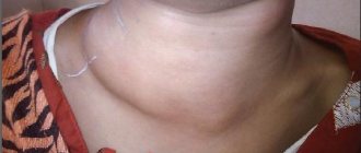
- redness and swelling of the skin behind the ear;
- smoothing the behind-the-ear fold;
- protrusion of the ear forward;
- sometimes - swelling of the neck and pain when turning the head.
It should be noted that at the first stage the disease is almost no different in clinical picture from acute otitis, since the main symptoms are pain in the ear, fever, headache, discharge from the ear, etc. Almost always, acute purulent otitis ends in perforation membranes (spontaneous, artificial) and drainage of the cavity, but the inflammatory process with concomitant mastoiditis continues to gain strength. The pus becomes thick, creamy, fills the cells inside the mastoid process and forms empyema, which gives more severe symptoms.
Symptoms of mastoiditis
Mastoiditis can appear simultaneously with the occurrence of purulent otitis media. But most often it develops 7-14 days from the onset of otitis media. In children of the first year of life, due to the structural features of the mastoid process, mastoiditis manifests itself in the form of otoanthritis. In adults, mastoiditis manifests itself as a pronounced deterioration in general condition with a rise in temperature to febrile levels, intoxication, and headache. sleep disturbance. Patients with mastoiditis complain of noise and pain in the ear, hearing loss, intense pain behind the ear, and a feeling of pulsation in the mastoid area. Pain radiates along the branches of the trigeminal nerve to the temporal and parietal region, orbit, and upper jaw. Less commonly, mastoiditis causes pain in the entire half of the head.
These symptoms of mastoiditis are usually accompanied by profuse suppuration from the external auditory canal. Moreover, the amount of pus is noticeably greater than the volume of the tympanic cavity, which indicates the spread of the purulent process beyond the middle ear. On the other hand, suppuration with mastoiditis may not be observed or may be insignificant. This occurs while maintaining the integrity of the eardrum, closing the perforation in it, and disrupting the outflow of pus from the mastoid process into the middle ear.
Objectively, with mastoiditis, redness and swelling of the area behind the ear, smoothness of the skin fold located behind the ear, and protrusion of the auricle are noted. When pus breaks through into the subcutaneous fatty tissue, a subperiosteal abscess forms, accompanied by severe pain when palpating the postauricular area and a symptom of fluctuation. From the area of the mastoid process, pus, exfoliating the soft tissues of the head, can spread to the occipital, parietal, and temporal regions. Thrombosis of the vessels supplying the cortical layer of the mastoid bone, which occurs as a result of inflammation, leads to necrosis of the periosteum with the breakthrough of pus to the surface of the scalp and the formation of an external fistula.
Main symptoms of inflammation
The acute form of mastoiditis is characterized by the following symptoms:
- Fever;
- General weakness;
- Painful sensations in the ear and behind the auricle;
- Noise in the ears and head;
- Hearing impairment;
- Discharge of pus from the ear.
Mastoiditis affects areas highlighted in red
Increased body temperature and weakness are also characteristic of acute otitis, but in this case these symptoms appear a couple of weeks after its onset. Depending on the stage of mastoiditis, certain signs appear. So at the exudative stage, ear pain, pus discharge, headaches occur, and the temperature can rise to forty degrees.
In an alternative stage, pus may enter the periosteum, under the skin, and come out. A fistula appears, from which purulent contents flow. If pus breaks into the inner ear, labyrinthitis develops. It is characterized by dizziness, tinnitus, and hearing loss.
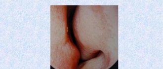
Photo of mastoiditis
The main symptoms of mastoiditis largely depend on the severity and stage of development of the disease. For example, at the initial stages it is very difficult to distinguish inflammation of the mastoid process from ordinary otitis media.
Patients complain of sharp, shooting pain in the ear. There is an increase in temperature, weakness and body aches, and headaches. Discharge appears from the ear canal.
In the absence of therapy or insufficient treatment (for example, stopping antibiotics too quickly), the clinical picture changes. The mastoid process of the ear gradually fills with pus, and under pressure the bone partitions between the cells are destroyed. The skin and subcutaneous tissues behind the auricle swell and turn red, become hard and hot to the touch. The ear pain becomes more severe, and thick purulent masses are discharged from the ear canal.
Inflammation from the mastoid cavities can spread under the periosteum - pus accumulates in the subcutaneous tissue layer. Quite often, the abscess ruptures on its own, resulting in the formation of a fistula on the skin.
Classification of mastoiditis
Depending on the cause, otolaryngology distinguishes between primary and secondary; otogenic, hematogenous and traumatic mastoiditis.
According to the stage of the inflammatory process, mastoiditis is classified as exudative and true (proliferative-alterative).
There are typical and atypical clinical forms of mastoiditis. The atypical (latent) form of mastoiditis is characterized by a slow and sluggish course without pronounced symptoms characteristic of mastoiditis. A separate group of apical mastoiditis is distinguished, which includes Bezold's mastoiditis, Orleans mastoiditis and Mouret's mastoiditis.
How dangerous can the disease be? The most common complications
An easier option for a purulent lesion to break through is to direct it under the skin, when a large swelling appears in the area of the mastoid process and the formation of a fistula. In this case, timely, complete treatment leads to complete recovery without consequences. But the spread of pus in other directions leads to various complications:
- in the area of the inner ear - to labyrinthitis and neuritis of the auditory nerve; Read more about inner ear inflammation
- in the zygomatic process - to phlegmon of the orbit, endophthalmitis, panophthalmitis;
- in the area of the interfascial space of the neck - to phlegmon of the neck, then - to purulent mediastinitis;
- into the cranial cavity - to meningitis, encephalitis, brain abscess, thrombosis of the sigmoid sinus;
- Often with mastoiditis, several paths for the outflow of purulent fluid are formed with serious consequences for humans.
After infection penetrates the sigmoid sinus, sepsis and acute cerebral circulatory disorders most often develop, and it is quite difficult to save a person from death in such a situation.
Mastoiditis can affect the functioning of the facial nerve. As a result, complications develop such as:
- Facial asymmetry.
- Drooping of the edges of the lips or eyes.
- Inability to close the eyelid.
Other possible complications of mastoiditis include inflammation of the brain, accumulation of fluid there, an abscess, and encephalitis. When microorganisms spread through the blood, sepsis occurs. Particles of pus can penetrate into the neck, eyeballs, temporal, parietal and occipital regions.
Why mastoiditis is really dangerous - possible complications
If we talk about visual errors, the most common are those associated with dysfunction of the facial nerve:
- Facial asymmetry.
- Drooping of the edge of the eye/mouth.
- Inability to close the eyelid/eyelids.
When pus gets inside the skull, the patient experiences a severe fever, sleeps poorly, eats little, has nausea and vomiting.
We also recommend
- Familial hypercholesterolemia: gene pathology, possible causes, symptoms, diagnostic tests and treatment
- Review of atrial fibrillation: causes, diagnosis and treatment, why it is dangerous Types of atrial fibrillation
- From the mesenchyme of the dental sac are formed
- Melanoma: clinical symptoms and signs
- Alzheimer's disease: causes and treatment, essence, initial symptoms, development, photo of Alzheimer's disease Who treats Alzheimer's
- What ECG indicators are considered normal: interpretation of examination results
Atypical course of mastoiditis
Atypical forms of mastoiditis pose the greatest risk of incorrect or delayed diagnosis, treatment and the development of complications. The appearance of this type of pathology depends on the pathogen, for example, its drug-resistant forms, on the state of the immune system, on age, the structure of the temporal bone, etc.
The main difference between atypical forms of mastoiditis is the absence of a characteristic clinical picture and a typical staged course. Most often, the patient does not develop pus, ear pain is not noticeable, and the temperature may remain normal. However, with atypical forms of pathology, severe destruction of bone tissue and the appearance of severe complications are most often observed.
The following atypical forms of mastoiditis are distinguished:
- Apical cervical mastoiditis, or Bezold's abscess. Leads to the formation of a purulent focus on the neck under the mastoid process.
- Zygomatitis. It is a lesion of the zygomatic process of the temporal bone.
- Squamite. This disease consists of purulent lesions of the scales of the temporal bone.
- Petrosit. This form of mastoiditis covers the pyramid of the temporal bone.
- Chitelevsky mastoiditis. The angular cells of the process, which come into contact with the posterior cranial fossa, become inflamed. It is a very dangerous disease, as it threatens complications from the brain.
- Dry mastoiditis. In this case, inflammation of the mastoid process passes without the release of pus.
- Other atypical forms caused by mucous streptococcus, actinomycete, fusospirochetosis. They can be blurry and lead to the development of chronic forms of the disease.
Diagnosis of mastoiditis
As a rule, diagnosing mastoiditis does not present any difficulties for an otolaryngologist. Difficulties arise in the case of a low-symptomatic atypical form of mastoiditis. Diagnosis of mastoiditis is based on the patient’s characteristic complaints, anamnestic information about trauma or inflammation of the middle ear, examination and palpation of the postauricular area, and otoscopy results. microotoscopy. audiometry. bacterial culture of ear discharge, computed tomography and x-ray examination.
Otoscopy for mastoiditis reveals inflammatory changes on the side of the eardrum, typical for otitis media; if there is a hole in it, profuse suppuration is noted. A pathognomonic otoscopic sign of mastoiditis is the overhang of the posterior superior wall of the auditory canal. Audiometry and hearing testing with a tuning fork can determine the degree of hearing loss in a patient with mastoiditis.
Sight radiography of the skull in the exudative stage of mastoiditis reveals cells veiled as a result of inflammation and unclearly distinguishable partitions between them. The X-ray picture of the proliferative-alterative stage of mastoiditis is characterized by the absence of a cellular structure of the mastoid process, instead of which one or several large cavities are determined. Better visualization is achieved by performing a CT scan of the skull in the area of the temporal bone.
Treatment of mastoiditis
Treatment of mastoiditis depends on the ethnology of the disease, the stage of development of mastoiditis and the presence of various complications. In case of mastoiditis that has developed against the background of exacerbation of chronic purulent otitis media, according to absolute indications, sanitizing surgery is performed on the middle ear.
Mastoiditis that develops as a result of acute otitis media is treated conservatively or surgically. In the first exudative and uncomplicated stage, conservative treatment is carried out in the first days of the disease, primarily paracentesis of the tympanic membrane and oral antibiotic therapy. When empirically determining the nature and scope of antibacterial therapy (it is considered appropriate to use amoxicillin, clavulanic acid (i-lactamase inhibitor) or cephalosporins of the II-III generation (cefaclor, cefixime, ceftibuten, cefuroxime, ceftriaxone, cefotaxime, etc.).
In the destructive stage of development of the inflammatory process and the mastoid process, especially in its complicated forms, urgent surgical intervention is indicated - anthromastoidotomy with the administration of fluoroquinolones (ciprofloxacin, levofloxacin, moxifloxacin) and parenteral cephalosporins in the postoperative period.
Fluoroquinolones are contraindicated for children under 10 years of age due to the possibility of a destructive effect on the skeletal system; they are predominantly treated with parenteral cephalosporins. In the postoperative period, detoxification intravenous therapy, immune drugs are used, and, if indicated, modern antifungal agents are used.
In the conservative management of initial forms of mastoiditis that developed against the background of acute otitis media, in its uncomplicated: sudsative stage, physiotherapy (UHF, microwave, etc.) is included in the complex of treatment. Warm or cold compresses are recommended for the area behind the ear.
Antibacterial therapy begins immediately after the diagnosis of mastoiditis is confirmed, in the initial stages of its development, and is carried out especially intensively in the postoperative period along with active detoxification, hyposensitizing and immunocorrective treatment and local therapeutic measures.
In case of thrombophlebitis of the sigmoid sinus, which has complicated the course of the Chitell form of mastoiditis, anticoagulants must be included in the complex of treatment. Of the direct anticoagulants, heparin sodium is used, of indirect ones - acenocoumarol, phenindione, etc., always under the control of a thromboelastogram, with simultaneous local use of the drug "Lioton-1000*", heparin or troxevasin ointment*.
In cases of mastoiditis that has complicated the course of chronic otitis media, radical surgery on the middle ear is performed according to urgent indications; for mastoiditis that developed during acute otitis media - anthromastoidotomy.
Further management
Monitoring the patient at the place of residence, carrying out therapeutic measures to prevent relapse of the disease, restorative therapy, correction of immunological disorders.
With timely and rational treatment, conservative and surgical, in the vast majority of cases the prognosis is favorable. With late diagnosis and unfavorable course of the disease, severe intracranial complications and facial nerve paresis may develop.
The choice of treatment method for the disease will depend on its form and severity. Conservative therapy is carried out in a hospital, is used at the very primary stage of the pathology after acute otitis, as well as at the exudative stage without complications and includes:
- UHF, microcurrents to the behind-the-ear area;
- antibacterial treatment using cephalosporins, penicillins and other antibiotics;
- anti-inflammatory and desensitizing therapy;
- taking anticoagulants if there is a tendency to develop thrombosis;
- taking immunomodulators, vitamins, restorative drugs;
- In parallel, it may be recommended to wash the middle ear with antiseptics through the hole in the eardrum.
The indication for surgery is the presence of any purulent complications, including abscesses and phlegmon. Surgical intervention is also necessary if, after conservative treatment, X-rays reveal the progress of bone tissue destruction. The operation will have to be performed for any form of mastoiditis, which has already entered the stage of destructive changes.
Among the operations practiced are anthromastoidectomy (removal of the mastoid process) or anthromastoidotomy (opening of the mastoid process), and the latter type of operation can even be done under local anesthesia. The affected layer of mastoid bone is removed, granulations are removed, and the pus-filled cave is opened.
Often, after opening the mastoid process, it is necessary to remove it with the apex - perform a mastoidectomy. The operation is performed under general anesthesia, and its goal is to completely remove pus, granulations, areas of necrosis and altered bone. In case of extensive purulent process, it is necessary to open all parts of the temporal bone, as well as perform an extended mastoidectomy (in case of intracranial complications).
Mastoiditis is very life-threatening, so it must be treated immediately as soon as the doctor makes this diagnosis.
At the exudative stage, when bone destruction has not yet occurred and the outflow of purulent contents has not been disrupted, treatment with medications in a hospital setting is indicated. In this case, antibiotics are prescribed that have a broad spectrum of action, for example, penicillins or cephalosporins, and local antibiotics. In addition, drainage of the auditory tube may be required to ensure the best outflow of purulent contents.
Indications for surgery (anthromastoidotomy):
- Intracranial complications;
- Abscess;
- Labyrinthitis;
- Breakthrough of pus through the mastoid process;
- Otogenic paresis and so on.
Antromastoidotomy is performed to remove pus from the mastoid process and drain the tympanic cavity. After the operation, the person is prescribed antibiotics, vitamins, immunomodulators, and local ultraviolet treatment. The wound is bandaged every day, the turundas are removed from it, dried and washed with an antiseptic.
The following complications may occur after anthromastoidotomy:
- Flow of pus from the ear.
- Changes in auditory perception.
- Inflammation of the meninges, abscess.
- Temporary dizziness.
- Temporary loss of taste on one side of the tongue.
- Damage to the facial nerve, paralysis of one side of the face.
Treatment
When treating mastoiditis, it is impossible to do without antibiotic therapy and the use of painkillers. In case of damage to the bone tissue of the mastoid process, surgical intervention is necessary. This operation is a puncture of the eardrum and is called myringotomy. This operation ensures the outflow of pus, the nature of the microflora of which is determined in the laboratory.
When the use of conservative methods does not bring positive results, and changes occur in the brain, mastoidectomy is used. During this operation, the posterior wall of the auditory canal is removed; the stapes and facial nerve are usually not affected during this operation. But it is worth remembering that this operation can lead to hearing impairment, loss of taste buds, and can also cause paralysis of half the face. Therefore, treatment of mastoiditis should begin as early as possible.
Treatment with folk remedies can only be used as an addition to complex therapy. One of the effective methods for getting rid of ear pain is rinsing with infusion of rose petals.
Traditional medicine says that you can get rid of ear pain by warming it up. To do this, you need to heat the salt, wrap it in a bag and apply it to the sore ear, this should relieve the pain.
Alternative medicine methods also include instilling a strong infusion of wormwood mixed with milk into the ear in a 1:1 ratio. But it is worth noting that the effectiveness of these methods has not been proven, and their use instead of qualified treatment by a competent doctor can lead to serious consequences.
Carrying out diagnostics
An experienced doctor, based on a typical clinic and during an examination and history taking, will make a diagnosis at the first appointment. If the form of the pathology is atypical, then additional examination will be required:
- otoscopy;
- audiometry;
- microotoscopy;
- clinical blood test;
- examination of discharge from the ear;
- X-ray of the head;
- CT scan of the temporal bone;
- MRI of the brain;
- ophthalmoscopy, etc.
Usually, during otoscopy, the patient reveals inflammation of the eardrum and copious discharge of pus. It is necessary to differentiate the disease from otitis, cysts and purulent fistulas in the ear area, with tuberculous manifestations. If mastoiditis has already caused complications, emergency surgery may be required.
Based on characteristic general and local otoscopic signs, data from palpation and percussion of the mastoid process, and radiography of the temporal bones in the Schüller projection; in doubtful cases, if differential diagnosis is necessary with lesions of the mastoid process of another etiology, CT or MRI is performed.
The medical history includes information about previous ear diseases, treatment, and the frequency of exacerbations of otitis media during its recurrent or chronic course; circumstances and reasons for the development of this disease, the severity of the disturbance in general condition, temperature reaction, the amount of previous emergency medical care.
Clinical blood test, pus smear from the ear canal and from the mastoid cavity for microflora and sensitivity to antibiotics.
Instrumental studies
Otoscopy, diagnostic paracentesis of the tympanic membrane for mastoiditis that developed against the background of acute otitis media.
Carry out with external otitis, furuncle of the auditory canal, purulent parotid lymphadenitis, suppuration of congenital parotid cysts and fistulas; with apical mastoiditis - with other sources of formation of phlegmon of the neck, with tuberculous leaks.
Diagnosis and treatment
Mastoiditis is an infectious inflammation that develops in the process of the cranial temporal bone, and therefore it needs to be identified as soon as possible. It is much easier to diagnose acute mastoiditis, but in this case it is necessary to act very quickly. Symptoms of the chronic form of the disease may remain invisible for a long time, and it is this feature that poses the greatest danger.
For diagnosis, it is necessary to check the patient’s hearing organs and examine the condition of the cells of the problem bone. To begin, the doctor does the following:
- Anamnesis collection. These are patient complaints and identification of superficial symptoms, such as swelling, hyperemia, and a significant amount of discharge from the ear.
- Palpation. Feeling the swelling behind the ear, identifying pain, fluctuating infiltrate (boil), etc. The penetration of bacteria into the fatty tissue increases the pain. Visually, in addition to swelling and the development of infiltration, protrusion of the auricle is observed. When an abscess develops, a hole with purulent discharge forms on the surface of the skin, and a fistula is formed.
- Otoscopy and microotoscopy. Examination of the ear to determine the extent of damage to the soft tissues of the organ.
- Bac sowing. By analyzing the secretions, the type of bacteria that provoked the inflammation is determined, as well as the degree of their virulence (pathogenicity).
After establishing a suspicion of mastoiditis, auxiliary hardware tests are performed:
- audiometry and tuning forks;
- radiography;
- MRI and CT.
An MRI image makes it possible to examine the internal filling of the temporal bone. In the purulent stage, the cells are almost completely destroyed and filled with turbid contents, which is determined by the blurriness of this area on an x-ray.
It is also necessary to undergo additional examinations by specialized specialists, from a dentist to a thoracic surgeon, to detect complications.
Treatment of mastoiditis consists of eliminating unpleasant symptoms, the consequences of bacterial activity and exudate from the bone cavity. This impact is carried out comprehensively. Drug therapy consists of taking the following groups of medications:
- broad spectrum antibiotics;
- anti-inflammatory and antipyretic drugs;
- analgesics;
- antihistamines to relieve swelling.
Additionally, symptoms of body intoxication are eliminated and immunocorrection is carried out to maintain natural defenses. In some cases, physiotherapy is indicated as a restorative course of health procedures.
Treatment of mastoiditis in most cases combines approaches such as drug therapy and surgery. It is the operation that eliminates the main problem - purulent exudate. To clean mastoiditis, the temporal bone is opened and subsequent shunting and sanitation of the cells are performed. Mastoidotomy in combination with drainage allows you to quickly eliminate the pathogenic environment and disinfect the affected tissue.
In some cases, at the initial stage of the disease, the problem can be solved by paracentesis of the ear membrane. This will reduce the pressure of the secretions on the bone cells. After paracentesis, the middle ear cavity and the process itself are treated with medications.
Prognosis and prevention
Prevention of mastoiditis is closely related to the prevention of acute otitis media, the need for qualified treatment of chronic otitis media, and, if necessary, with timely sanitizing hearing-preserving operations on the middle ear. It is important to increase the body's resistance, timely sanitation of the nasal cavity, nasopharynx and pharynx, care of the nasal and oral cavity and treatment of inflammatory diseases that arise in them, early diagnosis of inflammatory diseases of the middle ear and carrying out comprehensive rational anti-inflammatory therapy.
Mastoiditis is typical for children, elderly people, patients with diabetes mellitus and HIV infection.
In childhood, the mastoid process is a single cavity without any bridges. An infection can very easily get there, resulting in otitis media. Young children are most susceptible to various colds, which can also be the onset of mastoiditis.
In diabetics and older people, sensitivity is lost, so it is not always possible to notice the symptoms of mastoiditis. It is these groups of people who are susceptible to all sorts of complications. Most often, they take various painkillers, which makes diagnosing mastoiditis more difficult.
Mastoiditis can be prevented by timely diagnosis of otitis media. In addition, preventive measures include:
- Correct and timely treatment of inflammatory diseases of the ENT organs.
- Timely elimination of all foci of infection in the body.
- Boosting immunity.
- Proper nutrition.
- Hardening.
- Regular moderate physical activity.
With timely treatment, including surgery, the prognosis is favorable. Poor outcome is usually observed in the presence of sepsis and intracranial complications. To prevent mastoiditis, it is important to properly treat otitis media, prevent head and ear injuries, promptly eliminate all foci of infection from the nasopharynx, and increase immunity.
In the next video, Elena Malysheva will tell you how to treat mastoiditis and what to do if it hurts behind the ear.
This disease is understood as inflammation of the middle ear, which develops as a result of the transition of the inflammatory process from the nasopharynx to the mucous membrane of the auditory tube and the tympanic cavity. Synonyms for middle ear catarrh are exudative otitis media, salpingootitis, tubo-otitis, tubotympanitis, tubotympanic catarrh, secretory otitis.
Depending on the composition of the exudate, serous-catarrhal and purulent-catarrhal inflammation are distinguished.
Etiology and pathogenesis. The main cause of catarrhal inflammation of the middle ear is inflammation of the mucous membrane of the auditory tube and disruption of its ventilation function. Inflammation of the auditory tube, in turn, occurs as a result of the spread of infection into it from the nasopharynx (adenoiditis, nasopharyngitis, etc.).
The etiological factor of the inflammatory process in the nasopharynx can be streptococci, staphylococci, pneumococci or microbial mixtures. As a result of disruption of the ventilation function of the auditory tube and the occurrence of low pressure in the tympanic cavity, interstitial fluid leaks into the tympanic cavity.
Symptoms and clinical picture: ear congestion, tinnitus, autophony and hearing loss of varying degrees, ear pain. Otoscopic signs correspond to the stages of development of the inflammatory process (Fig. 1).
Rice. 1. Types of the tympanic membrane at different stages of development of acute catarrhal inflammation of the middle ear: 1 - transudate in the lower part of the tympanic cavity, 2 - injection of vessels in the relaxed part of the tympanic membrane and along the handle of the malleus, 3 - radial injection of vessels of the tympanic membrane
The stage of hyperemia is characterized by the injection of blood vessels along the handle of the malleus, retraction and radial injection of the vessels of the tympanic membrane, and shortening of the cone of light. At the stage of catarrhal inflammation, an effusion of a different nature (dull gray or xanthomatous) appears in the tympanic cavity. If the exudate is hemorrhagic, the eardrum becomes bluish or purple in color.
Characteristic signs of the disease: retraction of the eardrum, in which the handle of the malleus acquires an almost horizontal position, and its short process protrudes sharply into the lumen of the ear canal (index finger symptom); the relaxed part, if it is not protruded by effusion, is retracted and directly adjacent to the medial wall of the epitympanic space, the light cone is sharply shortened or completely absent.
A hearing test reveals a conductive type of hearing loss, predominantly at low frequencies. When the form is complicated by acute purulent otitis media, preceptive hearing loss also occurs due to intoxication of the inner ear. A hearing test using live speech reveals a decrease in hearing for low-octave words, while whispered speech can be perceived at the sink, or from a distance of no more than 1-2 m, spoken speech - 3-6 m.
Outcomes: self-healing, rapid recovery with targeted treatment, recovery with residual effects in the form of intratympanic scars and the transition of the process to tympanosclerosis, infection of the exudate and the development of acute purulent otitis media. Most often, with timely treatment, the disease is eliminated without a trace within 1-2 weeks.
The diagnosis is based on complaints and otoscopic picture. The disease should be differentiated from acute purulent inflammation of the middle ear in the pre-perforation phase, which is characterized by severe pain in the ear and a number of other clinical and otoscopic symptoms described below. It is more difficult to differentiate this disease from latent forms of otitis in infants and acute inflammation of the middle ear in the elderly.
The prognosis depends on the nature of the pathological condition of the nasopharynx and auditory tube, the general allergic background, virulence and the quality of treatment measures.
Treatment: elimination of chronic foci of infection in the upper respiratory tract; carrying out therapeutic measures in the presence of an allergic background and chronic inflammatory processes in the paranasal sinuses; normalization of nasal breathing in the presence of obstructive pathology (polyps, deviated nasal septum, hypertrophic rhinitis, etc.);
Local treatment: administration of vasoconstrictor solutions and aerosols (naphthyzin, sanorin, galazolin, etc.) into the nose; blowing the auditory tubes with preliminary anemization of their pharyngeal mouth; injection of a hydrocortisone suspension into the auditory tube; if there is viscous content in the tympanic cavity, a freshly prepared proteolytic enzyme is introduced into it through the auditory tube;
orally - antihistamines and decongestants (diphenhydramine, diazolin, pipolfen, etc.) in combination with ascorbic acid and calcium gluconate; if purulent complications are suspected (the appearance of throbbing pain in the ear, increased hyperemia of the eardrum and its protrusion), broad-spectrum antibiotics are prescribed orally.
To quickly resolve the contents of the tympanic cavity, various physiotherapeutic procedures are used (warming compress, Sollux, UHF, laser therapy, etc.).
The disease is characterized by inflammation of the mucous membrane of the tympanic cavity, including the cave and auditory tube. It occurs mainly when the infection spreads hematogenously from distant foci and in severe general infectious diseases during the period of rash. The infection can also enter the tympanic cavity from the external auditory canal, but only if the integrity of the eardrum is damaged. The disease most often occurs in childhood and adolescence.
Etiology and pathogenesis. The disease develops most often against the background of acute respiratory infections and influenza. The etiological factors are hemolytic streptococcus, staphylococcus, often in combination with Pseudomonas aeruginosa, Proteus vulgaris and various types of Escherichia coli.
The occurrence of the disease is facilitated by many reasons: adenoiditis, tubootitis, rhinosinusitis, chronic tonsillitis, ozena. Often the disease occurs when there is a “dry” perforation of the eardrum after washing the external auditory canal or bathing or showering.
A number of unfavorable conditions in the working environment also contribute to inflammatory diseases of the ear: changes in atmospheric pressure (for divers, pilots, submariners, caisson workers), dampness, cooling, fatigue, etc.
Pathological anatomy. At the onset of the disease, the mucous membrane of the tympanic cavity is hyperemic and infiltrated. With the development of inflammation, it thickens greatly and hemorrhages occur in it. At the same time, serous and purulent exudate accumulates in the tympanic cavity, protruding the eardrum (Fig. 2).
Rice. 2. Types of tympanic membrane in two forms of acute otitis media: 1 - diffuse acute otitis media (mesotympanic form); 2 - acute otitis media (epitympanic form)
Subsequently, at the height of clinical manifestations, a focus of softening appears in the eardrum, and due to the pressure of the exudate, a perforation occurs in this place, most often slit-like, which during otoscopy reveals itself as a pulsating reflex. During recovery, inflammatory phenomena in the mucous membrane subside, hyperemia decreases, exudate from the tympanic cavity resolves or is partially evacuated through the auditory tube.
The perforated hole is closed with a scar or transformed into a persistent perforation with a compacted connective tissue edge. A perforation that occurs in the stretched part of the eardrum is called rim or central. Perforation in the area of the relaxed part is called marginal (in the epitympanic form of otitis) (Fig. 3).
Prevention of mastoiditis
Prevention of otogenic mastoiditis comes down to timely diagnosis of inflammatory lesions of the middle ear and adequate treatment of otitis media.
timely paracentesis of the eardrum and sanitizing operations. Correct treatment of nasopharyngeal diseases and rapid elimination of infectious foci also help prevent mastoiditis. In addition, it is important to increase the efficiency of the body’s immune mechanisms, which is achieved by maintaining a healthy lifestyle, proper nutrition, and, if necessary, immunocorrective therapy. CONTACTS